Photoacoustic Tomography Detects Early Vessel Regression and Normalization During Ovarian Tumor Response to the Antiangiogenic Therapy Trebananib
- PMID: 26315834
- PMCID: PMC5612481
- DOI: 10.2967/jnumed.115.160002
Photoacoustic Tomography Detects Early Vessel Regression and Normalization During Ovarian Tumor Response to the Antiangiogenic Therapy Trebananib
Abstract
The primary aim of this study was to assess the potential of in vivo photoacoustic tomography for direct functional measurement of ovarian tumor response to antiangiogenic therapy.
Methods: In vivo studies were performed with institutional animal care and use committee approval. We used an orthotopic mouse model of ovarian cancer treated with trebananib (n = 9) or vehicle (n = 9). Tumor-bearing mice were randomized into trebananib or vehicle groups at day 10 and dosed on days 12, 15, and 18 after implantation. Photoacoustic tomography and blood draws were performed at day 10 and then 24 h after each drug dose. Tumors were excised for histopathology after the final studies on day 19. Data analysis to test for statistical significance was performed blinded.
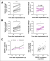
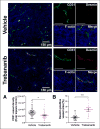
Results: Blockade of angiopoietin signaling using trebananib resulted in reduced total hemoglobin-weighted photoacoustic signal (n = 9, P = 0.01) and increased oxyhemoglobin-weighted photoacoustic signal (n = 9, P < 0.01). The latter observation indicated normalization of the residual tumor vessels, which was also implied by low levels of angiopoietin 1 in serum biomarker profiling (0.76 ± 0.12 ng/mL). These noninvasive measures reflected a 30% reduction in microvessel density and increased vessel maturation in ex vivo sections.
Conclusion: Photoacoustic tomography is able to evaluate both vessel regression and normalization in response to trebananib. Noninvasive imaging data were supported by modulation of serum markers in vitro and ex vivo histopathology.
Keywords: angiogenesis; angiopoietin; ovarian cancer; photoacoustics.
© 2015 by the Society of Nuclear Medicine and Molecular Imaging, Inc.
Conflict of interest statement
No other potential conflict of interest relevant to this article was reported.
Figures


Comment in
-
Commentary on Photoacoustic Tomography.J Nucl Med. 2015 Dec;56(12):1815-6. doi: 10.2967/jnumed.115.165183. Epub 2015 Sep 17. J Nucl Med. 2015. PMID: 26383150 No abstract available.
Similar articles
-
Commentary on Photoacoustic Tomography.J Nucl Med. 2015 Dec;56(12):1815-6. doi: 10.2967/jnumed.115.165183. Epub 2015 Sep 17. J Nucl Med. 2015. PMID: 26383150 No abstract available.
-
Anti-angiopoietin therapy with trebananib for recurrent ovarian cancer (TRINOVA-1): a randomised, multicentre, double-blind, placebo-controlled phase 3 trial.Lancet Oncol. 2014 Jul;15(8):799-808. doi: 10.1016/S1470-2045(14)70244-X. Epub 2014 Jun 17. Lancet Oncol. 2014. PMID: 24950985 Clinical Trial.
-
Efficacy of trebananib (AMG 386) in treating epithelial ovarian cancer.Expert Opin Pharmacother. 2016;17(6):853-60. doi: 10.1517/14656566.2016.1161027. Epub 2016 Mar 21. Expert Opin Pharmacother. 2016. PMID: 26933765 Review.
-
Trebananib or placebo plus carboplatin and paclitaxel as first-line treatment for advanced ovarian cancer (TRINOVA-3/ENGOT-ov2/GOG-3001): a randomised, double-blind, phase 3 trial.Lancet Oncol. 2019 Jun;20(6):862-876. doi: 10.1016/S1470-2045(19)30178-0. Epub 2019 May 7. Lancet Oncol. 2019. PMID: 31076365 Clinical Trial.
-
Advances in anti-angiogenic agents for ovarian cancer treatment: The role of trebananib (AMG 386).Crit Rev Oncol Hematol. 2015 Jun;94(3):302-10. doi: 10.1016/j.critrevonc.2015.02.001. Epub 2015 Mar 4. Crit Rev Oncol Hematol. 2015. PMID: 25783620 Review.
Cited by
-
Imaging methods to evaluate tumor microenvironment factors affecting nanoparticle drug delivery and antitumor response.Cancer Drug Resist. 2021;4(2):382-413. doi: 10.20517/cdr.2020.94. Epub 2021 Jun 19. Cancer Drug Resist. 2021. PMID: 34796317 Free PMC article.
-
Chemotherapeutic effects on breast tumor hemodynamics revealed by eigenspectra multispectral optoacoustic tomography (eMSOT).Theranostics. 2021 Jun 26;11(16):7813-7828. doi: 10.7150/thno.56173. eCollection 2021. Theranostics. 2021. PMID: 34335966 Free PMC article.
-
Multivalent fusion protein targeting VEGFR2 and DR5 receptors: assessing the antiangiogenic and antitumor effects via multimodal microangiography.J Transl Med. 2025 Aug 21;23(1):949. doi: 10.1186/s12967-025-06859-8. J Transl Med. 2025. PMID: 40841907 Free PMC article.
-
Copper Sulfide Nanodisks and Nanoprisms for Photoacoustic Ovarian Tumor Imaging.Part Part Syst Charact. 2019 Aug;36(8):1900171. doi: 10.1002/ppsc.201900171. Epub 2019 Jun 2. Part Part Syst Charact. 2019. PMID: 32863594 Free PMC article.
-
Intraoperative Imaging in Hepatopancreatobiliary Surgery.Cancers (Basel). 2023 Jul 20;15(14):3694. doi: 10.3390/cancers15143694. Cancers (Basel). 2023. PMID: 37509355 Free PMC article. Review.
References
-
- Laufer J, Johnson P, Zhang E, et al. In vivo preclinical photoacoustic imaging of tumor vasculature development and therapy. J Biomed Opt. 2012;17:056016. - PubMed
-
- Herzog E, Taruttis A, Beziere N, et al. Optical imaging of cancer heterogeneity with multispectral optoacoustic tomography. Radiology. 2012;263:461–468. - PubMed
Publication types
MeSH terms
Substances
Grants and funding
LinkOut - more resources
Full Text Sources
Other Literature Sources
Medical
