Review
. 2015 Oct;47(4):1189-202.
doi: 10.3892/ijo.2015.3119. Epub 2015 Aug 12.

Deficient mismatch repair: Read all about it (Review)

Affiliations
Review

Deficient mismatch repair: Read all about it (Review)

Susan Richman. Int J Oncol. 2015 Oct.

Abstract

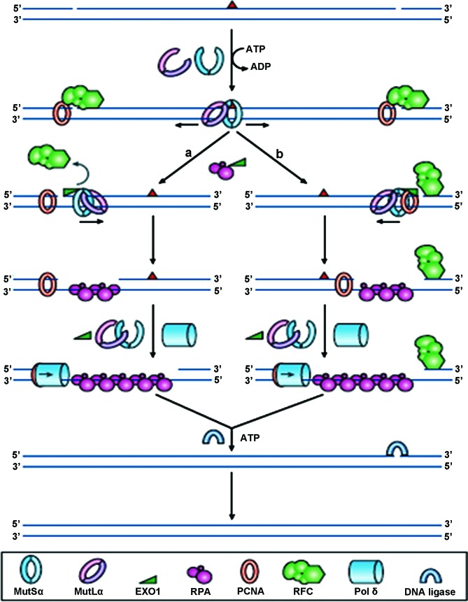
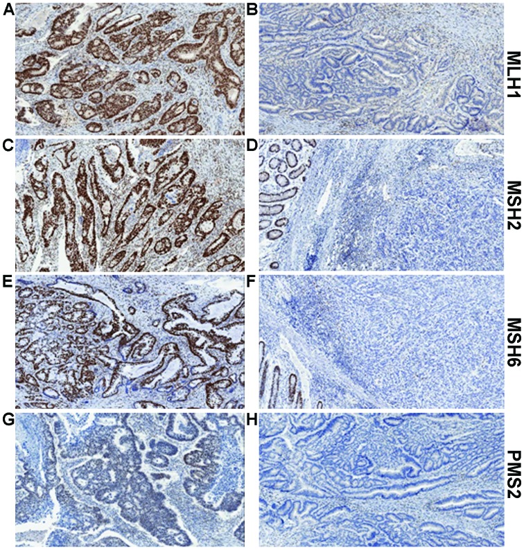
Defects in the DNA mismatch repair (MMR) proteins, result in a phenotype called microsatellite instability (MSI), occurring in up to 15% of sporadic colorectal cancers. Approximately one quarter of colon cancers with deficient MMR (dMMR) develop as a result of an inherited predisposition syndrome, Lynch syndrome (formerly known as HNPCC). It is essential to identify patients who potentially have Lynch syndrome, as not only they, but also family members, may require screening and monitoring. Diagnostic criteria have been developed, based primarily on Western populations, and several methodologies are available to identify dMMR tumours, including immunohistochemistry and microsatellite testing. These criteria have provided evidence supporting the introduction of reflex testing. Yet, it is becoming increasingly clear that tests have a limited sensitivity and specificity and may yet be superseded by next generation sequencing. In this review, the limitations of diagnostic criteria are discussed, and current and emerging screening technologies explained. There is now useful evidence supporting the prognostic and predictive value of dMMR status in colorectal tumours, but much less is known about their value in extracolonic tumours, that may also feature in Lynch syndrome. This review assesses current literature relating to dMMR in endometrial, ovarian, gastric and melanoma cancers, which it would seem, may benefit from large-scale clinical trials in order to further close the gap in knowledge between colorectal and extracolonic tumours.


Figures

Figure 1
Figure 1
Reprinted by permission from Macmillan Publishers Ltd., Nature Reviews Molecular Cell Biology, 7 (5): 335–346, copyright 2006. DNA mismatch repair. In normal cells, any mismatched base pairs (or incorrect insertion or deletion loops) are repaired by the complex machinery which forms the DNA mismatch repair process. MSH2 and MSH6 form a heterodimeric complex, called mutSα, which identifies and binds to the error, resulting in an ATP-dependent conformational change, which recruits mutLα, a heterodimer consisting of MLH1 and PMS2. The resultant complex undergoes an ATP-driven conformational alteration, releasing it from the error site. If it diffuses upstream, it displaces replication factor C (RFC) and loads exonuclease-1 (EXO1). This degrades the strand in the 5′→3′ direction. Replication factor A (RPA) then stabilises the single-stranded DNA, while a complex of DNA polymerase Pol δ (Pol δ) and proliferating cell nuclear antigen (PCNA) fills the gap and finally DNA ligase seals the remaining nick to finalise the repair. If the mutSα/mutLα complex diffuses downstream, EXO1 is recruited and degrades the region of the DNA strand, up to the RFC complex. As stated before, the single-strand is stabilised by bound RPA, which also inhibits EXO1 activity. Pol δ fills the gap and finally DNA ligase I seals the remaining nick to finalise the repair.
Figure 2
Figure 2
Examples of MLH1, MSH2, MSH6 and PMS2 immunohistochemistry. (A) Positive MLH1 staining and (B) absence of MLH1 staining in tumour epithelium yet showing the positive internal control staining of lymphocytes in the stroma. (C) Positive MLH2 staining and (B) absence of MLH2 staining in tumour epithelium, yet showing positive staining in the adjacent normal colonic epithelium. (E) Positive MSH6 staining and (F) absence of MSH6 staining in tumour epithelium yet with positive staining in the adjacent normal colonic epithelium. (G) Positive PMS2 staining and (H) absence of PMS2 staining in tumour epithelium yet with positive internal control staining of lymphocytes in the stroma.

Similar articles

Cited by

References

    1. Nilbert M, Planck M, Fernebro E, Borg A, Johnson A. Microsatellite instability is rare in rectal carcinomas and signifies hereditary cancer. Eur J Cancer. 1999;35:942–945. doi: 10.1016/S0959-8049(99)00045-3. - DOI - PubMed
    1. Sinicrope FA, Rego RL, Foster N, Sargent DJ, Windschitl HE, Burgart LJ, Witzig TE, Thibodeau SN. Microsatellite instability accounts for tumor site-related differences in clinicopathologic variables and prognosis in human colon cancers. Am J Gastroenterol. 2006;101:2818–2825. doi: 10.1111/j.1572-0241.2006.00845.x. - DOI - PubMed
    1. Jass JR. Classification of colorectal cancer based on correlation of clinical, morphological and molecular features. Histopathology. 2007;50:113–130. doi: 10.1111/j.1365-2559.2006.02549.x. - DOI - PubMed
    1. Fearon ER, Vogelstein B. A genetic model for colorectal tumorigenesis. Cell. 1990;61:759–767. doi: 10.1016/0092-8674(90)90186-I. - DOI - PubMed
    1. Thibodeau SN, French AJ, Cunningham JM, Tester D, Burgart LJ, Roche PC, McDonnell SK, Schaid DJ, Vockley CW, Michels VV, et al. Microsatellite instability in colorectal cancer: Different mutator phenotypes and the principal involvement of hMLH1. Cancer Res. 1998;58:1713–1718. - PubMed