. 2015 Oct;93(4):92.
doi: 10.1095/biolreprod.115.129098. Epub 2015 Aug 26.

In utero exposure to di-(2-ethylhexyl) phthalate induces testicular effects in neonatal rats that are antagonized by genistein cotreatment

Affiliations

In utero exposure to di-(2-ethylhexyl) phthalate induces testicular effects in neonatal rats that are antagonized by genistein cotreatment

Steven Jones et al. Biol Reprod. 2015 Oct.

Abstract

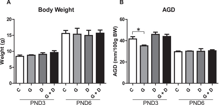
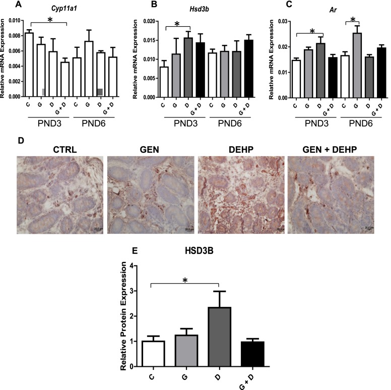
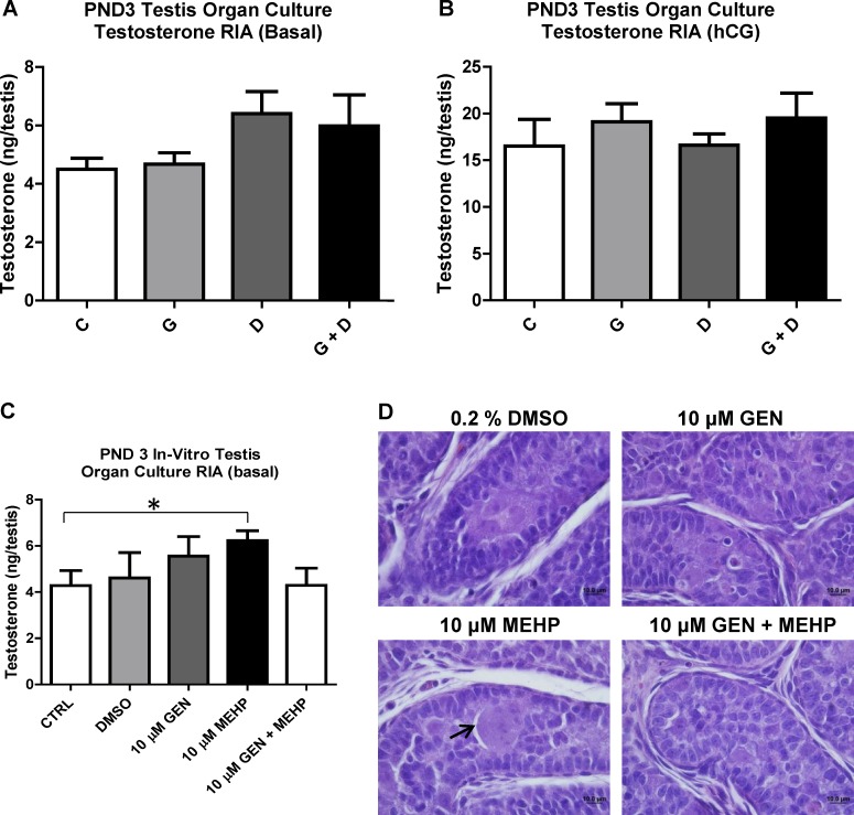
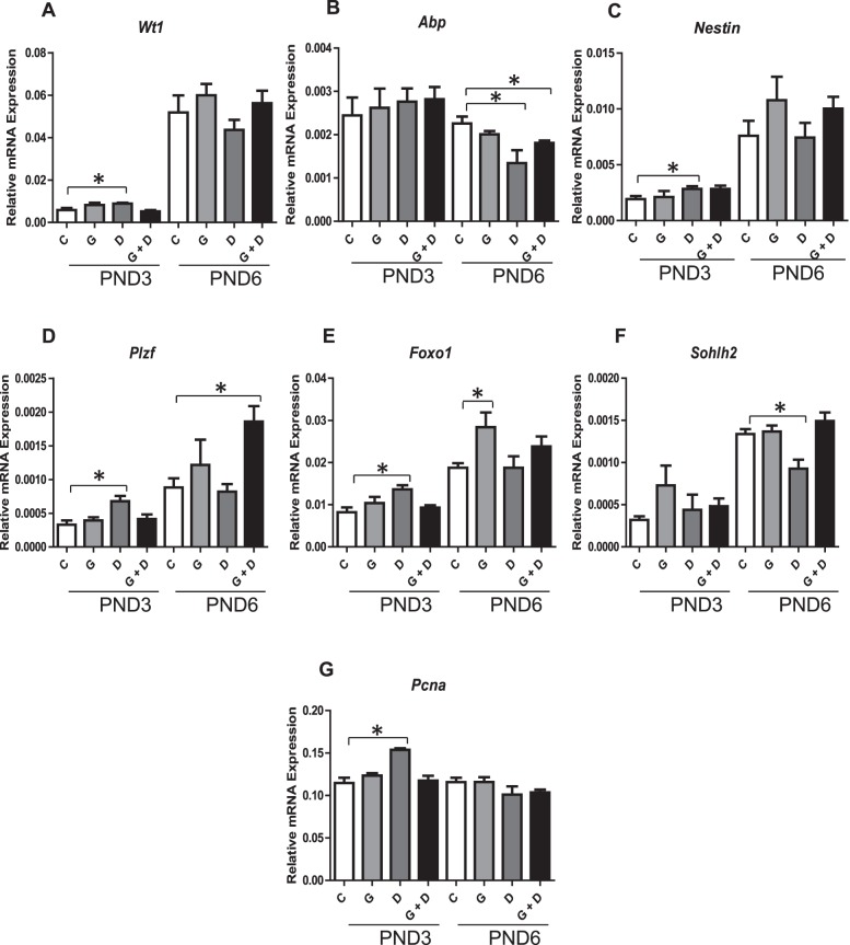
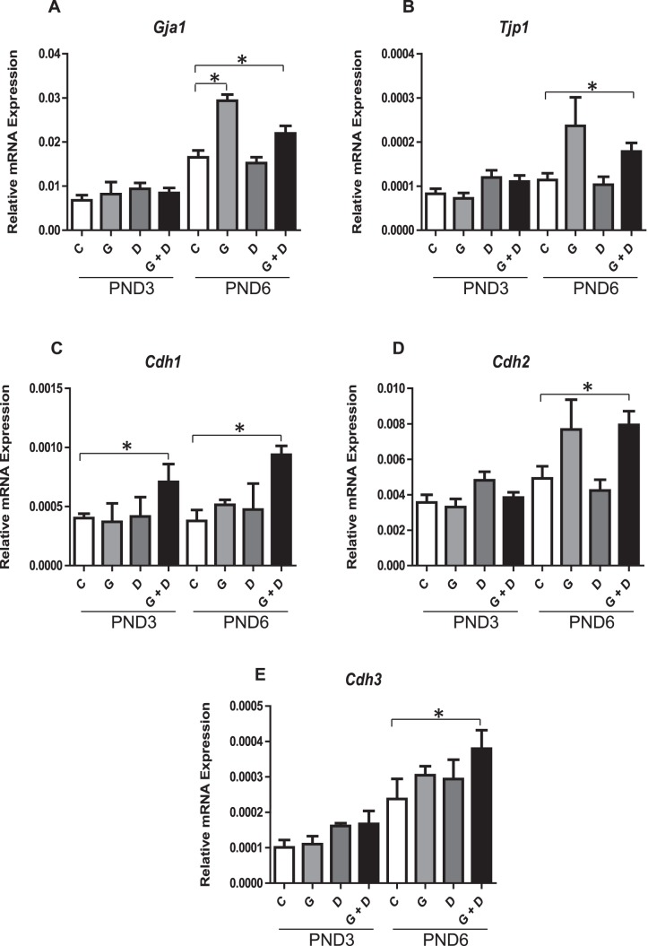
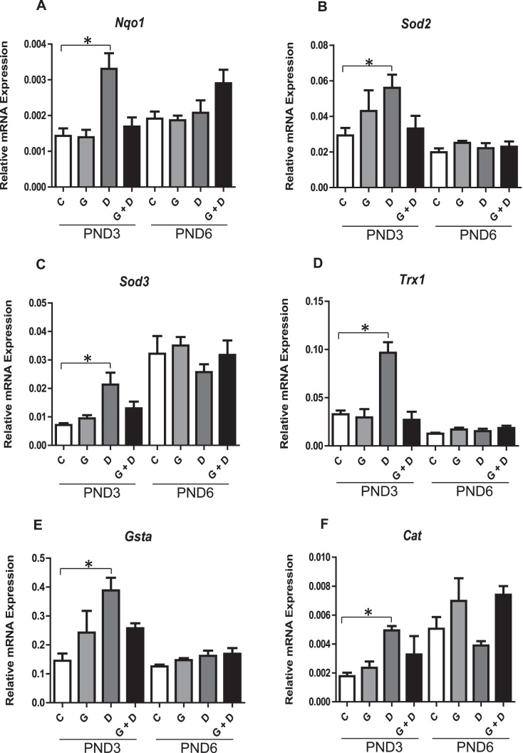
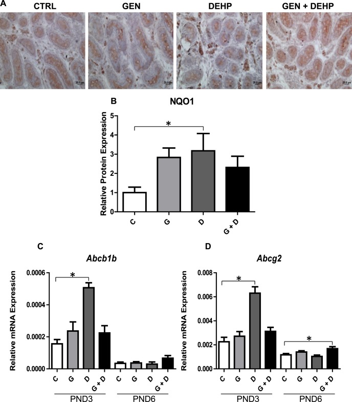
Fetal exposure to endocrine disruptors (EDs) is believed to predispose males to reproductive abnormalities. Although males are exposed to combinations of chemicals, few studies have evaluated the effects of ED mixtures at environmentally relevant doses. Our previous work showed that fetal exposure to a mixture of the phytoestrogen genistein (GEN) and the plasticizer di-(2-ethylhexyl) phthalate (DEHP) induced unique alterations in adult testis. In this follow-up study, we examined Postnatal Day 3 (PND3) and PND6 male offspring exposed from Gestational Day 14 to parturition to corn oil, 10mg/kg GEN, DEHP, or their combination, to gain insight into the early molecular events driving long-term alterations. DEHP stimulated the mRNA and protein expression of the steroidogenic enzyme HSD3B, uniquely at PND3. DEHP also increased the mRNA expression of Nestin, a Leydig progenitor/Sertoli cell marker, and markers of Sertoli cell (Wt1), gonocyte (Plzf, Foxo1), and proliferation (Pcna) at PND3, while these genes were unchanged by the mixture. Redox (Nqo1, Sod2, Sod3, Trx, Gst, Cat) and xenobiotic transporter (Abcb1b, Abcg2) gene expression was also increased by DEHP at PND3, while attenuated when combined with GEN, suggesting the involvement of cellular stress in short-term DEHP effects and a protective effect of GEN. The direct effects of GEN and mono-(2-ethylhexyl) phthalate, the principal bioactive metabolite of DEHP, on testis were investigated in PND3 organ cultures, showing a stimulatory effect of 10 μM mono-(2-ethylhexyl) phthalate on basal testosterone production that was normalized by GEN. These effects contrasted with previous reports of androgen suppression and decreased gene expression in perinatal rat testis by high DEHP doses, implying that neonatal effects are not predictive of adult effects. We propose that GEN, through an antioxidant action, normalizes reactive oxygen species-induced neonatal effects of DEHP. The notion that these EDs do not follow classical dose-response effects and involve different mechanisms of toxicity from perinatal ages to adulthood highlights the importance of assessing impacts across a range of doses and ages.

Keywords: Leydig; ROS; Sertoli; endocrine disruptor; gene expression; genistein; germ cells; gonadal function; mixture; phthalate; rodents (mice, guinea pigs, rats, voles); testis; toxicology.


Figures

FIG. 1
FIG. 1
Effects of in utero exposure to genistein (GEN) and DEHP on general and reproductive health parameters. PND3 and PND6 average body weight (A) and anogenital distance (AGD) normalized to body weight (B). Data are represented as mean measurements (± SEM) of parameters measured in the offspring of four dams per treatment. Asterisk indicates a significant difference relative to control in respective age groups (P ≤ 0.05); C, control; G, GEN; D, DEHP; G + D, GEN + DEHP.
FIG. 2
FIG. 2
Effects of in utero exposure to genistein (GEN) and DEHP on steroidogenic mediators. Relative mRNA expression of steroidogenic enzymes Cyp11a1 (A) and Hsd3b (B), and androgen receptor (Ar) (C) in PND3 and PND6 testes. Data are expressed as mean relative mRNA levels ± SEM normalized to alpha-tubulin (n = 4 per treatment). To further validate mRNA alterations, immunohistochemical analysis of HSD3B (D) was performed on PND3 testes and positive interstitial staining was quantified by image analysis (E); photos taken at 40× magnification. Immunohistochemical quantification is represented by mean fold change ± SEM of HSD3B staining relative to control (n ≥ 3). Omission of the primary antibody was used as a negative control (data not shown). Representative pictures are presented. Asterisks in both mRNA and protein analyses indicate a significant difference relative to control (P ≤ 0.05); C, control; G, GEN; D, DEHP; G + D, GEN + DEHP.
FIG. 3
FIG. 3
Effects of in utero and in vitro exposure to genistein (GEN) and DEHP on PND3 testes T production. Ex vivo testis organ culture was performed over 3 days in basal (A) or hCG-containing (B) medium using PND3 testes from in utero treated offspring. C) Control (untreated) PND3 testes were also treated in vitro over 3 days with either plain medium (control), vehicle (0.2% DMSO), 10 μM GEN, 10 μM MEHP, or combined 10 μM GEN + MEHP. Testosterone levels in supernatant medium for both ex vivo and in vitro organ cultures were determined by radioimmunoassay (RIA) and expressed in ng/testes. Graphs represent the sum of T produced over 3 days (supernatant collected once daily). Asterisk indicates a significant difference relative to control (P ≤ 0.05, n = 4). To assess histological alterations, testes from ex vivo (data not shown) and in vitro organ cultures (D) were collected for processing and hematoxylin and eosin staining (photos taken at 100× magnification). Arrow indicates multinucleated germ cell. Representative pictures are presented; C, control; G, GEN; D, DEHP; G + D, GEN + DEHP.
FIG. 4
FIG. 4
Effects of in utero exposure to genistein (GEN) and DEHP on Sertoli, proliferation, and early germ cell markers. Relative mRNA expression of Sertoli cell markers Wt1 (A) and Abp (B), Sertoli cell and Leydig marker Nestin (C), early germ cell markers Plzf (D), Foxo1 (E), and Sohlh2 (F), and proliferation marker Pcna (G) in PND3 and PND6 testes. Data are expressed as mean relative mRNA levels ± SEM normalized to alpha-tubulin (n = 4 per treatment). Asterisks indicate a significant difference relative to control (p ≤ 0.05); C, Control; G, GEN; D, DEHP; G + D, GEN + DEHP.
FIG. 5
FIG. 5
Effects of in utero exposure to genistein (GEN) and DEHP on cellular junctions. Relative mRNA expression of gap junction Gja1 (A), tight junction protein Tjp1 (B), and adherens junctions Cdh1 (C), Cdh2 (D), and Cdh3 (E) in PND3 and PND6 testes. Data are expressed as mean relative mRNA levels ± SEM normalized to alpha-tubulin (n = 4 per treatment). Asterisks indicate a significant difference relative to control (P ≤ 0.05); C, control; G, GEN; D, DEHP; G + D, GEN + DEHP.
FIG. 6
FIG. 6
Effects of in utero exposure to genistein (GEN) and DEHP on cellular defense mediators. Relative mRNA of downstream NRF2 antioxidant and detoxifying enzymes Nqo1 (A), Sod2 (B), Sod3 (C), Trx1 (D), Gsta (E), and Cat (F) in PND3 and PND6 testes. Data are expressed as mean relative mRNA levels ± SEM normalized to alpha-tubulin (n = 4 per treatment). Asterisks indicate a significant difference relative to control (P ≤ 0.05); C, control; G, GEN; D, DEHP; G + D, GEN + DEHP.
FIG. 7
FIG. 7
Effects of in utero exposure to genistein (GEN) and DEHP on antioxidant protein and xenobiotic transporter gene expression. Immunohistochemical analysis of NQO1 was performed on PND3 testes (A) and positive staining was quantified by image analysis (B) (photos taken at 40× magnification). Immunohistochemical quantification is represented by mean fold change ± SEM of NQO1 staining relative to control (n ≥ 3). Omission of the primary antibody was used as a negative control (data not shown). Representative pictures are presented. Relative mRNA expression of xenobiotic transporters Abcb1b (C) and Abcg2 (D) in PND3 and PND6 testes. Data are expressed as mean relative mRNA levels ± SEM normalized to alpha-tubulin (n = 4 per treatment). Asterisks in both mRNA and protein analyses indicate a significant difference relative to control (P ≤ 0.05); C, control; G, GEN; D, DEHP; G + D, GEN + DEHP.

Similar articles

Cited by

References

    1. de Boo HA, Harding JE. The developmental origins of adult disease (Barker) hypothesis. Aust N Z J Obstet Gynaecol. 2006;46:4–14. - PubMed
    1. Barker DJ, Osmond C. Infant mortality, childhood nutrition, and ischaemic heart disease in England and Wales. Lancet. 1986;1:1077–1081. - PubMed
    1. Carlsen E, Giwercman A, Keiding N, Skakkebaek NE. Evidence for decreasing quality of semen during past 50 years. BMJ. 1992;305:609–613. - PMC - PubMed
    1. Carlsen E, Giwercman AJ, Keiding N, Skakkebaek NE. Declining semen quality and increasing incidence of testicular cancer: is there a common cause? Environ Health Perspect. 1995;103 Suppl 7:137–139. - PMC - PubMed
    1. Toppari J, Virtanen HE, Main KM, Skakkebaek NE. Cryptorchidism and hypospadias as a sign of testicular dysgenesis syndrome (TDS): environmental connection. Birth Defects Res a Clin Mol Teratol. 2010;88:910–919. - PubMed

Publication types

MeSH terms