. 2016 Nov;104(8):1610-1621.
doi: 10.1002/jbm.b.33507. Epub 2015 Aug 28.

In vivo assessment of two endothelialization approaches on bioprosthetic valves for the treatment of chronic deep venous insufficiency

Affiliations

In vivo assessment of two endothelialization approaches on bioprosthetic valves for the treatment of chronic deep venous insufficiency

Jeremy J Glynn et al. J Biomed Mater Res B Appl Biomater. 2016 Nov.

Abstract

Chronic deep venous insufficiency is a debilitating disease with limited therapeutic interventions. A bioprosthetic venous valve could not only replace a diseased valve, but has the potential to fully integrate into the patient with a minimally invasive procedure. Previous work with valves constructed from small intestinal submucosa (SIS) showed improvements in patients' symptoms in clinical studies; however, substantial thickening of the implanted valve leaflets also occurred. As endothelial cells are key regulators of vascular homeostasis, their presence on the SIS valves may reduce the observed thickening. This work tested an off-the-shelf approach to capture circulating endothelial cells in vivo using biotinylated antikinase insert domain receptor antibodies in a suspended leaflet ovine model. The antibodies on SIS were oriented to promote cell capture and showed positive binding to endothelial cells in vitro; however, no differences were observed in leaflet thickness in vivo between antibody-modified and unmodified SIS. In an alternative approach, valves were pre-seeded with autologous endothelial cells and tested in vivo. Nearly all the implanted pre-seeded valves were patent and functioning; however, no statistical difference was observed in valve thickness with cell pre-seeding. Additional cell capture schemes or surface modifications should be examined to find an optimal method for encouraging SIS valve endothelialization to improve long-term valve function in vivo. © 2015 Wiley Periodicals, Inc. J Biomed Mater Res Part B: Appl Biomater, 104B: 1610-1621, 2016.

Keywords: endothelial cells; small intestinal submucosa (SIS); vascular endothelial growth factor receptor-2; venous insufficiency; venous valve.


Figures

Figure 1:
Figure 1:. SIS Device Construction and Deployment.
(a-d) Suspended SIS leaflet devices were modified with anti-KDR and implanted in the inferior vena cava of a sheep to capture circulating EOCs in vivo. (a and b) The non-functioning valve-like devices were suspended from a steel frame designed to suspend the valve in the sheep inferior vena cava to test cell capture in the absence of cell migration onto the devices. (d) Angiography was used to confirm the placement of the suspended devices. (e-h) SIS venous valves were pre-seeded with EOCs and implanted in sheep jugular veins. (e) SIS valves were mounted onto a circular frame for uniform seeding of EOCs. (f and g)The valves could then be pulled into a catheter using a suture loop. (h) Valves were placed in the jugular veins of sheep and the position was confirmed using angiography.
Figure 2:
Figure 2:. Flow cytometry characterization of sheep EOCs.
Compared to controls (filled histograms), multicolor stained sheep EOCs (unfilled) demonstrated strong positive staining for CD146, and weak positive staining for CD31 and KDR.
Figure 3:
Figure 3:. Biotinylation of anti-KDR antibody.
(a) Ponceau stain indicates the total protein of the heavy and light chains for both the unmodified anti-KDR antibody (column 1) and the biotinylated anti-KDR antibody (column 2). (b) Streptavidin HRP preferentially bound to the heavy chain of the biotinylated anti-KDR antibody. Activity of the biotinylated antibodies was confirmed with staining of EOCs. (c) The biotinylated anti-KDR antibody adhered to fixed EOCs as detected by AlexaFluor 568-conjugated streptavidin (top) with DAPI indicating total cells (bottom). Inset contains negative control of unmodified anti-KDR stained EOCs. Scale bars = 100 μm.
Figure 4:
Figure 4:. Confirmation of SIS modification.
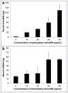
(a) SIS punches modified with biotin and streptavidin were exposed to I125 radiolabeled biotinylated KDR. Radioactivity of the punches was measured to give a quantification of bound biotinylated anti-KDR. (b) Modified SIS punches were incubated with increasing concentrations of biotinylated anti-KDR and then exposed to I125 radiolabeled recombinant human KDR. Radioactivity of the punches was measured to give a quantification of bound KDR protein. All data is presented as the mean ± standard deviation, n=4.
Figure 5:
Figure 5:. Thickness of suspended SIS devices post-implantation.
(a) Suspended devices that were constructed with leaflets of either unmodified SIS or SIS modified with anti-KDR were implanted into sheep inferior vena cava for 2 weeks, and the leaflet thickness was measured at explantation. Data is presented as the mean + standard deviation, n=3. Panels b and c show a representative device constructed with unmodified SIS, while d and e show an anti-KDR modified SIS device. (c and e) Mason’s trichrome staining was used to highlight the collagenous SIS material (blue) surrounded by a cellular remodeling response. Scale bar = 100 μm.
Figure 6:
Figure 6:. Representative angiography of EOC-seeded SIS devices in sheep jugular veins and of a valve post-explantation.
(a) Contrast dye injected distally to the valve demonstrated that both valves were patent. (b and c) Contrast dye injected proximally to the valves did not regurgitate through the valve, suggesting competent valves were able to prevent retrograde blood flow. (d) Representative photograph of an SIS valve explanted after 3 months.
Figure 7:
Figure 7:. Thickness of explanted SIS valves post-implantation.
(a) Bioartificial SIS venous valves that were either unmodified or pre-seeded with EOCs were implanted in sheep jugular veins for 3 months, and the thickness of the valve leaflets were measured at explantation. Data is presented as the mean + standard deviation, n=3. (b) A plot comparing the thickness of unmodified SIS valves compared with the contralaterally implanted EOC-seeded valves indicates that the degree of thickening varied greatly between animals but was similar between treatment groups.
Figure 8:
Figure 8:. Immunohistochemical staining of SIS valves.
(a and b) EOC-seeded valves were stained for smooth muscle actin; this stain showed that some valves had relatively little smooth muscle cell infiltration (a), while others had robust smooth muscle cell infiltration and proliferation on the valve leaflet (b). (c and d) Von Willebrand factor staining of unmodified SIS valves (c) and EOC-seeded valves (d) demonstrated that all valves regardless of treatment developed an endothelial cell lining on the luminal surface of the leaflet by the end of the 3 month implant period. Scale bars = 500 μm (a and b) or 50 μm (c and d).

Similar articles

Cited by

References

    1. Kaplan RM, Criqui MH, Denenberg JO, Bergan J, Fronek A. Quality of life in patients with chronic venous disease: San Diego population study. J Vasc Surg. 2003;37:1047–53. - PubMed
    1. Eberhardt RT, Raffetto JD. Chronic venous insufficiency. Circulation. 2005;111:2398–409. - PubMed
    1. Gohel MS, Barwell JR, Taylor M, Chant T, Foy C, Earnshaw JJ, Heather BP, Mitchell DC, Whyman MR, Poskitt KR. Long term results of compression therapy alone versus compression plus surgery in chronic venous ulceration (ESCHAR): Randomised controlled trial. Br Med J. 2007;335:83–7. - PMC - PubMed
    1. Gloviczki P, Comerota AJ, Dalsing MC, Eklof BG, Gillespie DL, Gloviczki ML, Lohr JM, McLafferty RB, Meissner MH, Murad MH, Padberg FT, Pappas PJ, Passman MA, Raffetto JD, Vasquez MA, Wakefield TW. The care of patients with varicose veins and associated chronic venous diseases: Clinical practice guidelines of the Society for Vascular Surgery and the American Venous Forum. J Vasc Surg. 2011;53:2S–48S. - PubMed
    1. Geerts W, Bergqvist D. Prevention of Venous Thromboembolism American College of Chest Physicians Evidence-Based Clinical Practice Guidelines. CHEST. 2008;133:381S–453S. - PubMed

Publication types

LinkOut - more resources