. 2015 Oct:87:171-9.
doi: 10.1016/j.yjmcc.2015.08.009. Epub 2015 Aug 24.

Cardiomyocytes induce macrophage receptor shedding to suppress phagocytosis

Affiliations

Cardiomyocytes induce macrophage receptor shedding to suppress phagocytosis

Shuang Zhang et al. J Mol Cell Cardiol. 2015 Oct.

Abstract

Background: Mobilization of the innate immune response to clear and metabolize necrotic and apoptotic cardiomyocytes is a prerequisite to heart repair after cardiac injury. Suboptimal kinetics of dying myocyte clearance leads to secondary necrosis, and in the case of the heart, increased potential for collateral loss of neighboring non-regenerative myocytes. Despite the importance of myocyte phagocytic clearance during heart repair, surprisingly little is known about its underlying cell and molecular biology.

Objective: To determine if phagocytic receptor MERTK is expressed in human hearts and to elucidate key sequential steps and phagocytosis efficiency of dying adult cardiomyocytes, by macrophages.

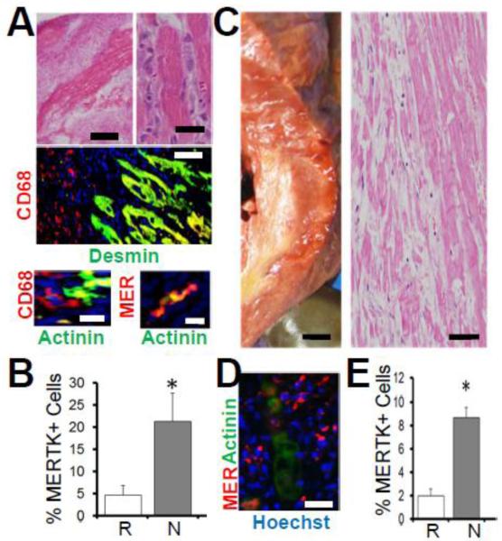
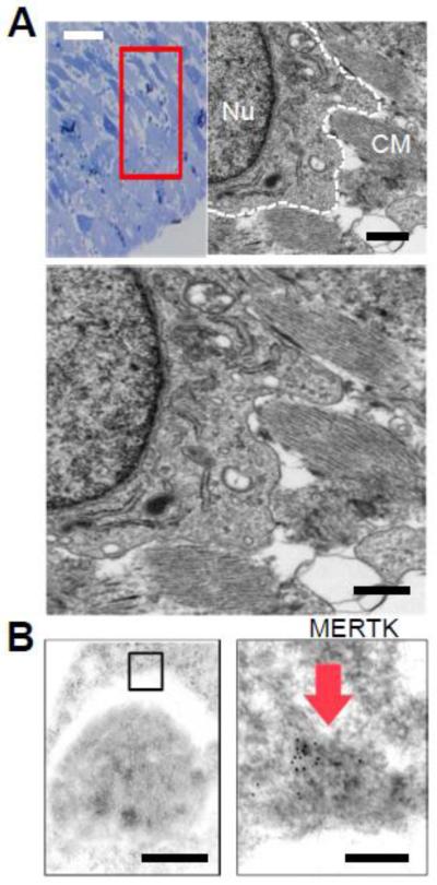
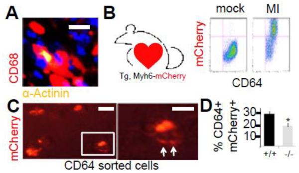
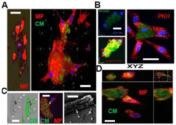
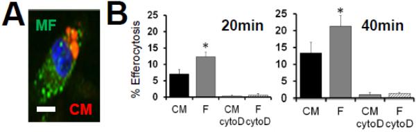
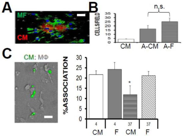
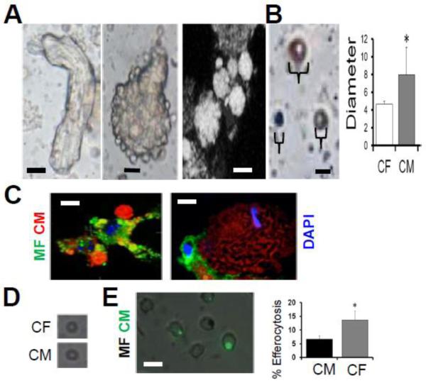
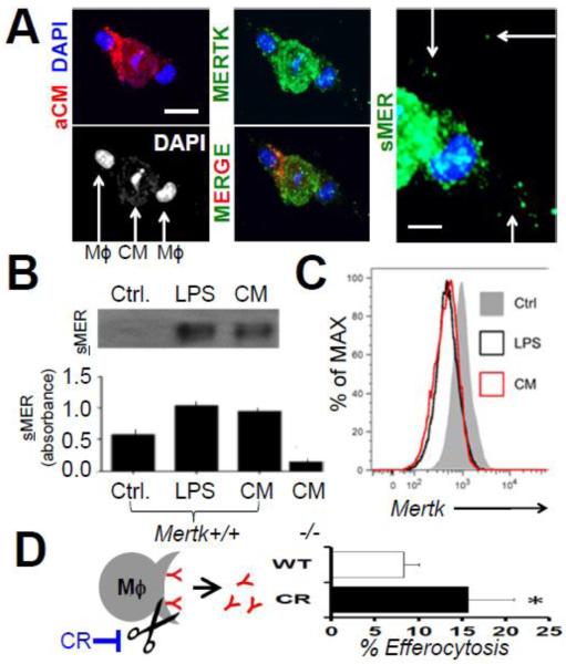
Results: In infarcted human hearts, expression profiles of the phagocytic receptor MER-tyrosine kinase (MERTK) mimicked that found in experimental ischemic mouse hearts. Electron micrographs of myocardium identified MERTK signal along macrophage phagocytic cups and Mertk-/- macrophages contained reduced digested myocyte debris after myocardial infarction. Ex vivo co-culture of primary macrophages and adult cardiomyocyte apoptotic bodies revealed reduced engulfment relative to resident cardiac fibroblasts. Inefficient clearance was not due to the larger size of myocyte apoptotic bodies, nor were other key steps preceding the formation of phagocytic synapses significantly affected; this included macrophage chemotaxis and direct binding of phagocytes to myocytes. Instead, suppressed phagocytosis was directly associated with myocyte-induced inactivation of MERTK, which was partially rescued by genetic deletion of a MERTK proteolytic susceptibility site.

Conclusion: Utilizing an ex vivo co-cultivation approach to model key cellular and molecular events found in vivo during infarction, cardiomyocyte phagocytosis was found to be inefficient, in part due to myocyte-induced shedding of macrophage MERTK. These findings warrant future studies to identify other cofactors of macrophage-cardiomyocyte cross-talk that contribute to cardiac pathophysiology.

Keywords: Acute myocardial infarction; Animal models of human disease; Cardiomyocyte; Phagocytosis.


Figures

Fig. 1
Fig. 1. Evidence for MERTK in human myocardium
A and B is mouse ischemic myocardium and C,D,E is human. (A) Hematoxylin and Eosin (H&E) images of mouse myocardium showing hematoxylin+ mono-nuclear cells juxtaposed to hyper-eosinophilic cardiomyocytes. Left scale bar is 500μm and right is 20μm. Below H&E micrographs are Immunohistochemistry (IHC) of mouse heart with indicated markers for macrophages (CD68 and MER-TK) and cardiomyocytes (Desmin & Actinin). CD68 vs Desmin bar = 50μm. Bottom images = 10μm. In (B), results of IHC quantification in Remote (R) vs inflammatory Necrotic (N) myocardial ROIs (regions of interest). (C, D, E) Human Myocardium. (C) Left is cross-section of myocardium from gross autopsy showing yellow infarct and right is H&E histology of same heart. Left is 0.5cm scale and right is 50μm scale bar. (D) IHC (100μm) with indicated markers (hoechst are nuclei) and (E) quantitation of MERTK positive signal in healthy/Remote versus inflammatory/Necrotic myocardial ROIs.
Fig. 2
Fig. 2. Evidence for MERTK at the phagocytic cup in myocardium
(A) Toluidine blue staining of cardiac sections after experimental infarction and selection of region of interest with mono-nuclear infiltration for transmission electron microscopy. TEM image to the right shows putative cardiac macrophage (outlined in white dotted line) and its nucleus (Nu) with extended pseudopods proximal to remnants of striated cardiomyocyte (CM) debris. Image is magnified below. Black bar = 2 micrometers. (B) Phagocytic pseudopod on top surrounding an apoptotic body that was found to be immunopositive for CM markers Desmin and Actinin. Right: MERTK immunogold staining identified on the phagocytic pseudopod. Black bars = 500 nanometers to the left and 50 nanometers to the right.
Fig. 3
Fig. 3. MERTK-dependent internalization of cardiomyocytes by macrophages in myocardium
(A) Immunohistochemistry shows co-localization of CD68+ macrophages with actinin+ cardiomyocytes (yellow). Scale bar = 15 micrometers. (B) Transgenic Myh6-driven mCherrry mouse and flow cytograms from myocardial extracts post left anterior descending artery ligation (MI) to induce infarction. MI induces CD64, mCherry double positive cells. (C) CD64+ sorted cells were imaged by fluorescent microscopy for macrophages containing myocardial mCherry signal. Cells were trypsinzed to dissociate cell-cell interactions and reveal only internalized mCherry signal. Image to the right is an enlargement of the boxedin cell, displaying evidence of mCherry digestion (arrows). Scale bar = 15 micrometers. (D) Quantitation of myocardial mCherry internalization in Mertk−/− mice vs. Mertk+/+ mice after MI.
Fig. 4
Fig. 4. Ex vivo co-cultivations reveal macrophage attraction to dying cardiomyocytes (CMs) and piece-meal phagocytic processing
(A) Left epi-fluorescent and brightfield merged image displays red pseudo-colored (Calcein-AM/CAM) macrophages (MF) and CM that are solely labeled with DAPI for nuclei. Multiple macrophages engage the singular adult CM. To the right, confocal image shows multiple (nuclei are blue from DAPI) red-immunostained macrophages (F4/80) enveloping green a CM. Scale bars = 30μm. (B) Evidence of membrane remodeling in MFs upon binding to CMs. Top left shows multiple DAPI-labeled MFs bound to a CAM labelled CM. Below cells are stained for Filamentous Actin (F-actin) dye phalloidin in red. Phalloidin reveals actin polymerization of phagocytes surrounding the CM core. To the right, MFs labelled with membrane dye PKH and CMs are green. MFs directly interacting with CMs show redistribution of PKH dye. Scale bars = 20 μm. (C) Evidence of piece-meal phagocytosis. Left images show green-labeled CMs transferring green dye to attached macrophages (MF, arrowheads). Middle confocal image shows phagocyte appearing to internalize green signal from CM in a phagosome. Right image is scanning electron micrograph of CM and attached MFs appearing to internalize bright/refractile apoptotic bodies. Scale bars = 15 μm. (D) Internalization of green CM apoptotic bodies into green MFs confirmed by confocal microscopy Z sections. Scale bar = 20 μm.
Fig. 5
Fig. 5. Inefficient Phagocytic Uptake of CM apoptotic bodies (ABs)
(A) Micrograph of epiflourescent macrophage (green) engulfing red cardiomyocyte (CM) apoptotic bodies. Scale bar = 7μm. (B) Primary resident cardiac fibroblasts (F) and adult ventricular myocytes were adhered onto tissue culture plates and induced to apoptosis (1 hour 1μM staurosporine/STS followed by 6hr treatment for CF and 18hr CF to achieve ~80% morphological cell death). Non-adherent/floating apoptotic bodies were harvested and separated from larger-CM-core bodies by low-speed centrifugation at 50 x g to isolate apoptotic bodies from cells and overlaid under saturating conditions onto adherent primary bone-marrow derived macrophages for efferocytosis enumeration. Cytochalasin D (cytoD) was added to macrophages to inhibit actin polymerization and control for engulfment-specificity vs. non-specific binding.
Fig. 6
Fig. 6. Steps Leading (Chemotaxis) and Preceding (Binding) Engulfment of Cardiomyocytes do Not Differ Significantly Relative to Cardiac Fibroblasts
(A) Macrophages (green) swarm to apoptotic cardiomyocytes (CM) (red). Scale bar = 20μm. (B) Bar graph displays quantification of macrophage chemotaxis to live CMs vs apoptotic cardiomyocytes (A-CMs) versus apoptotic cardiac fibroblasts (A-F). n.s.=not statistically different. (C) Micrograph shows green CM apoptotic bodies bound to non-labeled macrophages. Binding assay bar graph for affinity of macrophages to dying CMs versus dying cardiac fibroblasts (F) at temperature of 4C. After thermoshift to 37C and rinsing away of non-engulfed cells, % association in an indication of engulfment. Scale bar is 15 μm.
Fig. 7
Fig. 7. Inefficient Cardiomyocyte (CM) efferocytosis is largely independent of the size of CM apoptotic bodies
(A) Leftmost bright-field image depicts rod-shaped CM in the beginning stages of cell death. To the immediate right is a collapsed rounded CM exhibiting protruding apoptotic body blebs. Next image is confocal microscopy and shows apoptotic bodies of varying sizes forming on a dying CM. Scale bars = 15μm. (B) Soluble apoptotic bodies (ABs) of various sizes are depicted in photomicrograph. Scale bar = 4 μms. Quantification of CM vs cardiac fibroblast (CF) apoptotic body size. (C) Phagocytes engage CM ABs of various sizes. Scale bar is 10μm. (D) Average size of largest CM and CF apoptotic bodies after filtration was <5 μms. (E) Normalized efferocytosis after filtration with size exclusion filter. Scale bar is 10μm.
Fig. 8
Fig. 8. Dying Cardiomyocytes (CMs) Induce MERTK Cleavage in Macrophages to Inhibit Efferocytosis
(A) Confocal images of dying Red (PKH) labeled CMs were cocultivated with MERTK-labeled (green) macrophages. DAPI shows macrophage nuclei flanking condensed apoptotic CM nuclei. Scale bar = 20μm. Rightmost panel depicts higher magnification image (white bar = 10 μms) of extracellular (arrows) or soluble MER (sMER) signal. (B) Western blots (above) and ELISA (below) for solMER after Mertk+/+ or Mertk−/− macrophage incubation with positive control lipopolysaccharide (LPS) or CMs. (C) Flow cytometry histograms for cell-surface MERTK after co-cultivation with LPS or CMs (D). Schematic representation of inhibition of sMER cleavage with mutant Cleavage-Resistant (CR) MERTK. Bar graph is enumeration of % CM efferocytosis by HEK293 cells after transfection with equal masses of wild type (WT) vs CR-Mertk cDNAs.

Similar articles

Cited by

References

    1. Aderem A. How to eat something bigger than your head. Cell. 2002;110(1):5–8. - PubMed
    1. Briassouli P, Komissarova EV, et al. Role of the urokinase plasminogen activator receptor in mediating impaired efferocytosis of anti-SSA/Ro-bound apoptotic cardiocytes: Implications in the pathogenesis of congenital heart block. Circ Res. 2010;107(3):374–387. - PMC - PubMed
    1. Bujak M, Kweon HJ, et al. Aging-related defects are associated with adverse cardiac remodeling in a mouse model of reperfused myocardial infarction. J Am Coll Cardiol. 2008;51(14):1384–1392. - PMC - PubMed
    1. Camenisch TD, Koller BH, et al. A novel receptor tyrosine kinase, Mer, inhibits TNF-alpha production and lipopolysaccharide-induced endotoxic shock. J Immunol. 1999;162(6):3498–3503. - PubMed
    1. Cannon GJ, Swanson JA. The macrophage capacity for phagocytosis. J Cell Sci. 1992;101(Pt 4):907–913. - PubMed

Publication types

MeSH terms

Substances