. 2015 Nov:83:75-89.
doi: 10.1016/j.nbd.2015.08.021. Epub 2015 Aug 24.

Genetic targeting of protease activated receptor 2 reduces inflammatory astrogliosis and improves recovery of function after spinal cord injury

Affiliations

Genetic targeting of protease activated receptor 2 reduces inflammatory astrogliosis and improves recovery of function after spinal cord injury

Maja Radulovic et al. Neurobiol Dis. 2015 Nov.

Abstract

Inflammatory-astrogliosis exacerbates damage in the injured spinal cord and limits repair. Here we identify Protease Activated Receptor 2 (PAR2) as an essential regulator of these events with mice lacking the PAR2 gene showing greater improvements in motor coordination and strength after compression-spinal cord injury (SCI) compared to wild type littermates. Molecular profiling of the injury epicenter, and spinal segments above and below, demonstrated that mice lacking PAR2 had significantly attenuated elevations in key hallmarks of astrogliosis (glial fibrillary acidic protein (GFAP), vimentin and neurocan) and in expression of pro-inflammatory cytokines (interleukin-6 (IL-6), tumor necrosis factor (TNF) and interleukin-1 beta (IL-1β)). SCI in PAR2-/- mice was also accompanied by improved preservation of protein kinase C gamma (PKCγ)-immunopositive corticospinal axons and reductions in GFAP-immunoreactivity, expression of the pro-apoptotic marker BCL2-interacting mediator of cell death (BIM), and in signal transducer and activator of transcription 3 (STAT3). The potential mechanistic link between PAR2, STAT3 and astrogliosis was further investigated in primary astrocytes to reveal that the SCI-related serine protease, neurosin (kallikrein 6) promotes IL-6 secretion in a PAR2 and STAT3-dependent manner. Data point to a signaling circuit in primary astrocytes in which neurosin signaling at PAR2 promotes IL-6 secretion and canonical STAT3 signaling. IL-6 promotes expression of GFAP, vimentin, additional IL-6 and robust increases in both neurosin and PAR2, thereby driving the PAR2-signaling circuit forward. Given the significant reductions in astrogliosis and inflammation as well as superior neuromotor recovery observed in PAR2 knockout mice after SCI, we suggest that this receptor and its agonists represent new drug targets to foster neuromotor recovery.

Keywords: Astrogliosis; Cytokine; GPCR; Inflammation; Serine protease; Traumatic spinal cord injury.


Conflict of interest statement

Conflict of Interest: The authors declare no competing financial interests.

Figures

Figure 1
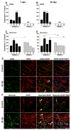
Figure 1. The expression of PAR2 and neurosin (Klk6) are elevated in response to contusion-compression SCI
Histograms show transcriptional changes in RNA encoding PAR2 in the spinal cord of uninjured control (C) PAR2+/+ mice, or at the level of the injury epicenter (E), above (A), or below (B), at 7 and 30 dpi (A, B). PAR2 RNA was not detected (ND) in PAR2−/− mice. In PAR2+/+ mice, co-ordinate elevations were seen in the SCI-related secreted serine protease neurosin, already established to activate nervous system PAR2 (Vandell et al., 2008; Yoon et al., 2013). By contrast, in mice lacking the PAR2 gene, significant elevations in neurosin in response to SCI were not observed (C, D). (*P < 0.03; **P = 0.009, ***P ≤ 0.001 NK; ND, not detected). Photomicrographs show co-localization of PAR2 and GFAP (E), or neurosin and GFAP (F), in the uninjured spinal cord (Control) and within the injury epicenter at 30 dpi. Increases in GFAP-positive cells co-labeled for PAR2 or neurosin were observed at 30 dpi and a selection of double labeled cells in each case is shown at arrowheads (Scale bar = 20 um).
Figure 2
Figure 2. PAR2 gene deficient mice show improved locomotor recovery after experimental contusion-compression SCI
(A) Basso Mouse Scale (BMS) scores evaluated in PAR2+/+ and PAR2−/− mice from 0 to 31 d after SCI, demonstrate significant improvements in over-ground locomotion. Significantly higher levels of recovery on the BMS were observed in PAR2−/− mice at 7 dpi (P = 0.045) and at each time point thereafter (P < 0.001). (B) BMS subscores were significantly higher in PAR2−/− compared to PAR2+/+ mice starting on 14 dpi and continuing through the last time point examined (P < 0.001). (C, D) Improvements in stepping accuracy in the easy and hard rung spacing ladder walk tests were observed by 15 dpi and persisted when examined at 22 and 31 dpi. Improvements in ladder walk accuracy were also observed at 8 dpi in PAR2−/− relative to PAR2+/+ mice in the hard set up. (E) PAR2−/− mice showed significantly better motor strength in the incline plane test at 30 dpi relative to that observed in PAR2+/+ mice. Data shown represents the mean and standard error of results obtained across two independent experiments, PAR2+/+, n=17; PAR2−/− n=20. (Two Way Repeated Measures ANOVA, NK *P < 0.05, **P ≤ .008, ***P < 0.001).
Figure 3
Figure 3. PAR2 gene deletion is associated with reduced inflammatory astrogliosis, tissue sparring and greater preservation of PKCγ-immunopositive corticospinal tract axons after experimental contusion-compression SCI
Photomicrographs and associated histograms show measurements of spinal cord areas taken from H&E stained sections (A to I), or the percent of spinal cord area immunoreactive for GFAP (J to N), or Isolectin B-IR (IsoB) microglia/macrophages (Q to U), at the 32d endpoint of each experiment. In addition, the % area at the base of the dorsal column white matter stained for PKCγ, as a measure of corticospinal axon function (V to Z), was evaluated. In each case, measurements were made in spinal cord sections taken from the level of the injury epicenter (E), above (A), or below (B). Arrows J to Y indicate an example of significant immunostaining in each case. In mice lacking PAR2 there was tissue sparring at the level of the injury epicenter (I), reductions in GFAP-IR at the injury epicenter, above and below (N), reductions in IsoB-IR below and preservation of PKCγ-IR in spinal segments above the site of lesion (PAR2+/+, n=13 (4 Control, 9 SCI); PAR2−/−, n=12 (4 Control, 8 SCI); *P < 0.05, **P ≤ 0.01, ***P ≤ 0.001 NK). Scale Bar A–H = 250 μm; J–Y = 100 μm.
Figure 4
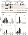
Figure 4. Spinal cord injury-associated elevations in GFAP, vimentin and STAT3 signaling were reduced in PAR2 gene deficient mice
Western blots and corresponding histograms show differential expression of GFAP, vimentin (VIM), pSTAT3 and STAT3 in the uninjured (C, Control) or injured PAR2+/+ or PAR2−/− spinal cord at 3 or 30 dpi. Protein samples isolated from the injury epicenter (E), above (A), or below (B) from PAR2+/+ or PAR2−/− mice were examined in parallel with uninjured samples in each case and results developed on the same film. SCI-induced elevations in GFAP, vimentin and STAT3 were all significantly reduced in mice lacking the PAR2 gene. In some cases, multiple bands for GFAP or vimentin were detected after SCI and the band used for quantification is shown at the arrowhead in each case. Histograms show the mean and standard error of ROD readings across multiple membranes (n = 3 to 4) normalized to Actin as loading control. (*P < 0.05, **P ≤ 0.01, ***P ≤ 0.001 NK).
Figure 5
Figure 5. Spinal cord injury-induced elevations in markers of astrogliosis and apoptosis were reduced in mice lacking the PAR2 gene
Histograms show transcriptional changes in RNA encoding GFAP (A), vimentin (VIM, (B)), neurocan (NCAN, (C)), and BIM (F) in the spinal cord of PAR2+/+ or PAR2−/− uninjured controls (C), or in the 3 mm of spinal cord surrounding the injury epicenter (E), or that above (A), or below (B), at 30 dpi. The RNA expression levels shown are expressed as a percent of the uninjured genotype control and these data were used for statistical comparisons. In addition, to facilitate interpretation of any impact of PAR2 gene deletion on gene expression in the uninjured spinal cord, and to permit statistical comparisons, expression levels shown for uninjured control PAR2−/− mice are expressed as a percent of uninjured PAR2+/+ controls. Baseline levels of neurocan were higher in PAR2−/− compared to PAR2+/+ mice. Western blots (D) and corresponding histogram (E) show that elevations in the pro-apoptotic protein BIM observed at 30 dpi were significantly attenuated in mice lacking PAR2. (*P < 0.05, **P ≤ 0.01, ***P ≤ 0.001 NK).
Figure 6
Figure 6. Spinal cord injury-induced elevations in pro-inflammatory cytokines were reduced in mice lacking the PAR2 gene
Histograms show transcriptional changes in RNA encoding pro-inflammatory (TNF, IL1-β, IL-6), or anti-inflammatory cytokines (TGF-β, IL-10), in the spinal cord of PAR2+/+ or PAR2−/− uninjured controls (C), or in the 3 mm of spinal cord surrounding the injury epicenter (E), or that above (A), or below (B), at 30 dpi. The RNA expression levels shown are expressed as a percent of the uninjured genotype control and these data were used for statistical comparisons. In addition, to facilitate interpretation of any impact of PAR2 gene deletion on cytokine expression in the uninjured spinal cord, and to permit statistical comparisons, expression levels shown for uninjured control PAR2−/− mice are expressed as a percent of uninjured PAR2+/+ controls. No significant differences in cytokine RNA expression were observed in the uninjured spinal cord between PAR2+/+ and PAR2−/− mice. (*P < 0.05, **P ≤ 0.01, ***P ≤ 0.001 NK).
Figure 7
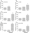
Figure 7. IL-6 is a positive driver of astroglial intermediate filament proteins and the PAR2-signaling axis in primary astrocytes
(A) Treatment of astrocytes with neurosin (150 ng/ml) for 24 h resulted in a significant increase in IL-6 secretion. (B) Astrocyte cultures treated with recombinant IL-6 (20 ng/ml) showed increased IL-6 RNA expression. Recombinant IL-6 also promoted significant increases in the expression of GFAP (C), vimentin (D), PAR2 (E), and neurosin (F) RNA. (*P < 0.05, **P ≤ 0.01, ***P ≤ 0.001, Students t-test).
Figure 8
Figure 8. PAR2-neurosin dependent mechanism of IL-6 secretion in primary astrocytes
(A) Neurosin (NRS), (150 nM, 24 h)-mediated IL-6 secretion is reduced in astrocytes derived from PAR2 knockout mice. Neurosin also stimulates increases in pSTAT3 (B), and total STAT3 (C), and these signaling responses were blocked in astrocytes lacking PAR2. A small molecule inhibitor of STAT3 (Stattic, 5 or 20 μM), or of MEK1/2 (U0126, 150 μM), each significantly attenuated neurosin-mediated IL-6 secretion (D, E). IL-6 secretion was measured by ELISA in cell culture supernatants. (*P < 0.05, **P ≤ 0.01, ***P ≤ 0.001 NK).
Figure 9
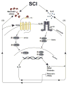
Figure 9. Model depicting how activation of PAR2 in astrocytes may promote IL-6-mediated STAT3 signaling and astrogliosis
Data presented suggest that SCI promotes elevations in PAR2 and its CNS endogenous agonist neurosin (Klk6). Prior studies demonstrate neurosin is produced by reactive astrocytes and activated microglia/macrophages at sites of spinal cord trauma (Scarisbrick et al., 2006b; Scarisbrick et al., 2012a; Radulovic et al., 2013). Neurosin cleaves thereby activating PAR2 (Vandell et al., 2008) to elicit IL-6 secretion at least in part by a MEK1/2-dependent signaling mechanism. Astrocyte derived IL-6, along with that generated by immune cells at sites of SCI is a well studied activator of STAT3 signaling (Darnell, 1997) and elicits expression of GFAP, vimentin (VIM) and additional IL-6 in primary astrocyte cultures. Data presented also suggest that IL-6 can increase expression of PAR2 (330-fold) and neurosin (2.5-fold) thereby contributing to a reverberating signaling circuit in astrocytes that will ultimately promote astrogliosis.

References

    1. Asher RA, Morgenstern DA, Moon LD, Fawcett JW. Chondroitin sulphate proteoglycans: inhibitory components of the glial scar. Prog Brain Res. 2001;132:611–619. - PubMed
    1. Basso DM, Fisher LC, Anderson AJ, Jakeman LB, McTigue DM, Popovich PG. Basso mouse scale for locomotion detects differences in recovery after spinal cord injury in five common mouse strains. J Neurotrauma. 2006;23:635–659. - PubMed
    1. Bastien D, Lacroix S. Cytokine pathways regulating glial and leukocyte function after spinal cord and peripheral nerve injury. Exp Neurol. 2014;258:62–77. - PubMed
    1. Blaber SI, Ciric B, Christophi GP, Bernett MJ, Blaber M, Rodriguez M, Scarisbrick IA. Targeting kallikrein 6-proteolysis attenuates CNS inflammatory disease. FASEB J. 2004;19:920–922. - PubMed
    1. Bradbury EJ, Moon LD, Popat RJ, King VR, Bennett GS, Patel PN, Fawcett JW, McMahon SB. Chondroitinase ABC promotes functional recovery after spinal cord injury. Nature. 2002;416:636–640. - PubMed

Publication types

MeSH terms