

The Evaluation of FDG PET/CT Scan Findings in Patients with Organizing Pneumonia Mimicking Lung Cancer
- PMID: 26316470
- PMCID: PMC4563171
- DOI: 10.4274/mirt.03016
The Evaluation of FDG PET/CT Scan Findings in Patients with Organizing Pneumonia Mimicking Lung Cancer
Abstract
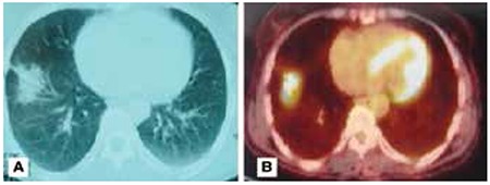
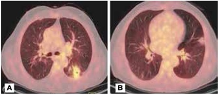
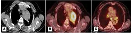
Objective: Organizing pneumonia (OP) is a rare lung condition that is characterized by the presence of polypoid tissues due to fibroblastic plugs within respiratory bronchioles, alveolar ducts and sacs. The three main radiologic patterns of OP include typical, solitary-focal and infiltrative forms. The maximum standardized uptake value (SUVmax) on positron emission tomography-computed tomography (PET/CT) may be high in benign conditions such as OP as well as malignant diseases. The aim of our study was to investigate PET-CT characteristics of OP in patients mimicking lung cancer.
Methods: The clinical and radiologic characteristics of 50 patients who were referred to our hospital for PET/CT evaluation due to suspicion of lung malignancy, and who were pathologically diagnosed as OP between 2009 and 2013 were retrospectively reviewed.
Results: The mean age of the patients was 58.2 years. Ninety-six percent of patients (48) were male. Radiologic evaluation revealed 27 (54%) focal involvement, 10 (20%) consolidation with air-bronchogram (typical), 1 (2%) infiltrative and 12 (24%) other types of involvement (multiple nodules and cavitary lesions). The mean SUVmax value of the lesions on PET/CT was calculated as 6.5. Mediastinal lymph node involvement (at least one station) was detected in 76% of our study group with a mean SUVmax value of 3.27.
Conclusion: OP may cause false positive results on PET/CT. However, PET/CT results may be used as a guide for invasive procedures that should be performed when there is suspicion of malignancy.
Amaç: Organize pnömoni (OP), respiratuvar bronşiyoller, alveoler kanallar ve alveollerde fibroblastik tıkaçların oluşturduğu polipoid yapılarla karakterize histopatolojik bir durumdur. Radyolojik olarak OP’nin tipik, soliter-fokal ve infiltratif olmak üzere 3 karakteristik görünümü vardır. Pozitron Emisyon Tomografi-Bilgisayarlı Tomografisi’de (PET-BT) yüksek FDG tutulumu malin hastalıklarda olduğu gibi OP gibi benin hastalıklarda da görülebilir. Bu çalışmada maliniteyi taklit eden OP’li olgulardaki PET-BT bulgularını değerlendirmeyi amaçladık. Yöntem: Çalışmamızda 2009-2013 yılları arasında Atatürk Göğüs Hastalıkları ve Göğüs Cerrahisi Eğitim Araştırma Hastanesine dış merkezlerden akciğer grafisi veya toraks bilgisayarlı tomografisinde akciğer malinitesinden şüphelenilerek, ileri tetkik ve tedavi amaçlı yönlendirilen ve malinite ön tanısı ile PET-BT çekilen 50 OP’li olgu dahil edildi. Retrospektif olarak radyolojik ve klinik özellikleri kayıt edildi. Bulgular: Çalışmaya dahil edilen olguların yaş ortalaması 58,2 idi. Yüzde 96’sını (48) erkek hastalar oluşturmaktaydı. Radyolojik olarak PET- BT’de 27 (%54) olguda fokal, 10 (%20) olguda hava bronkogramı içeren konsolidasyon (tipik), 1 (%2) olguda infiltratif ve 12 (%24) olguda da diğer görünümler (multiple nodüller, multiple kaviter lezyonlar) saptandı. PET- BT’de lezyonların maksimum standart tutulum değerlerinin (SUVmax) ortalaması 6,5 olarak hesaplandı. Çalışma grubunun %76’sında en az bir istasyonda mediastinal lenf nodu tutulumu saptandı. Lenf nodlarının ortalama SUVmax değeri 3,27 idi. Sonuç: Organize pnömoniler PET-BT’de yanlış pozitif sonuçlara yol açabilir. Ancak PET-BT maliniteden şüphe ediliyorsa yapılması gereken invaziv yöntemlere yol gösterici olarak kullanılabilir.
Figures
References
-
- Maldonado F, Daniels CE, Hoffman EA, Yi ES, Ryu JH. Focal organizing pneumonia on surgical lung biopsy: causes, clinicoradiologic features, and outcomes. Chest. 2007;132:1579–1583. - PubMed
-
- Oikonomou A, Hansell DM. Organizing pneumonia: the many morphological faces. Eur Radiol. 2002;12:1486–1496. - PubMed
-
- Shin L, Katz DS, Yung E. Hypermetabolism on F-18 FDG PET of multiple pulmonary nodules resulting from bronchiolitis obliterans organizing pneumonia. Clin Nucl Med. 2004;29:654–656. - PubMed
-
- Orino K, Kawamura M, Hatazawa J, Suzuki I, Sazawa Y. Efficacy of F-18 fluorodeoxyglucose positron emission tomography (FDG-PET) scans in diagnosis of pulmonary nodules. Jpn J Thorac Cardiovasc Surg. 1998;46:1267–1274. - PubMed
-
- Marques G, Annweiler T, Raoux D, Tiffet O, Vergnon JM, Bertoletti L. Nodular presentation of a cryptogenic organizing pneumonia. Rev Pneumol Clin. 2011;67:314–317. - PubMed
LinkOut - more resources
Full Text Sources
Other Literature Sources
Miscellaneous