

Detection of Nasopharyngeal Carcinoma by MR Imaging: Diagnostic Accuracy of MRI Compared with Endoscopy and Endoscopic Biopsy Based on Long-Term Follow-Up
- PMID: 26316564
- PMCID: PMC7964287
- DOI: 10.3174/ajnr.A4456
Detection of Nasopharyngeal Carcinoma by MR Imaging: Diagnostic Accuracy of MRI Compared with Endoscopy and Endoscopic Biopsy Based on Long-Term Follow-Up
Abstract
Background and purpose: Our previous nasopharyngeal carcinoma detection study, comparing MR imaging, endoscopy, and endoscopic biopsy, showed that MR imaging is a highly sensitive test that identifies nasopharyngeal carcinomas missed by endoscopy. However, at the close of that study, patients without biopsy-proved nasopharyngeal carcinoma nevertheless had shown suspicious abnormalities on endoscopy and/or MR imaging. The aim of this study was to determine whether there were any patients with undiagnosed nasopharyngeal carcinoma by obtaining long-term follow-up and to use these data to re-evaluate the diagnostic performance of MR imaging.
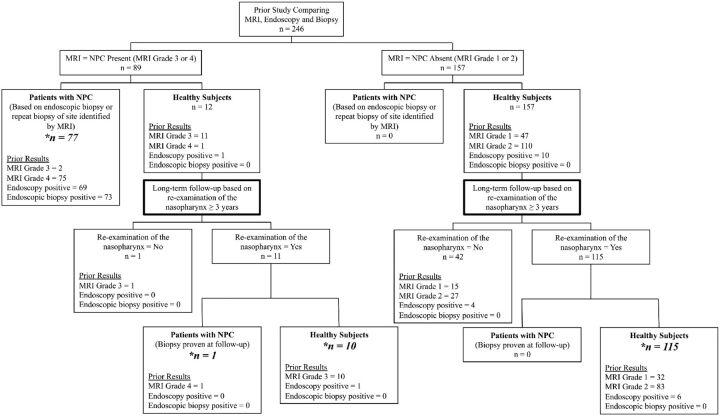
Materials and methods: In the previous study, 246 patients referred to a hospital ear, nose, and throat clinic with suspected nasopharyngeal carcinoma, based on a wide range of clinical indications, had undergone MR imaging, endoscopy, and endoscopic biopsy, and 77 had biopsy-proved nasopharyngeal carcinoma. One hundred twenty-six of 169 patients without biopsy-proved nasopharyngeal carcinoma underwent re-examination of the nasopharynx after a minimum of 3 years, including 17 patients in whom a previous examination (MR imaging = 11; endoscopy = 7) had been positive for nasopharyngeal carcinoma, but the biopsy had been negative for it. Patients with nasopharyngeal carcinoma were identified by biopsy obtained in the previous and this follow-up study; patients without nasopharyngeal carcinoma were identified by the absence of a tumor on re-examination of the nasopharynx. The sensitivity and specificity of the previous investigations were updated and compared by using the Fisher exact test.
Results: One patient with a previous positive MR imaging finding was subsequently proved to have nasopharyngeal carcinoma. Nasopharyngeal carcinomas were not found in the remaining 125 patients at follow-up, and the previous positive findings for nasopharyngeal carcinoma on MR imaging and endoscopy were attributed to benign lymphoid hyperplasia. The diagnostic performances for the previous MR imaging, endoscopy, and endoscopic biopsy were 100%, 88%, and 94%, respectively, for sensitivity, and 92%, 94%, and 100%, respectively, for specificity; the differences between MR imaging and endoscopy were significant for sensitivity (P = .003) but not specificity (P = .617).
Conclusions: MR imaging detected the 12% of nasopharyngeal carcinomas that were endoscopically invisible, including 1 cancer that remained endoscopically occult for several years. Lymphoid hyperplasia reduced the specificity of MR imaging.
© 2015 by American Journal of Neuroradiology.
Figures
References
Publication types
MeSH terms
LinkOut - more resources
Full Text Sources
Medical