. 2015 Dec;36(12):2296-302.
doi: 10.3174/ajnr.A4436. Epub 2015 Aug 27.

Brain Magnetic Susceptibility Changes in Patients with Natalizumab-Associated Progressive Multifocal Leukoencephalopathy

Affiliations

Brain Magnetic Susceptibility Changes in Patients with Natalizumab-Associated Progressive Multifocal Leukoencephalopathy

J Hodel et al. AJNR Am J Neuroradiol. 2015 Dec.

Abstract

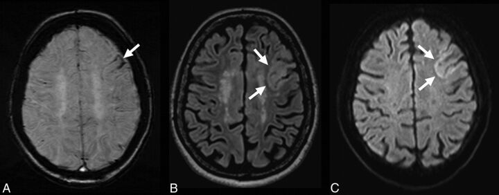
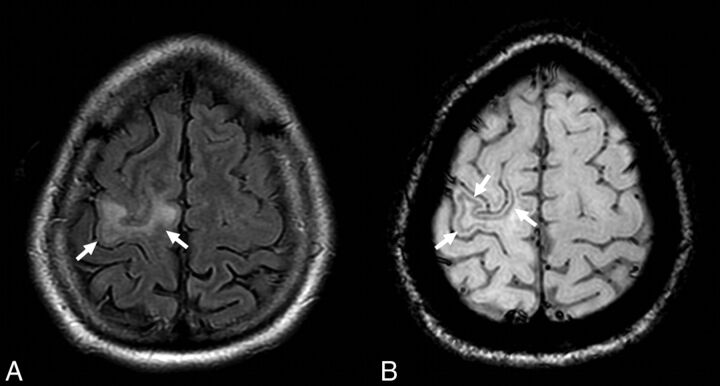
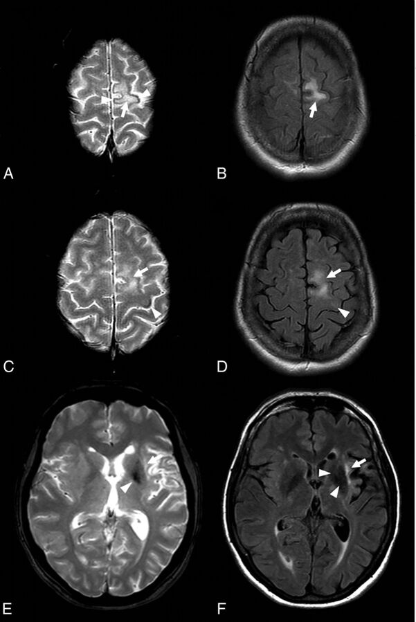
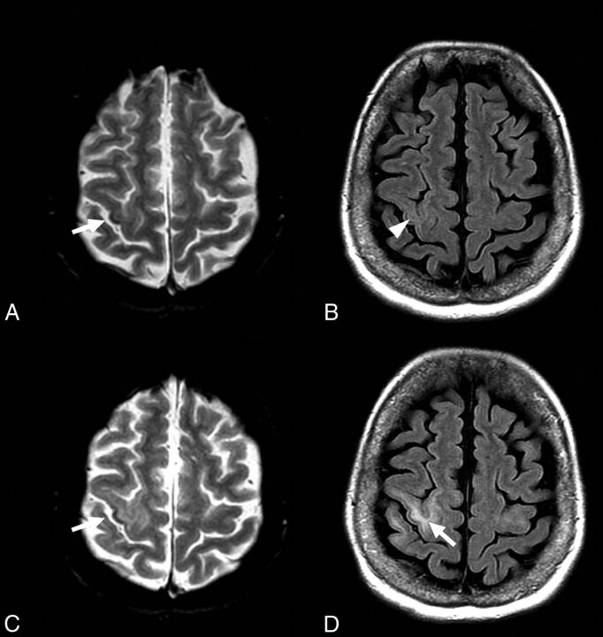
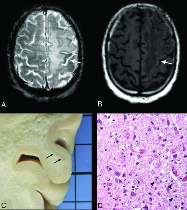
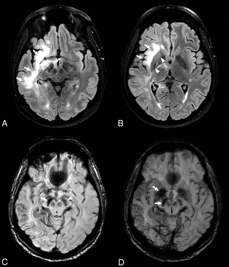
We investigated the brain magnetic susceptibility changes induced by natalizumab-associated progressive multifocal leukoencephalopathy. We retrospectively included 12 patients with natalizumab-progressive multifocal leukoencephalopathy, 5 with progressive multifocal leukoencephalopathy from other causes, and 55 patients with MS without progressive multifocal leukoencephalopathy for comparison. MR imaging examinations included T2* or SWI sequences in patients with progressive multifocal leukoencephalopathy (86 examinations) and SWI in all patients with MS without progressive multifocal leukoencephalopathy. Signal abnormalities on T2* and SWI were defined as low signal intensity within the cortex and/or U-fibers and the basal ganglia. We observed T2* or SWI signal abnormalities at the chronic stage in all patients with progressive multifocal leukoencephalopathy, whereas no area of low SWI signal intensity was detected in patients without progressive multifocal leukoencephalopathy. Among the 8 patients with asymptomatic natalizumab-progressive multifocal leukoencephalopathy, susceptibility changes were observed in 6 (75%). The basal ganglia adjacent to progressive multifocal leukoencephalopathy lesions systematically appeared hypointense by using T2* and/or SWI. Brain magnetic susceptibility changes may be explained by the increased iron deposition and constitute a useful tool for the diagnosis of progressive multifocal leukoencephalopathy.


Figures

Fig 1.
Fig 1.
Susceptibility-weighted, FLAIR, and diffusion images in patient 9 with unilobar left frontal NTZ-PML at the asymptomatic stage. A hypointense rim involving the cortex and the U-fibers of the left frontal lobe is visible on SWI (A, arrow). The NTZ-PML lesion appears hyperintense on FLAIR (B, arrow) and diffusion (C, arrows) images.
Fig 2.
Fig 2.
In patient 11 at the symptomatic stage, the NTZ-PML lesion appears hyperintense on the FLAIR image involving the right precentral gyrus (A, arrows). The SWI sequence reveals a hypointense rim involving the U-fibers adjacent to the PML lesion (B, arrows).
Fig 3.
Fig 3.
FLAIR and T2* images in patient 7 at the symptomatic stage. Cortical low signal intensity is visible on T2* images (A, arrowheads) adjacent to the hyperintense PML lesion (A–C, arrows). Such cortical T2* signal abnormality is also visible (C, arrowhead) with only subtle adjacent hyperintensity on FLAIR images (D, arrowhead). Note the low signal intensity of the left basal ganglia on both T2* and FLAIR images (E and F, arrowheads) adjacent to the insular PML lesion (E and F, arrows).
Fig 4.
Fig 4.
FLAIR and T2* images in patient 12 with NTZ-PML at asymptomatic and symptomatic stages. At the asymptomatic stage, a small area of cortical low signal intensity is visible on T2* image within the right central sulcus (A, arrow), with faint signal abnormalities on FLAIR image (B, arrowhead). At the symptomatic stage, the cortical hypointensity on T2* image is more prominent (C, arrow), while subcortical FLAIR hyperintensity is obvious (D, arrow).
Fig 5.
Fig 5.
T2* and T1WI at the chronic stage and pathologic specimen (hematoxylin-eosin staining) of patient 2. An extensive PML lesion is visible involving the left frontal and parietal white matter. Low signal intensity of the left precentral cortex is visible on T2* image (A, arrow), matching the hyperintensity on T1WI (B, arrow). Pathologic specimen reveals a loss of distinction between GM and WM (C, arrows) as well as microglial and macrophage infiltrate of the cortical neuropile (D, arrowheads; hematoxylin-eosin staining, high-power objective).
Fig 6.
Fig 6.
FLAIR and SWI in patient 8 at symptomatic and chronic stages. At the symptomatic stage, an extensive PML lesion is visible on FLAIR images involving the right frontal and temporal white matter and the brain stem (A and B, arrows). Note the low signal intensity of the right lenticular nucleus on FLAIR images (B, arrowheads). At the chronic stage, the SWI sequence revealed decreased signal-intensity of the right substantia nigra (C, arrow) compared with the left (C, arrowhead). SWI minimal-intensity-projection view confirms this pattern of asymmetric hypointensity involving the right lenticular nucleus and substantia nigra, adjacent to the PML lesion (D, arrows).

Comment in

  • REPLY.
    Hodel J, Outteryck O, Verclytte S, Deramecourt V, Lacour A, Pruvo JP, Vermersch P, Leclerc X. Hodel J, et al. AJNR Am J Neuroradiol. 2016 Feb;37(2):E12. doi: 10.3174/ajnr.A4634. Epub 2015 Dec 3. AJNR Am J Neuroradiol. 2016. PMID: 26635281 Free PMC article. No abstract available.
  • Low Signals on T2* and SWI Sequences in Patients with MS with Progressive Multifocal Leukoencephalopathy.
    Labauge P, Carra-Dalliere C, Ayrignac X, Menjot de Champfleur N. Labauge P, et al. AJNR Am J Neuroradiol. 2016 Feb;37(2):E11. doi: 10.3174/ajnr.A4632. Epub 2015 Dec 3. AJNR Am J Neuroradiol. 2016. PMID: 26635282 Free PMC article. No abstract available.

Similar articles

Cited by

References

    1. Clifford DB, De Luca A, Simpson DM, et al. . Natalizumab-associated progressive multifocal leukoencephalopathy in patients with multiple sclerosis: lessons from 28 cases. Lancet Neurol 2010;9:438–46 10.1016/S1474-4422(10)70028-4 - DOI - PubMed
    1. Dong-Si T, Richman S, Wattjes MP, et al. . Outcome and survival of asymptomatic PML in natalizumab-treated MS patients. Ann Clin Transl Neurol 2014;1:755–64 10.1002/acn3.114 - DOI - PMC - PubMed
    1. Wattjes MP, Richert ND, Killestein J, et al. . The chameleon of neuroinflammation: magnetic resonance imaging characteristics of natalizumab-associated progressive multifocal leukoencephalopathy. Mult Scler 2013;19:1826–40 10.1177/1352458513510224 - DOI - PubMed
    1. Wattjes MP, Vennegoor A, Steenwijk MD, et al. . MRI pattern in asymptomatic natalizumab-associated PML. J Neurol Neurosurg Psychiatry 2015;86:793–98 10.1136/jnnp-2014-308630 - DOI - PubMed
    1. Yousry TA, Pelletier D, Cadavid D, et al. . Magnetic resonance imaging pattern in natalizumab-associated progressive multifocal leukoencephalopathy. Ann Neurol 2012;72:779–87 10.1002/ana.23676 - DOI - PubMed

MeSH terms

LinkOut - more resources