. 2013:2013:407152.
doi: 10.1155/2013/407152. Epub 2013 Nov 21.

Neuroprotection by Exendin-4 Is GLP-1 Receptor Specific but DA D3 Receptor Dependent, Causing Altered BrdU Incorporation in Subventricular Zone and Substantia Nigra

Affiliations

Neuroprotection by Exendin-4 Is GLP-1 Receptor Specific but DA D3 Receptor Dependent, Causing Altered BrdU Incorporation in Subventricular Zone and Substantia Nigra

A Harkavyi et al. J Neurodegener Dis. 2013.

Abstract

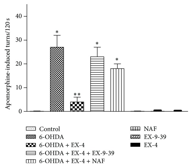
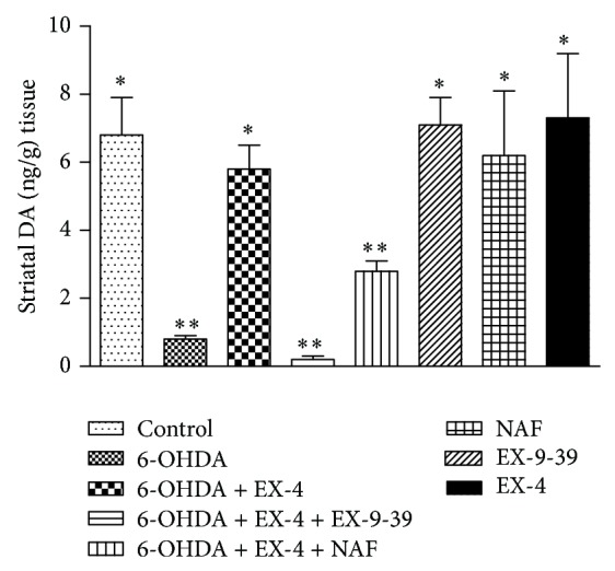
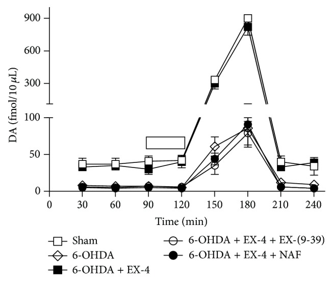
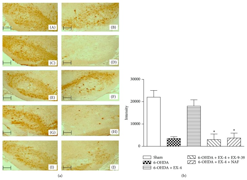
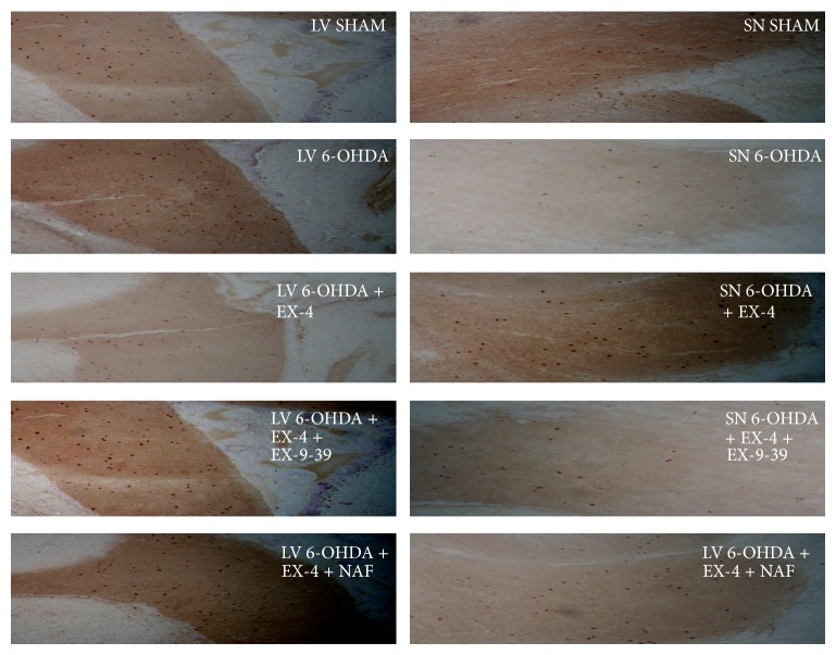
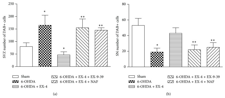
Glucagon-like peptide-1 receptor (GLP-1R) activation by exendin-4 (EX-4) is effective in preclinical models of Parkinson's disease (PD) and appears to promote neurogenesis even in severely lesioned rats. In the present study, we determined the effects of EX-4 on cellular BrdU incorporation in the rat subventricular zone (SVZ) and substantia nigra (SN). We also determined the specificity of this effect with the GLP-1R antagonist EX-(9-39) as well as the potential role of dopamine (DA) D3 receptors. Rats were administered 6-OHDA and 1 week later given EX-4 alone, with EX-(9-39) or nafadotride (D3 antagonist) and BrdU. Seven days later, rats were challenged with apomorphine to evaluate circling. Extracellular DA was measured using striatal microdialysis and subsequently tissue DA measured. Tyrosine hydroxylase and BrdU were verified using immunohistochemistry. Apomorphine circling was reversed by EX-4 in lesioned rats, an effect reduced by EX-4, while both EX-(9-39) and NAF attenuated this. 6-OHDA decreased extracellular and tissue DA, both reversed by EX-4 but again attenuated by EX-(9-39) or NAF. Analysis of BrdU+ cells in the SVZ revealed increases in 6-OHDA-treated rats which were reversed by EX-4 and antagonised by either EX-(9-39) or NAF, while in the SN the opposite profile was seen.


Figures

Figure 1
Figure 1
Effect of EX-4 on apomorphine-induced circling in the presence of 1 μg kg−1 EX-9-39 and 1 mg kg−1 NAF. Both antagonists were coadministered with EX-4 twice daily for seven days, one week after toxin injection. Circling was counted for 120 s 15 min after 0.5 mg kg−1 apomorphine injection. Results were analyzed using one-way ANOVA (F = 34.27; P < 0.0001) and a post hoc Bonferroni test to compare differences between groups. ∗P < 0.001 compared to shams, EX-4, NAF, and 6-OHDA + EX-4 groups; ∗∗P < 0.01 compared to shams, 6-OHDA, EX-4, EX-(9-39), NAF, 6-OHDA + EX-4 + EX-(9-39), and 6-OHDA + EX-4 + EX-(9-39).
Figure 2
Figure 2
Effect of 0.5 μg kg−1 EX-4 alone or in the presence of EX-9-39 or NAF on striatal tissue DA. Both antagonists were coadministered with EX-4 twice daily for seven days, one week after toxin injection. Results were analyzed using one-way ANOVA (F = 17.13; P < 0.001) and a post hoc Bonferroni test to compare differences between groups. Results were analyzed using one-way ANOVA (F = 34.27; P < 0.0001) and a post hoc Bonferroni test to compare differences between groups. ∗P < 0.001 compared to 6-OHDA, 6-OHDA + EX-4 + EX-(3-39), and 6-OHDA + EX-4 + NAF groups; ∗∗P < 0.01 compared to shams, 6-OHDA, 6-OHDA + EX-4, EX-4, EX-(9-39), and NAF.
Figure 3
Figure 3
Effect of EX-(9-39) or NAF on recovery of striatal extracellular DA by EX-4 in 6-OHDA-lesioned rats. Both antagonists were coadministered with EX-4 twice daily for seven days, one week after toxin. Bar at 90–120 min indicates infusion of 100 mM K+. Results were analyzed using two-way ANOVA (F = 13.32 between treatments and 43.52 overtime) and a post hoc Bonferroni multiple comparisons test. All basal and evoked 6-OHDA and EX-4 + either EX-(9-39) or NAF groups were significantly different (P < 0.01) from shams or 6-OHDA + EX-4 at all time points.
Figure 4
Figure 4
(a) Representative photomicrographs of 12 μm cryostat sections through the rat SN immunoassayed for TH. Nigral TH cell bodies appear dark brown in colour visualised using DAB staining; (A) and (B) represent right and left untreated SN, respectively; (D) represents 6-OHDA lesion assessed after two weeks compared to untreated side (C); (F) shows the effect of 0.5 μg/kg exendin-4 in a 6-OHDA-treated animal one week after lesion compared to untreated SN (E); (H) and (J) represent the effect of EX-9-39 or NAF coadministration, respectively, in 6-OHDA- and EX-4-treated animals compared to corresponding untreated sides (G) and (I). Scale bars—100 μm. (b) represents quantified data from immunohistochemical analysis of SN for TH. DAB staining was quantified using Lucia G image analysis software based on colour intensity. Results analyzed using one-way ANOVA and a post hoc Bonferroni test to compare differences between groups. ∗indicates significant differences compared to 6-OHDA + EX-4 group (ANOVA F[4, 25] = 16.99, ∗P < 0.05, n = 6 per group, confidence interval set at 95%).
Figure 5
Figure 5
Representative examples of selected 12 μm cryostat sections through the rat SVZ and SN immunoassayed for BrdU. BrdU+ cells appear dark brown visualised using DAB staining. SVZ sections were taken from the lateral ventricle (LV).
Figure 6
Figure 6
Quantified histograms of DAB-stained cell counts from 6 sections from each of the brains in the treatment group (n = 6) were used for analysis. DAB cell counts along the wall of the SVZ (a) or SN (b) were carried out manually. Results were analyzed using one-way ANOVA (SVZ F = 5.83; SN F = 3.847) and a post hoc Bonferroni test to compare relevant treatment groups. ∗P < 0.01 indicates significant difference compared to sham and 6-OHDA + EX-4. ∗∗P < 0.01 indicates significant difference compared to 6-OHDA and EX-4 treatment.

Similar articles

Cited by

References

    1. Bertilsson G., Patrone C., Zachrisson O., et al. Peptide hormone exendin-4 stimulates subventricular zone neurogenesis in the adult rodent brain and induces recovery in an animal model of Parkinson's disease. Journal of Neuroscience Research. 2008;86(2):326–338. doi: 10.1002/jnr.21483. - DOI - PubMed
    1. Harkavyi A., Abuirmeileh A., Lever R., Kingsbury A. E., Biggs C. S., Whitton P. S. Glucagon-like peptide 1 receptor stimulation reverses key deficits in distinct rodent models of Parkinson's disease. Journal of Neuroinflammation. 2008;5, article 19 doi: 10.1186/1742-2094-5-19. - DOI - PMC - PubMed
    1. Harkavyi A., Whitton P. S. Glucagon-like peptide 1 receptor stimulation as a means of neuroprotection. British Journal of Pharmacology. 2010;159(3):495–501. doi: 10.1111/j.1476-5381.2009.00486.x. - DOI - PMC - PubMed
    1. Kim S., Moon M., Park S. Exendin-4 protects dopaminergic neurons by inhibition of microglial activation and matrix metalloproteinase-3 expression in an animal model of Parkinson's disease. Journal of Endocrinology. 2009;202(3):431–439. doi: 10.1677/JOE-09-0132. - DOI - PubMed
    1. Li Y., Perry T., Kindy M. S., et al. GLP-1 receptor stimulation preserves primary cortical and dopaminergic neurons in cellular and rodent models of stroke and Parkinsonism. Proceedings of the National Academy of Sciences of the United States of America. 2009;106(4):1285–1290. doi: 10.1073/pnas.0806720106. - DOI - PMC - PubMed

LinkOut - more resources