

Review
doi: 10.1007/s40496-015-0045-z.
Application of Optical Coherence Tomography (OCT) for Diagnosis of Caries, Cracks, and Defects of Restorations
Affiliations
- PMID: 26317064
- PMCID: PMC4544493
- DOI: 10.1007/s40496-015-0045-z
Item in Clipboard
Review
Application of Optical Coherence Tomography (OCT) for Diagnosis of Caries, Cracks, and Defects of Restorations
Curr Oral Health Rep.
2015.
Abstract
Optical coherence tomography (OCT) is a noninvasive technique providing cross-sectional images of a tooth structure. This review describes the use of OCT for detecting dental caries, tooth fractures, and interfacial gaps in intraoral restorations. OCT can be a reliable and an accurate method and a safer alternative to X-ray radiography.
Keywords: Caries; Diagnosis; Interfacial gap; Optical coherence tomography (OCT); Tooth fracture.
Figures
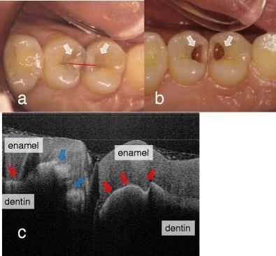
Dental caries in first and second premolars. a Occlusal view before the surgical treatment. Underlying dark shadows were visually observed at the first and second premolars (arrow). SS-OCT observation was performed along red line. b Occlusal view during the cavity preparation. Presence of deep lesions with softened dentin was obvious (white arrow). c SS-OCT image at red line in (a) before cavity preparation. Bright zone indicates the increased light scattering in porous demineralized tissue (blue arrow). A strong reflection penetrating along the DEJ indicates the lesion is “cavitated” (red arrow)
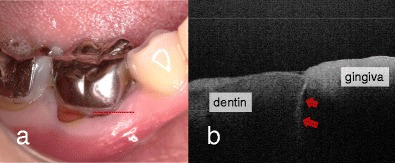
Mandibular first molar with occlusal pain. a Buccal view. Full coverage metal crown complicated the visual inspection and clinical diagnosis. SS-OCT observation was performed along the red line in order to image the cross-sectional view of cervical zone horizontally. b SS-OCT image at red line in (a). A strong reflection penetrating into dentin indicates presence of vertical crack in root dentin (red arrow)
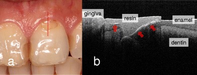
Clinical assessment of resin composite restoration. a Buccal view. Class V composite restoration was present with slight marginal discoloration but without any clinical symptoms. SS-OCT observation was performed along the red line. b SS-OCT image at red line in (a). Distinct white border between resin and enamel/dentin indicates “gap” (red arrow)
Similar articles
-
Optical Coherence Tomography.Dent Clin North Am. 2018 Jul;62(3):421-434. doi: 10.1016/j.cden.2018.03.004. Dent Clin North Am. 2018. PMID: 29903559 Review.
-
Evaluation of dental caries, tooth crack, and age-related changes in tooth structure using optical coherence tomography.Jpn Dent Sci Rev. 2020 Nov;56(1):109-118. doi: 10.1016/j.jdsr.2020.08.001. Epub 2020 Oct 2. Jpn Dent Sci Rev. 2020. PMID: 33033549 Free PMC article. Review.
-
Assessment of interfacial defects at composite restorations by swept source optical coherence tomography.J Biomed Opt. 2013 Jul;18(7):076018. doi: 10.1117/1.JBO.18.7.076018. J Biomed Opt. 2013. PMID: 23877771
-
Comparison of caries diagnostic modalities: A clinical study in 40 subjects.Lasers Surg Med. 2016 Dec;48(10):924-928. doi: 10.1002/lsm.22460. Epub 2016 Mar 21. Lasers Surg Med. 2016. PMID: 26997616 Free PMC article.
-
Assessment of enamel cracks at adhesive cavosurface margin using three-dimensional swept-source optical coherence tomography.J Dent. 2017 Jun;61:28-32. doi: 10.1016/j.jdent.2017.04.005. Epub 2017 Apr 19. J Dent. 2017. PMID: 28433536
Cited by
-
Tomographic Evaluation of the Internal Adaptation for Recent Calcium Silicate-Based Pulp Capping Materials in Primary Teeth.Biomed Res Int. 2021 May 8;2021:5523145. doi: 10.1155/2021/5523145. eCollection 2021. Biomed Res Int. 2021. PMID: 34046496 Free PMC article.
-
Exploiting Nanomaterials for Optical Coherence Tomography and Photoacoustic Imaging in Nanodentistry.Nanomaterials (Basel). 2022 Feb 1;12(3):506. doi: 10.3390/nano12030506. Nanomaterials (Basel). 2022. PMID: 35159853 Free PMC article. Review.
-
Optical Coherence Tomography for Patients with Developmental Disabilities: A Preliminary Study.Sensors (Basel). 2021 Nov 28;21(23):7940. doi: 10.3390/s21237940. Sensors (Basel). 2021. PMID: 34883945 Free PMC article.
-
Visualization of the pulp chamber roof and residual dentin thickness by spectral-domain optical coherence tomography in vitro.Lasers Med Sci. 2019 Jul;34(5):973-980. doi: 10.1007/s10103-018-2686-3. Epub 2018 Nov 13. Lasers Med Sci. 2019. PMID: 30426356
-
Use of optical coherence tomography in orthodontics.Exp Ther Med. 2021 Dec;22(6):1424. doi: 10.3892/etm.2021.10859. Epub 2021 Oct 11. Exp Ther Med. 2021. PMID: 34707705 Free PMC article.
References
-
- Fujimoto JG, Drexler W. Introduction to optical coherence tomography. In: Drexler W, Fujimoto JG, editors. Optical Coherence Tomography. Springer; 2008. pp. 1–45.
Publication types
LinkOut - more resources
Full Text Sources
Other Literature Sources
Miscellaneous