. 2013:2013:859257.
doi: 10.1155/2013/859257. Epub 2013 Jan 20.

Roles of Integrins and Intracellular Molecules in the Migration and Neuritogenesis of Fetal Cortical Neurons: MEK Regulates Only the Neuritogenesis

Affiliations

Roles of Integrins and Intracellular Molecules in the Migration and Neuritogenesis of Fetal Cortical Neurons: MEK Regulates Only the Neuritogenesis

Ujjwal K Rout. Neurosci J. 2013.

Abstract

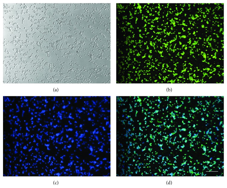
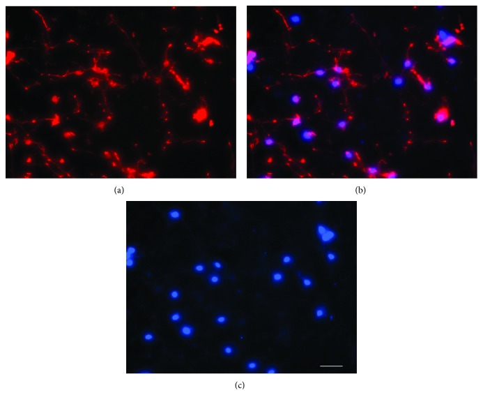
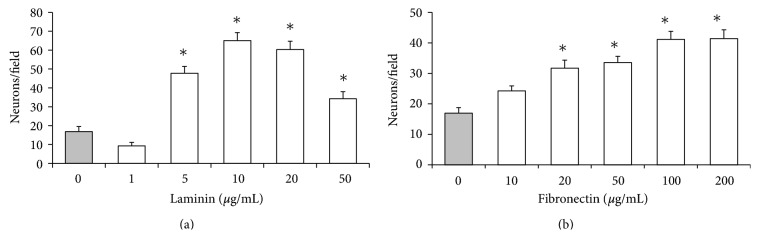
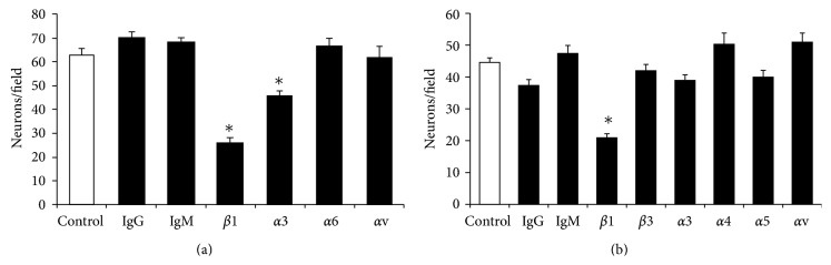
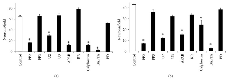
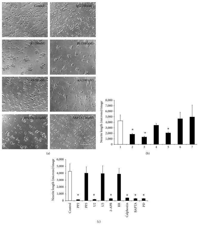
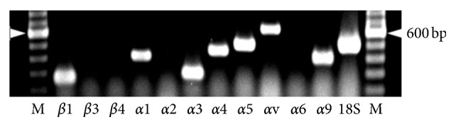
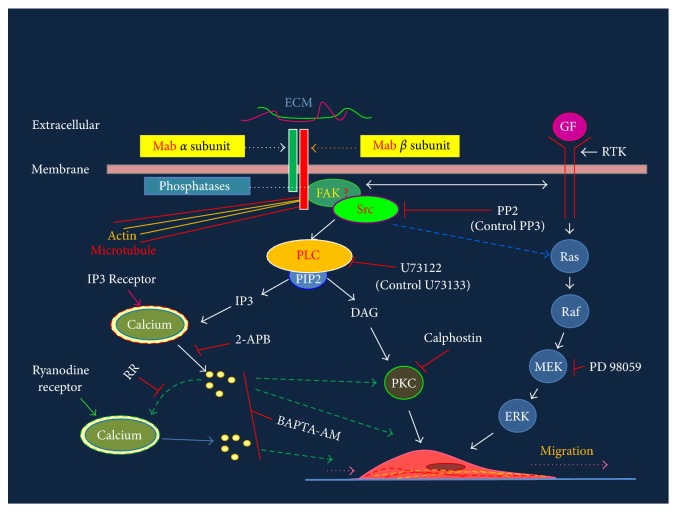
The roles of integrin subunits and intracellular molecules in regulating the migration and neuritogenesis of neurons isolated from 16.5 gestation days rat fetal cortices were examined using in vitro assays. Results showed that laminin supported the migration of fetal cortical neurons better than fibronectin and that the fetal cortical neurons migrated on laminin using β1 and α3 integrin subunits which make up the α3β1 integrin receptor. On fibronectin, the migration was mediated by β1 integrin subunit. Perturbation of src kinase, phospholipase C, or protein kinase C activity, inhibition of IP3 receptor mediated calcium release, or chelation of intracellular calcium inhibited both migration and neuritogenesis, whereas inhibition of growth factor signaling via MEK inhibited only the neuritogenesis. The detection of α1 and α9 transcripts suggested that the migration of fetal cortical neurons may also be mediated by α1β1 and α9β1 integrin receptors. Results showed that calcium may regulate migration and neuritogenesis by maintaining optimum levels of microtubules in the fetal cortical neurons. It is concluded that the fetal cortical neurons are fully equipped with the integrin signaling cascade required for their migration and neuritogenesis, whereas crosstalk between the integrin and growth-factor signaling regulate only the neuritogenesis.


Figures

Figure 1
Figure 1
Dissociated neurons in culture. Dissociated neurons at 2 h of plating on laminin-coated plates (a) immunostained for the neuronal marker NeuN (b) and nuclear stain Hoechst (c). Image (d) shows the overlaps of images (b) and (c). Bar100 microns. Note, Hoechst stained nuclei are out of focus because all images are captured at the same plane as (a).
Figure 2
Figure 2
Neurons at the undersurface of membrane. Neurons migrated at the undersurface of membrane expressed neuronal marker MAP2 (a). Neuronal nuclei stained with Hoechst (c). Overlapping images (a) and (c) are shown in (b). Bar 100 microns.
Figure 3
Figure 3
ECM effects on the migration of neurons. Neurons per field of Boyden membrane coated with laminin (a) or fibronectin (b). ∗Migration of cortical neurons were significantly (P < 0.05) higher on membranes coated with laminin at 5, 10, 20, and 50 μM or 20 to 200 μM fibronectin compared to those coated with only Poly-D lysine (shaded bar). Migration of neurons was highest at 10 μM laminin but it was not significantly different than those at 20 μM laminin. Migration of neurons on fibronectin-coated membranes increased with concentration to reach its maximum at 100 μM that was not significantly (P > 0.05) different than membranes coated with 200 μM fibronectin.
Figure 4
Figure 4
Effects of antibodies on the migration of fetal cortical neurons. Monoclonal antibodies (shown below) against β1 and α3 integrin subunits significantly (* P < 0.05) inhibited the migration of neurons (Neurons/field) on membranes coated with laminin (10 μg/mL) (a). The migrations of neurons on laminin-coated membranes were not significantly (P > 0.05) altered by antibody against α6 or αv subunit (the negative control) and control antibodies (IgG or IgM). The migrations of neurons on fibronectin (100 μg/mL) coated membranes were significantly (* P < 0.05) inhibited by the antibody against β1 integrin subunit only (b). The migrations of neurons on fibronectin-coated membranes were not altered by control antibodies (IgG or IgM) at P > 0.05.
Figure 5
Figure 5
Effects of inhibitors on the migration of cortical neurons. Inhibitors (shown below) of Src kinase (PP2) and Phospholipase Cγ activity (U2) inhibited the migration of fetal cortical neurons (Neurons/field) on laminin-(a) or fibronectin-(b) coated membranes (* P < 0.05). No significant changes in the migration of neurons occurred in the presence of control compound PP3 or U3 (P > 0.05). Inhibitor of protein kinase C (light-activated Calphostin), inhibitor of IP3 mediated calcium release (2-APB), and intracellular calcium chelator (BAPTA-AM) inhibited the migration of neurons significantly (* P < 0.05) on laminin-(a) or fibronectin-(b) coated membranes. Ruthenium Red (inhibitor calcium-induced calcium release) and PD (inhibitor of Mitogen activated kinase kinase) did not alter the migration of neurons on laminin-(a) or fibronectin-(b) coated membranes (P > 0.05).
Figure 6
Figure 6
Effects of antibodies, calcium modulators, and pharmacological inhibitors on neuritogenesis. (a) Representative images showing neuritogenesis in the absence (control) or presence of control IgG (50 nMole), antibodies against β1 (50 or 100 nMole) or α3 (50 or 100 nMole) integrin subunits on laminin. BAPTA-AM at low concentration (2.5 μM) inhibited neuritogenesis and at higher concentration (10 μM) totally abolished neuritogenesis. Bar150 microns. (b) Mean + standard error of mean values of neurite lengths/image in untreated neurons (1) and those treated with control IgG, monoclonal antibody against β1 or α3 integrin subunit at 50 nMole or 100 nMole concentration. Neurite lengths reduced significantly (* P < 0.05) in neurons treated with antibody against β1 integrin subunit at both 50 nMole (2) and 100 nMole (3) concentrations compared to control (1). Neurons treated with monoclonal antibody against α3 integrin subunit at 50 nMole (4) were lower than the control (1) but was not statistically significant (P > 0.05). Neurons treated with monoclonal antibody against α3 integrin subunit at 100 nMole (5) significantly inhibited the neuritogenesis (* P < 0.055). Neuritogenesis was not altered in presence of 50 (6) or 100 nMole (7) control IgG (P > 0.05). (c) Mean + standard error of mean values of neurite lengths/image of neurons treated with PP2, PP3, U2, U3, 2-APB, activated Calphostin, BAPTA-AM (2.5 μM), and PD for 22 h in culture (filled bars) were significantly different (* P < 0.05) from untreated neurons (blank bar) and negative controls (PP3 or U3) (filled bars). No significant changes in neurite lengths (P > 0.05) per image were recorded in neurons treated with RR.
Figure 7
Figure 7
BAPTA-AM reduced microtubule expression. Microtubule expression in the untreated neurons (a) and those treated with BAPTA-AM (10 μM) (b) in culture. Panel (c) and (d) shows nuclear stain in the control and BAPTA-AM treated neurons. Bar 100 microns. Note, Hoechst stained nuclei in (c) and (d) are out of focus because images are captured at the same plane as (a) or (b), respectively. Bar 100 microns.
Figure 8
Figure 8
Western blotting detection of phosphorylated PLC-γ1 in neurons. Phosphorylated form of PLC-γ1 protein at tyrosine 783 was detected in the neurons at 6 h of culture on laminin simultaneously with β-actin. Lanes 1 and 2 represent lysate loaded from different neuronal preparations. Molecular weight sizes of phosphorylated form of PLC-γ isoform and β-actin are shown in parentheses.
Figure 9
Figure 9
Transcripts of integrin subunits in the fetal cortical neurons. Ethidium bromide stained PCR products after agarose gel electrophoresis. Target mRNA species are shown at the bottom of the image. The 600 bp band of the 100 bp marker (M) is shown by arrows heads on sides.
Figure 10
Figure 10
Schematic of integrin signaling cascade and its perturbation with antibodies and pharmacological agents. Integrin subunits (red and green bars on top) are shown intercalated in the membrane and interacting with ECM molecule (red and green horizontal lines). Molecules involved in signaling events are labeled and arrows point to the directions of signaling that starts with the engagement of integrin subunit with the extracellular matrix. Directions of calcium mediated signaling are shown by dashed green arrows. Inhibitors (see Table 2) used for blocking signaling molecules and paths are shown by red blocks. Cross-talk between integrin and GF signaling is shown by double headed horizontal arrow close to membrane. Mab: monoclonal antibody, GF: growth factor, RTK: receptor tyrosine kinase, MEK: MAP kinase kinase, PLC: phospholipase C, PIP2: phosphotidal inositol biphosphate, IP3: inositol (1,4,5)-triphosphate (IP3), DAG: diacylglycerol and PKC: protein kinase C, Ras: G protein, Raf: MAPKKK, MEK: MAPKK, ERK: extracellular signal regulated kinase (MAPK).

References

    1. Métin C., Baudoin J. P., Rakić S., Parnavelas J. G. Cell and molecular mechanisms involved in the migration of cortical interneurons. European The Journal of Neuroscience. 2006;23(4):894–900. doi: 10.1111/j.1460-9568.2006.04630.x. - DOI - PubMed
    1. Gupton S. L., Gertler F. B. Integrin signaling switches the cytoskeletal and exocytic machinery that drives neuritogenesis. Developmental Cell. 2010;18(5):725–736. doi: 10.1016/j.devcel.2010.02.017. - DOI - PMC - PubMed
    1. Anton E. S., Kreidberg J. A., Rakic P. Distinct functions of α3 and α(v) integrin receptors in neuronal migration and laminar organization of the cerebral cortex. Neuron. 1999;22(2):277–289. - PubMed
    1. Campbell I. D., Humphries M. J. Integrin structure, activation, and interactions. Cold Spring Harbor Perspectives in Biology. 2011;3(3):1–14. doi: 10.1101/cshperspect.a004994. - DOI - PMC - PubMed
    1. Qin J., Vinogradova O., Plow E. F. Integrin bidirectional signaling: a molecular view. PLoS Biology. 2004;2(6, article e169) doi: 10.1371/journal.pbio.0020169. - DOI - PMC - PubMed

LinkOut - more resources