. 2015 Aug 28;10(8):e0136888.
doi: 10.1371/journal.pone.0136888. eCollection 2015.

A Multiple Antigenic Peptide Mimicking Peptidoglycan Induced T Cell Responses to Protect Mice from Systemic Infection with Staphylococcus aureus

Affiliations

A Multiple Antigenic Peptide Mimicking Peptidoglycan Induced T Cell Responses to Protect Mice from Systemic Infection with Staphylococcus aureus

Xiang-Yu Wang et al. PLoS One. .

Abstract

Due to the enormous capacity of Staphylococcus aureus to acquire antibiotic resistance, it becomes imperative to develop vaccines for decreasing the risk of its life-threatening infections. Peptidoglycan (PGN) is a conserved and major component of S. aureus cell wall. However, it has not been used as a vaccine candidate since it is a thymus-independent antigen. In this study, we synthesized a multiple antigenic peptide, named MAP27, which comprised four copies of a peptide that mimics the epitope of PGN. After immunization with MAP27 five times and boosting with heat-inactivated bacterium one time, anti-MAP27 serum bound directly to S. aureus or PGN. Immunization with MAP27 decreased the bacterial burden in organs of BALB/c mice and significantly prolonged their survival time after S. aureus lethal-challenge. The percentage of IFN-γ(+)CD3(+) T cells and IL-17(+)CD4(+) T cells in spleen, as well as the levels of IFN-γ, IL-17A/F and CCL3 in spleen and lung, significantly increased in the MAP27-immunized mice after infection. Moreover, in vitro incubation of heat-inactivated S. aureus with splenocytes isolated from MAP27-immunized mice stimulated the production of IFN-γ and IL-17A/F. Our findings demonstrated that MAP27, as a thymus-dependent antigen, is efficient at eliciting T cell-mediated responses to protect mice from S. aureus infection. This study sheds light on a possible strategy to design vaccines against S. aureus.


Conflict of interest statement

Competing Interests: The authors have declared that no competing interests exist.

Figures

Fig 1
Fig 1. Both SP27 and MAP27 bind to anti-PGN mAb specifically.
(A) The sequences of SP27 (Biotin-SASPHHHSRLRSESGG) and SP27’ (Biotin-SPHHHSRLRSESSAGG). Underlined letters represent nonspecific flanking amino acids. (B) Both SP27 and SP27’ bind to anti-PGN mAb in a dose-dependent manner. (C) Both SP27 and SP27’ (100 μg/ml) specifically bind to anti-PGN mAb, but not to anti-LTA mAb. For ELISA assays in panel B and C, anti-PGN mAb or anti-LTA mAb was used to coat the wells at a concentration of 5 μg/ml, SP27, SP27’ or non-specific peptide L2 (in panel B, sequence: Biotin-HSGHWDFRQWWQPSGG) was then added at indicated concentrations and incubated at 37°C for 40 min, followed by detection with HRP-labeled streptavidin. (D) The structure diagram of MAP27. MAP27 was synthesized in a tetra-branched form that contains four copies of a sequence (SASPHHHSRLRSESGG) that mimics PGN epitope. (E) MAP27 binds to anti-PGN mAb specifically. (F) MAP27 binds to anti-S. aureus polyclonal antibodies specifically. For ELISA assays in panel E and F, MAP27 or MAPctrl was used to coat the wells of a microplate. Anti-PGN mAb or anti-S. aureus polyclonal antibodies was added, followed by detection with HRP-labeled antibodies. The absorbance was measured at OD450nm. The results are shown as means ±SEM. * P<0.05, ** P<0.01, *** P<0.001.
Fig 2
Fig 2. BALB/c mice immunized with MAP27 produced anti-MAP27, anti-PGN and anti-S.aureus antibodies.
(A) Titers of anti-MAP27 serum during the period of immunization. 96-well plates were coated with MAP27. Serum samples from mice immunized with MAP27, MAPctrl or blank were pooled and added in a 10-fold serial dilution, followed by incubation with HRP-conjunctive goat anti-mouse IgG. (B) Sera from MAP27-immunized mice bind to PGN after the fifth MAP immunization. (C) Sera from MAP27-immunized mice bind to S. aureus after boosting with heat-killed S.aureus. For ELISA assays in panel B and C, 96-well plates were coated with PGN or sonicated S. aureus fragments. Serum samples were added in a 1/200 dilution as primary antibodies, followed by incubation with HRP-conjunctive goat anti-mouse IgG. The absorbance was measured at OD450nm. The results are presented as means ±SEM. * P<0.05, ** P<0.01, n = 5–9 mice/group.
Fig 3
Fig 3. Immunization with MAP27 protects mice against S. aureus infection.
(A) Survival analysis of mice with or without immunization after S. aureus infection. Five days after boosting with heat-inactivated S. aureus, all of BALB/c mice (including MAP27-immunized, MAPctrl-immunized and blank control, n = 8–9 mice/group) were challenged with S. aureus (ATCC 25923, 5×10⁸ CFU/mouse) by intravenous injection. Survival rate were monitored for seven days after infection. (B) Bacterial numbers in kidney from three groups of mice (n = 4 mice per group) were measured after three days infection of S. aureus (2×10⁷ CFU/mouse). (C) Bacterial numbers in lung from different groups of mice (n = 5 mice per group) were measured after three days infection. * P<0.05, ** P<0.01, *** P<0.001.
Fig 4
Fig 4. Immunization with MAP27 promoted IFN-γ, IL-17A/F and CCL3 production in organs post infection with S. aureus.
Five days after boosting with heat-inactivated S. aureus, all of the mice (n = 5 mice per group) were infected with S. aureus (2×10⁷ CFU/mouse) for three days. Spleen and lung were aseptically taken and homogenized with sterile PBS. The supernatants were pooled and analyzed by ELISA. The concentrations of IFN-γ (panel A and D), IL-17A/F (panel B and E), and chemokine ligand 3 (CCL3, panel C and F) were measured in spleen and lung, respectively. * P<0.05, ** P<0.01, *** P<0.001.
Fig 5
Fig 5. Vaccination with MAP27 primed IFN-γ+CD3+ T cells and IL-17+CD4+ T cells after S. aureus infection.
Five days after the last boost with heat-inactivated S. aureus, all of the mice were infected with S. aureus (2×10⁷ CFU/mouse). Spleen cells (n = 5 mice per group) were harvested three days post infection and incubated with cocktail (including PMA and ionomycin) and BFA for 6 h. Cells were then fixed, permeabilized and stained with FITC-anti CD3, APC-anti CD4, PE-anti IFN-γ and PE-anti IL-17A mAb, respectively. Intracellular cytokine analysis was performed by flow cytometry. All lymphocytes were first gated based on FSC and SSC. CD3+ lymphocytes were then further gated from the lymphocyte cluster according to the fluorescence signal intensities. The results were presented as mean±SEM. (A) The percentage of IFN-γ+CD3+CD4+ cells. (B) The percentage of IFN-γ+CD3+CD4- cells. (C) Representative flow cytometry plots showing the percentage of IFN-γ+CD3+ cells in spleen. (D) The percentage of IL-17A+CD3+CD4+ cells. (E) The percentage of IL-17A+CD3+CD4- cells. (F) Representative flow cytometry plots showing the percentage of IL-17A+CD3+ cells in spleen. * P<0.05. ns: not significantly different between two groups (p>0.05).
Fig 6
Fig 6. In vitro MAP27 treatment promoted the production of IFN-γ in splenocytes from MAP27-immunized mice.
Five days after the last boost immunization, splenocytes isolated from the mice (n = 5 mice per group) were cultured at 2×10⁵/well in U-bottom 96-well plates and stimulated with MAP27 (100 μg/ml) for 24 h. The concentrations of IL-2 (A), IFN-γ (B), and IL-4 (C) in supernatant were measured by ELISA. (D) Frequency of IFN-γ-producing cells induced by MAP27. Splenocytes isolated from the mice (n = 5 mice per group) were cultured at 4×10⁵/well in pre-coated plates, and stimulated with MAP27 (10 μg/ml) plus anti-mouse CD28 antibody (1 μg/ml) for 24 h. The number of IFN-γ-producing cells was then measured by ELISPOT and shown in box and whisker plots. (E) Representation of one of the ELISPOT assays. ** P<0.01; *** P<0.001; ns: P>0.05.
Fig 7
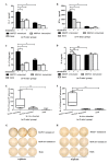
Fig 7. Heat-inactivated S. aureus promoted Th1 and Th17 cell responses in the spleen of MAP27-immunized mice.
Five days after the last booster with heat-inactivated bacteria, the splenocytes isolated from the mice (n = 5 mice per group) were cultured (2×10⁵/well), and stimulated with 2×10⁴ CFU/well heated-inactivated S. aureus for 72 h. The concentrations of IL-2 (A), IFN-γ (B), IL-17A/F (C) and IL-4 (D) were measured by ELISA. The isolated splenocytes were also cultured at 4×10⁵/well in pre-coated plates and stimulated with heat-inactivated S. aureus for 24 h. Frequency of IFN-γ-producing (E) and IL-17A-producing cells (F) was determined by ELISPOT. Cell numbers were shown in box and Whisker plots. (G) Representation of one of the ELISPOT assays for IFN-γ-producing cells. (H) Representation of one of the ELISPOT assays for IL-17A-producing cells. * P<0.05; ** P<0.01; ns: P>0.05.

Similar articles

Cited by

References

    1. Foster TJ. Colonization and infection of the human host by staphylococci: adhesion, survival and immune evasion. Vet Dermatol. 2009; 20(5–6): 456–470. 10.1111/j.1365-3164.2009.00825.x - DOI - PubMed
    1. Lowy FD. Staphylococcus aureus infections. N Engl J Med. 1998; 339(8): 520–532. - PubMed
    1. DeLeo FR, Otto M, Kreiswirth BN, Chambers HF. Community-associated meticillin-resistant Staphylococcus aureus. Lancet. 2010; 375(9725): 1557–1568. 10.1016/S0140-6736(09)61999-1 - DOI - PMC - PubMed
    1. Fowler VJ, Proctor RA. Where does a Staphylococcus aureus vaccine stand? Clin Microbiol Infect. 2014; 20 Suppl 5: 66–75. 10.1111/1469-0691.12570 - DOI - PMC - PubMed
    1. Broughan J, Anderson R, Anderson AS. Strategies for and advances in the development of Staphylococcus aureus prophylactic vaccines. Expert Rev Vaccines. 2011; 10(5): 695–708. 10.1586/erv.11.54 - DOI - PubMed

Publication types