. 2015 Aug 28;9(8):e0003990.
doi: 10.1371/journal.pntd.0003990. eCollection 2015.

Increased Nucleosomes and Neutrophil Activation Link to Disease Progression in Patients with Scrub Typhus but Not Murine Typhus in Laos

Affiliations

Increased Nucleosomes and Neutrophil Activation Link to Disease Progression in Patients with Scrub Typhus but Not Murine Typhus in Laos

Daniel H Paris et al. PLoS Negl Trop Dis. .

Abstract

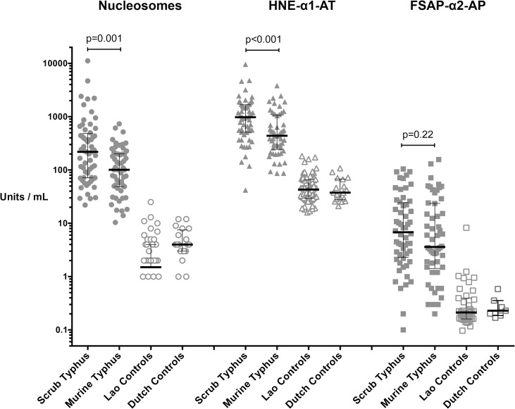
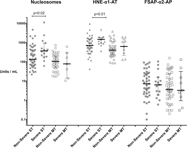
Cell-mediated immunity is essential in protection against rickettsial illnesses, but the role of neutrophils in these intracellular vasculotropic infections remains unclear. This study analyzed the plasma levels of nucleosomes, FSAP-activation (nucleosome-releasing factor), and neutrophil activation, as evidenced by neutrophil-elastase (ELA) complexes, in sympatric Lao patients with scrub typhus and murine typhus. In acute scrub typhus elevated nucleosome levels correlated with lower GCS scores, raised respiratory rate, jaundice and impaired liver function, whereas neutrophil activation correlated with fibrinolysis and high IL-8 plasma levels, a recently identified predictor of severe disease and mortality. Nucleosome and ELA complex levels were associated with a 4.8-fold and 4-fold increased risk of developing severe scrub typhus, beyond cut off values of 1,040 U/ml for nucleosomes and 275 U/ml for ELA complexes respectively. In murine typhus, nucleosome levels associated with pro-inflammatory cytokines and the duration of illness, while ELA complexes correlated strongly with inflammation markers, jaundice and increased respiratory rates. This study found strong correlations between circulating nucleosomes and neutrophil activation in patients with scrub typhus, but not murine typhus, providing indirect evidence that nucleosomes could originate from neutrophil extracellular trap (NET) degradation. High circulating plasma nucleosomes and ELA complexes represent independent risk factors for developing severe complications in scrub typhus. As nucleosomes and histones exposed on NETs are highly cytotoxic to endothelial cells and are strongly pro-coagulant, neutrophil-derived nucleosomes could contribute to vascular damage, the pro-coagulant state and exacerbation of disease in scrub typhus, thus indicating a detrimental role of neutrophil activation. The data suggest that increased neutrophil activation relates to disease progression and severe complications, and increased plasma levels of nucleosomes and ELA complexes represent independent risk factors for developing severe scrub typhus.


Conflict of interest statement

The authors have declared that no competing interests exist.

Figures

Fig 1
Fig 1. Markers of cell death and neutrophil activation in patients with scrub typhus, murine typhus and controls.
Markers of cell death and neutrophil activation. Plasma levels of nucleosomes, human neutrophil elastase (ELA) complexes and Factor VII activating protease anti-plasmin complexes (FSAP) were significantly higher in patients with scrub typhus (ST) and murine typhus (MT) than in Asian (Laos) and Caucasian (Dutch) controls (p-values all <0.0001). On comparison between ST and MT, the nucleosome and ELA-complex plasma levels were significantly higher in ST than in MT, but not the FSAP levels. Data are expressed as median with interquartile range (whiskers) of admission samples.
Fig 2
Fig 2. Neutrophils are prominent in the eschars of scrub typhus patients.
The necrotic zone of a human eschar biopsy in a patient with scrub typhus is characterized by high-density neutrophil infiltrates with partially karyorrhectic neutrophils. Panel A: The granulocyte-dense zones are predominantly located adjacent to the central necrotic zone (N), at the dermal-epidermal junction and in superficial dermal infiltrates (anti-neutrophil elastase stained red, patient TM2193, magnification x400). Panel B: Immunofluorescence image of a neutrophil rich infiltrate of the upper dermis containing Orientia tsutsugamushi stained bright apple green (in-house anti-56kDa Orientia mAb) and neutrophils stained red (anti-CD15 antibody, Abcam By87), patient TM2193, magnification x600). Panels C and D: Immunohistochemical staining of neutrophils in brown, using anti-CD15 antibodies (Abcam By87). Panel D is an enlargement and corresponds to the red square insert, demonstrating the typical granulation of neutrophils. Patient TM 2644, magnification x100, insert x600. Panels E-H: Controls included anti-56kDa Orientia mAb positive control in heavily-infected L929 cells (immunohistochemistry, x600, panel E); anti-CD15 antibody (Abcam By87) positive control in uninfected human tonsil (immunohistochemistry, x400, panel F) and skin (immunofluorescence with anti-CD15 in red, x100, panel G); and anti-56kDa Orientia mAb negative control in chronically inflamed (psoriasis) human skin (immunofluorescence with HLADR in red and anti-56kDa in green, x400, panel F).
Fig 3
Fig 3. Nucleosomes and neutrophil elastase (ELA) complex plasma levels are raised in severe scrub typhus.
Plasma levels of nucleosomes, ELA complexes and FSAP complexes were significantly higher in patients with severe scrub typhus than in non-severe cases (p = 0.02 and p = 0.01 respectively), but not FSAP complexes. Increased disease severity was not reflected by higher levels of nucleosomes or ELA complexes in murine typhus patients, although the p-values of p = 0.06 and p = 0.08, respectively, are suggestive of a trend. Scrub typhus data are bold, while murine typhus data are outlined. Data are expressed as median with inter- quartile range (whiskers) of admission samples.

Similar articles

Cited by

References

    1. Suttinont C, Losuwanaluk K, Niwatayakul K, Hoontrakul S, Intaranongpai W, et al. (2006) Causes of acute, undifferentiated, febrile illness in rural Thailand: results of a prospective observational study. Ann Trop Med Parasitol 100: 363–370. - PubMed
    1. Mayxay M, Castonguay-Vanier J, Chansamouth V, Dubot-Pérès A, Paris DH, et al. (2013) Causes of non-malarial fever in Laos: a prospective study. Lancet Glob Health 1: e46–e54. - PMC - PubMed
    1. Phongmany S, Rolain J-M, Phetsouvanh R, Blacksell SD, Soukkhaseum V, et al. (2006) Rickettsial infections and fever, Vientiane, Laos. Emerg Infect Dis 12: 256–262. - PMC - PubMed
    1. Chheng K, Carter MJ, Emary K, Chanpheaktra N, Moore CE, et al. (2013) A prospective study of the causes of febrile illness requiring hospitalization in children in cambodia. PLoS One 8: e60634 10.1371/journal.pone.0060634 - DOI - PMC - PubMed
    1. WHO (1999) World Health Organization. Recommended surveillance standards WHO/CDS/CDR/ISR/99.2, second edition.

Publication types