

Novel Preparation Methods of (52)Mn for ImmunoPET Imaging
- PMID: 26317429
- PMCID: PMC4618027
- DOI: 10.1021/acs.bioconjchem.5b00414
Novel Preparation Methods of (52)Mn for ImmunoPET Imaging
Abstract
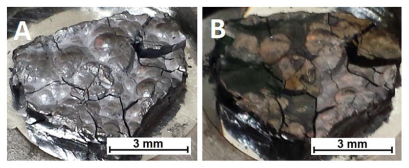
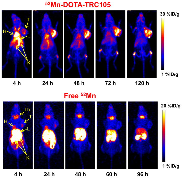
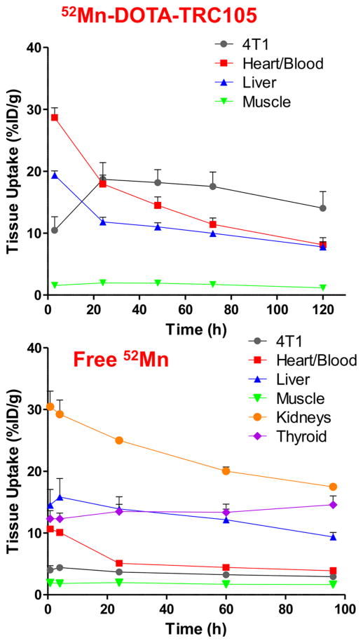
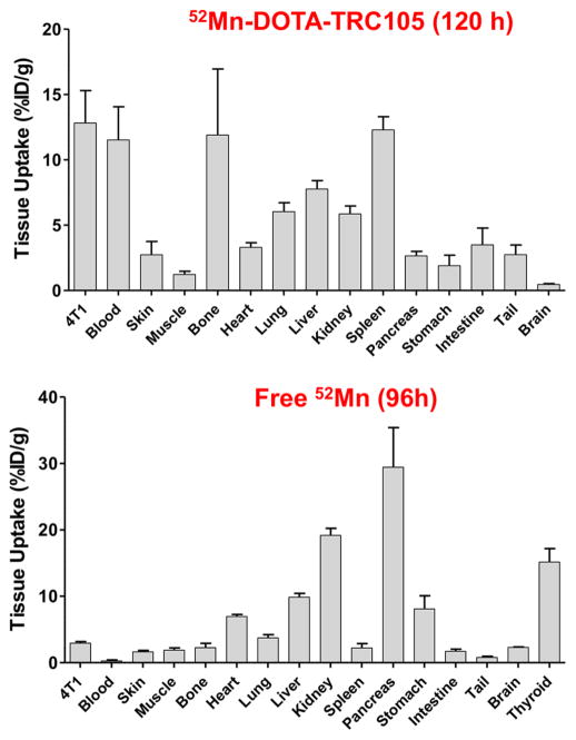
(52)Mn (t1/2 = 5.59 d, β(+) = 29.6%, Eβave = 0.24 MeV) shows promise in positron emission tomography (PET) and in dual-modality manganese-enhanced magnetic resonance imaging (MEMRI) applications including neural tractography, stem cell tracking, and biological toxicity studies. The extension to bioconjugate application requires high-specific-activity (52)Mn in a state suitable for macromolecule labeling. To that end a (52)Mn production, purification, and labeling system is presented, and its applicability in preclinical, macromolecule PET is shown using the conjugate (52)Mn-DOTA-TRC105. (52)Mn is produced by 60 μA, 16 MeV proton irradiation of natural chromium metal pressed into a silver disc support. Radiochemical separation proceeds by strong anion exchange chromatography of the dissolved Cr target, employing a semiorganic mobile phase, 97:3 (v:v) ethanol:HCl (11 M, aqueous). The method is 62 ± 14% efficient (n = 7) in (52)Mn recovery, leading to a separation factor from Cr of (1.6 ± 1.0) × 10(6) (n = 4), and an average effective specific activity of 0.8 GBq/μmol (n = 4) in titration against DOTA. (52)Mn-DOTA-TRC105 conjugation and labeling demonstrate the potential for chelation applications. In vivo images acquired using PET/CT in mice bearing 4T1 xenograft tumors are presented. Peak tumor uptake is 18.7 ± 2.7%ID/g at 24 h post injection and ex vivo (52)Mn biodistribution validates the in vivo PET data. Free (52)Mn(2+) (as chloride or acetate) is used as a control in additional mice to evaluate the nontargeted biodistribution in the tumor model.
Conflict of interest statement
The authors declare the following competing financial interest(s): Charles P. Theuer is an employee of TRACON Pharmaceuticals. The other authors have no competing interests to declare.
Figures
References
-
- Pirko I, Gamez J, Shearier E, Raman M, Johnson A, Macura S. Manganese Enhanced MRI (MEMRI) in Murine CNS Inflammatory Disease Models (S21. 007) Neurology. 2012;78:S21.007.
-
- Yamada M, Momoshima S, Masutani Y, Fujiyoshi K, Abe O, Nakamura M, Aoki S, Tamaoki N, Okano H. Diffusion-Tensor Neuronal Fiber Tractography and Manganese-enhanced MR Imaging of Primate Visual Pathway in the Common Marmoset: Preliminary Results. Radiology. 2008;249:855–864. - PubMed
Publication types
MeSH terms
Substances
Grants and funding
LinkOut - more resources
Full Text Sources
Other Literature Sources
