. 2015 Aug 27;162(5):987-1002.
doi: 10.1016/j.cell.2015.07.058.

Viral and Cellular Genomes Activate Distinct DNA Damage Responses

Affiliations

Viral and Cellular Genomes Activate Distinct DNA Damage Responses

Govind A Shah et al. Cell. .

Abstract

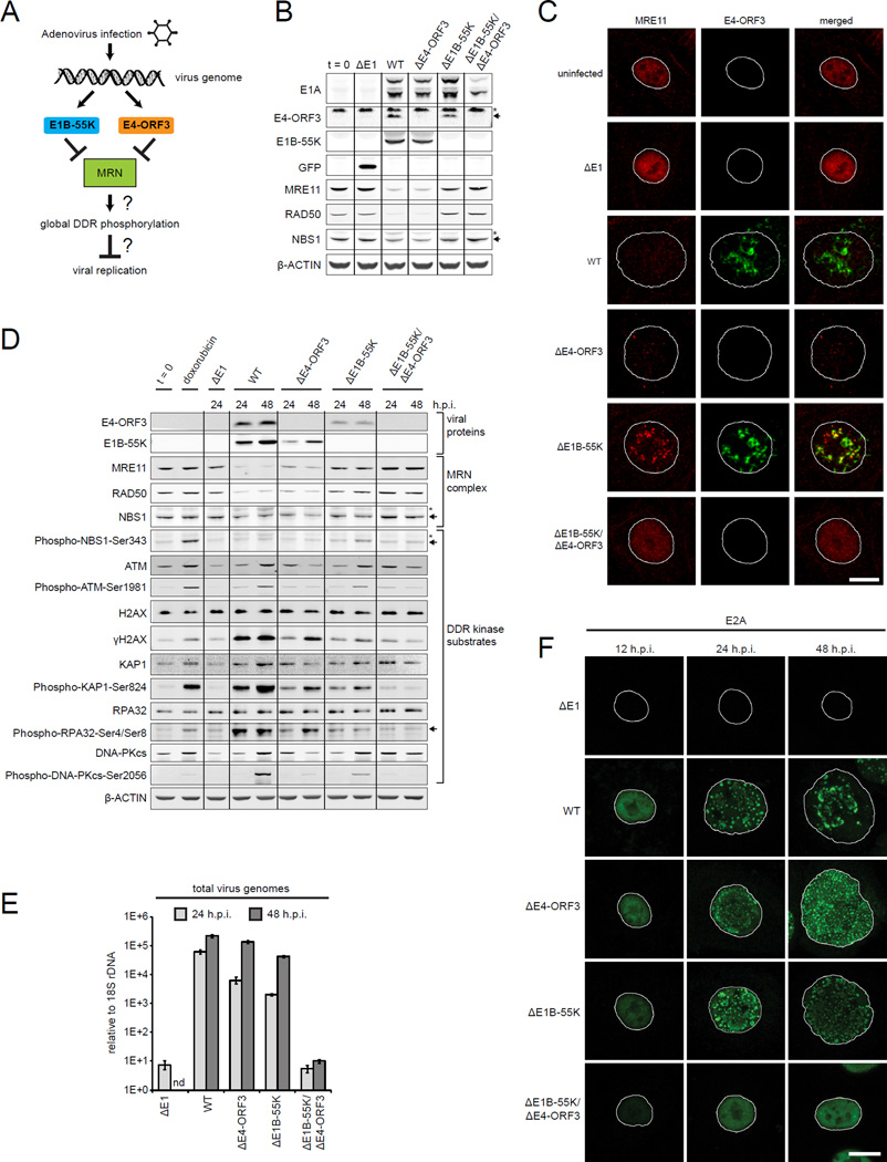
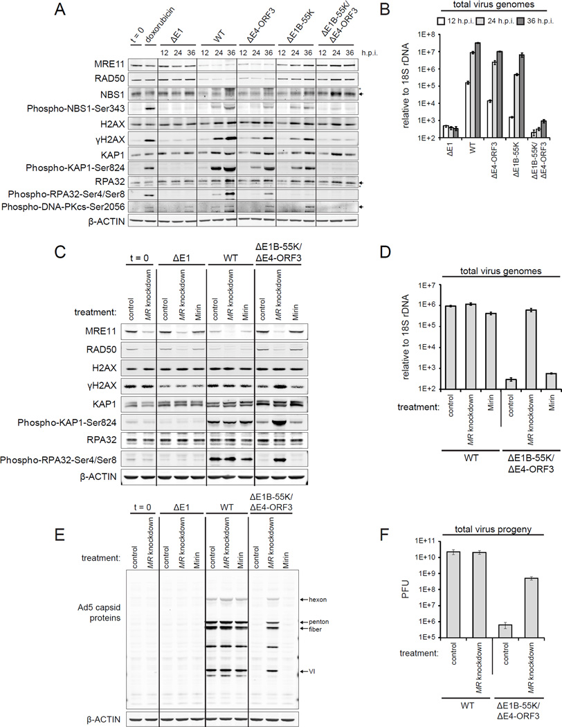
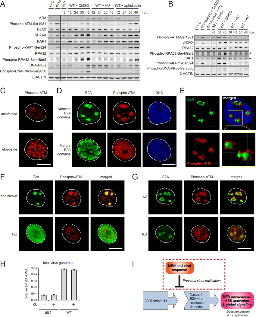
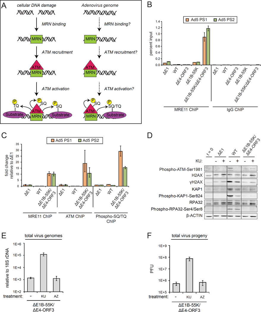
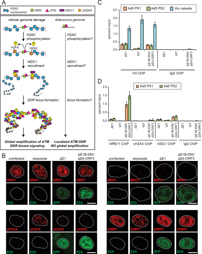
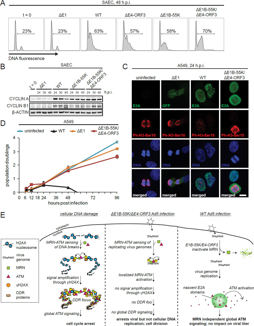
In response to cellular genome breaks, MRE11/RAD50/NBS1 (MRN) activates a global ATM DNA damage response (DDR) that prevents cellular replication. Here, we show that MRN-ATM also has critical functions in defending the cell against DNA viruses. We reveal temporally distinct responses to adenovirus genomes: a critical MRN-ATM DDR that must be inactivated by E1B-55K/E4-ORF3 viral oncoproteins and a global MRN-independent ATM DDR to viral nuclear domains that does not impact viral replication. We show that MRN binds to adenovirus genomes and activates a localized ATM response that specifically prevents viral DNA replication. In contrast to chromosomal breaks, ATM activation is not amplified by H2AX across megabases of chromatin to induce global signaling and replicative arrest. Thus, γH2AX foci discriminate "self" and "non-self" genomes and determine whether a localized anti-viral or global ATM response is appropriate. This provides an elegant mechanism to neutralize viral genomes without jeopardizing cellular viability.


Figures

Figure 1
Figure 1. E1B-55K and E4-ORF3 inactivate MRN and are critical for viral genome replication but do not prevent global DDR kinase signaling
(A) Adenovirus 5 (Ad5) early proteins target MRN through two independent mechanisms. (B) Human small airway epithelial cells (SAECs) were infected with mock (ΔE1), wild type Ad5 (WT), ΔE4-ORF3, ΔE1B-55K or ΔE1B-55K/ΔE4-ORF3 viruses. Protein lysates were collected at 24 h.p.i. and immunoblotted as indicated. “t = 0” indicates uninfected cells. Arrows and asterisks indicate specific and non-specific bands, respectively. (C–E) SAECs were infected as indicated. (C) SAECs were fixed at 24 h.p.i. and stained for E4-ORF3 (green) and MRE11 (red). DNA was counterstained with Hoechst. Nuclei are outlined in white. Scale bar: 10 μm. (D) Protein lysates were immunoblotted as indicated. Doxorubicin was used as a positive control for cellular DNA damage. (E) Viral genomes were quantified by Q-PCR and normalized to 18S rDNA; error bars indicate standard deviation (nd: not done). (F) Virus genome replication domains were visualized in infected SAECs by E2A immunofluorescence (green). Scale bar: 10 µm. (See also Figure S1.)
Figure 2
Figure 2. The inactivation of the MRN complex is critical for viral genome replication
(A and B) A549 cells were infected as indicated and harvested at 12, 24, and 36 h.p.i. (A) Protein lysates were immunoblotted as indicated. (B) Viral genomes were quantified by Q-PCR; error bars indicate standard deviation. (C–F) A549 cells were transfected with either control siRNA (control) or MRE11 and RAD50 siRNAs (MR knockdown). Cells were infected 48 hours post transfection as indicated. Mirin was added at 2 h.p.i. (Mirin) to control siRNA treated cells. Protein lysates and DNA were harvested at 24 h.p.i. (C and E) Protein lysates were immunoblotted as indicated. (D) Viral genomes were quantified by Q-PCR. (F) Total virus plaque forming units (PFU) were quantified at 48 h.p.i. Error bars indicate standard deviation of triplicates. (See also Figure S2.)
Figure 3
Figure 3. The assembly of viral genome domains activates global ATM phosphorylation independently of MRN
(A) SAECs were infected as indicated and treated with DMSO, hydroxyurea (HU) or aphidicolin at 2 h.p.i. Protein lysates were immunoblotted as indicated. (B) SAECs were infected as indicated and treated with DMSO, HU or the ATM kinase inhibitor KU-55933 (KU) at 2 h.p.i. Doxorubicin treatment was used as a positive control. Protein lysates were harvested and immunoblotted as indicated. (C) SAECs were treated with DMSO or etoposide for 12 hours and stained for Phospho-ATM-Ser1981 (red). Scale bar: 10 μm. (D–G) WT virus infected cells were treated with DMSO, HU, aphidicolin, KU or AZ20 (AZ) at 2 h.p.i. Cells were fixed at 18 h.p.i. and stained for E2A (green) and Phospho-ATM-Ser1981 (red). DNA was counterstained with Hoechst (blue). (D) Representative images of WT virus infected cells with nascent (upper) and more mature E2A domains (lower). (E) 3D rendering, merge and zoom of nascent E2A domains. (F) Aphidicolin and HU treated WT virus infected cells. (G) KU and AZ treated WT virus infected cells. Scale bar: 10 μm. (H) SAECs were infected as indicated and treated with DMSO or KU at 2 h.p.i. Virus genomes were quantified by Q-PCR at 48 h.p.i. Error bars indicate standard deviation of triplicates. (I) Model: In WT Ad5 infection E1B-55K/E4-ORF3 inactivate MRN and enable logarithmic viral genome replication. The assembly of viral genomes in nuclear domains activates global ATM phosphorylation but does not impact viral replication. (See also Figure S3 and Movie S1.)
Figure 4
Figure 4. MRN senses replicating virus genomes and recruits ATM that activates a local DDR to prevent viral DNA replication
(A) Cellular DSBs are sensed by MRN that activates ATM phosphorylation of DDR substrates at SQ/TQ motifs. (B) A549 cells were infected as indicated and harvested at 12 h.p.i. ChIP was performed using MRE11 antibodies and an IgG control. The left and right ends of the virus genome were quantified by Q-PCR (Ad5 PS1 and PS2). Error bars indicate the standard deviation of quadruplicates. (C) A549 cells were infected as indicated and harvested at 12 h.p.i. ChIP was performed using MRE11, ATM and Phospho-SQ/TQ substrate motif antibodies and plotted as fold enrichment relative to ΔE1 samples. (D–F) A549 cells were infected as indicated and treated with DMSO (−), KU or AZ20 (AZ) at 2 h.p.i. (D) Protein lysates at 24 h.p.i. were immunoblotted as indicated. (E) Virus genomes at 24 h.p.i. were quantified by Q-PCR. (F) Total virus plaque forming units (PFU) at 48 h.p.i. Error bars indicate standard deviation of triplicates. (See also Figure S4.)
Figure 5
Figure 5. MRN-ATM activation at viral genomes is not amplified by H2AX to induce DDR foci and global signaling
(A) At cellular genomes ATM activation is amplified by H2AX phosphorylation that recruits MDC1 and DDR proteins into nuclear foci. A key question is if small viral genomes meet the threshold for amplifying ATM activation through H2AX. (B) A549 cells were treated as indicated, fixed at 12 h.p.i. and stained for E2A (green) and NBS1 (red), γH2AX (red), MDC1 (red) or 53BP1 (red). ΔE1 infected cells were identified by GFP expression. Scale bar: 10 μm. (C) A549 cells were infected as indicated and harvested at 12 h.p.i. ChIP was performed using histone H3 antibodies or an IgG control. Virus genomes and cellular Alu sequences were quantified by Q-PCR. (D) A549 cells were infected as indicated and harvested at 12 h.p.i. ChIP was performed using MRE11, γH2AX, MDC1 and IgG control antibodies. Virus genomes were quantified by Q-PCR. (See also Figure S5.)
Figure 6
Figure 6. Cellular DNA damage prevents MRN sensing and restriction of viral genome replication
(A) Model: Cellular DSBs compete for MRN binding and prevent MRN restriction of viral genome replication. (B) A549 cells were infected with ΔE1B-55K/ΔE4-ORF3 and treated with DMSO or 10 μg/ml etoposide at 2 h.p.i. Cells were fixed at 12 h.p.i. and co-stained for E2A (green) and γH2AX or 53BP1 (red). Scale bar: 10 μm. (C) A549 cells were infected with ΔE1B-55K/ΔE4-ORF3 and treated with different concentrations of etoposide at 6 h.p.i. and harvested at 24 h.p.i. Virus genomes were quantified by Q-PCR. (D–E) A549 cells were infected as indicated and treated with DMSO (−), 30 μg/ml etoposide or 10 μM bleomycin at 6 h.p.i. Protein lysates and DNA were harvested at 24 h.p.i, (D) Virus genomes were quantified by Q-PCR. (E) Protein lysates were immunoblotted as indicated. (F) A549 cells were infected and treated as indicated. Cells were harvested for MRE11 ChIP analysis at 12 h.p.i. Virus genomes were quantified by Q-PCR. Error bars indicate standard deviation of quadruplicates. (See also Figure S6.)
Figure 7
Figure 7. The localized MRN-ATM anti-viral DDR specifically prevents viral but not cellular replication
(A) SAECs were infected as indicated, fixed at 48 h.p.i., stained with propidium iodide and analyzed by flow cytometry. The % of cells with DNA content > 2N is indicated. (B) SAECs were infected and immunoblotted as indicated. (C) A549 cells were treated as indicated, fixed at 24 h.p.i. and stained for the mitotic marker, Phospho-H3-Ser10 (P-H3-Ser10) (red). Infected cells were identified by E2A staining or GFP (green). DNA was counterstained with Hoechst (blue). Scale bar: 10 μm. (D) A549 cells were untreated or infected as indicated. Population doublings are plotted against h.p.i. Error bars indicate standard deviation of triplicates. (E) Model: At cellular DSBs MRN-ATM activation is amplified by H2AX to induce global DDR signaling and arrest (left). At small viral genomes MRN activates a local ATM DDR that prevents viral but not cellular genome replication (middle). In WT Ad5 infection, MRN is inactivated by E1B-55K and E4-ORF3. The assembly of virus genomes in nuclear domains activates ATM independently of MRN. ATM phosphorylates global DDR substrates throughout the nucleus but does not impact virus replication.

Similar articles

Cited by

References

    1. Ayrapetov MK, Gursoy-Yuzugullu O, Xu C, Xu Y, Price BD. DNA double-strand breaks promote methylation of histone H3 on lysine 9 and transient formation of repressive chromatin. Proceedings of the National Academy of Sciences of the United States of America. 2014;111:9169–9174. - PMC - PubMed
    1. Bakkenist CJ, Kastan MB. DNA damage activates ATM through intermolecular autophosphorylation and dimer dissociation. Nature. 2003;421:499–506. - PubMed
    1. Bassing CH, Chua KF, Sekiguchi J, Suh H, Whitlow SR, Fleming JC, Monroe BC, Ciccone DN, Yan C, Vlasakova K, et al. Increased ionizing radiation sensitivity and genomic instability in the absence of histone H2AX. Proceedings of the National Academy of Sciences of the United States of America. 2002;99:8173–8178. - PMC - PubMed
    1. Bennett CB, Lewis AL, Baldwin KK, Resnick MA. Lethality induced by a single site-specific double-strand break in a dispensable yeast plasmid. Proceedings of the National Academy of Sciences of the United States of America. 1993;90:5613–5617. - PMC - PubMed
    1. Blackford AN, Bruton RK, Dirlik O, Stewart GS, Taylor AM, Dobner T, Grand RJ, Turnell AS. A role for E1B–AP5 in ATR signaling pathways during adenovirus infection. Journal of virology. 2008;82:7640–7652. - PMC - PubMed

Publication types