. 2015 Aug 28;10(8):e0136143.
doi: 10.1371/journal.pone.0136143. eCollection 2015.

Effects of Estrogens on Adipokines and Glucose Homeostasis in Female Aromatase Knockout Mice

Affiliations

Effects of Estrogens on Adipokines and Glucose Homeostasis in Female Aromatase Knockout Mice

Michelle L Van Sinderen et al. PLoS One. .

Abstract

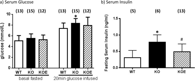
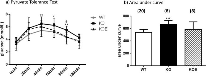
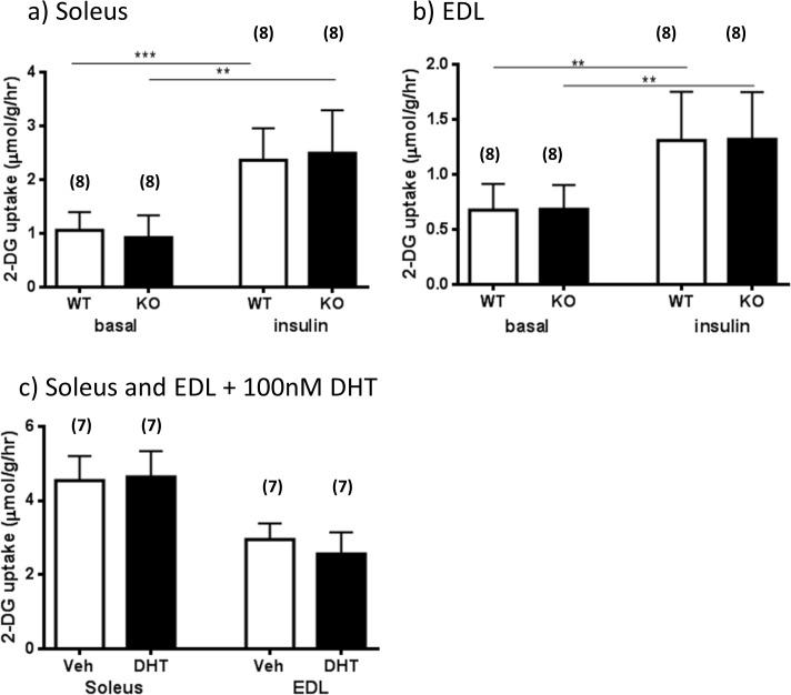
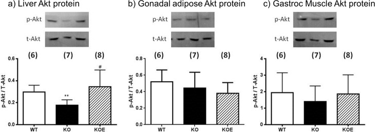
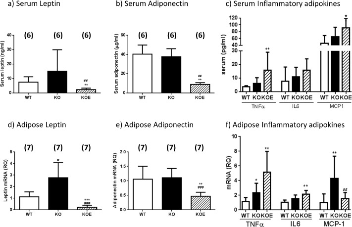
The maintenance of glucose homeostasis within the body is crucial for constant and precise performance of energy balance and is sustained by a number of peripheral organs. Estrogens are known to play a role in the maintenance of glucose homeostasis. Aromatase knockout (ArKO) mice are estrogen-deficient and display symptoms of dysregulated glucose metabolism. We aim to investigate the effects of estrogen ablation and exogenous estrogen administration on glucose homeostasis regulation. Six month-old female wildtype, ArKO, and 17β-estradiol (E2) treated ArKO mice were subjected to whole body tolerance tests, serum examination of estrogen, glucose and insulin, ex-vivo muscle glucose uptake, and insulin signaling pathway analyses. Female ArKO mice display increased body weight, gonadal (omental) adiposity, hyperinsulinemia, and liver triglycerides, which were ameliorated upon estrogen treatment. Tolerance tests revealed that estrogen-deficient ArKO mice were pyruvate intolerant hence reflecting dysregulated hepatic gluconeogenesis. Analyses of skeletal muscle, liver, and adipose tissues supported a hepatic-based glucose dysregulation, with a down-regulation of Akt phosphorylation (a key insulin signaling pathway molecule) in the ArKO liver, which was improved with E2 treatment. Concurrently, estrogen treatment lowered ArKO serum leptin and adiponectin levels and increased inflammatory adipokines such as tumour necrosis factor alpha (TNFα) and interleukin 6 (IL6). Furthermore, estrogen deficiency resulted in the infiltration of CD45 macrophages into gonadal adipose tissues, which cannot be reversed by E2 treatment. This study describes the effects of estrogens on glucose homeostasis in female ArKO mice and highlights a primary phenotype of hepatic glucose dysregulation and a parallel estrogen modified adipokine profile.


Conflict of interest statement

Competing Interests: The authors have declared that no competing interests exist. SBJ conducted the experiments in St Vincent's Institute, before employment in Novo Nordis.

Figures

Fig 1
Fig 1. Serum glucose and insulin levels.
(a) Fasted basal serum glucose and 20 min after glucose challenge serum glucose levels; (b) Fasted basal serum insulin (from 6 month-old female WT (wild type), KO (aromatase knockout) and KOE (aromatase knockout treated with 2.5µg/day 17β-estradiol) mice. Data are expressed as mean ± SD (n = 5–13 per group).
Fig 2
Fig 2. Whole body glucose, insulin and pyruvate tolerance.
(a) Whole body tolerance tests were completed on fasted 6 month-old female wildtype (WT) and aromatase knockout (KO) and 2.5μg/day 17β-estradiol-treated KO (KOE) pyruvate tolerance test and (b) corresponding area under curve. Data are presented as the mean ± SD. (n = shown on corresponding bar/ group) *p<0.05, **p<0.01, and ***p<0.001 versus expression in WT samples and # p<0.05 and ## p<0.01 versus KO samples.
Fig 3
Fig 3. The role of estrogens and androgens in ex-vivo muscle 2-deoxy-glucose (2-DG) uptake.
Six month-old wildtype (WT) and aromatase knockout (KO) female ex vivo 2-DG uptake in basal or insulin stimulated (a) soleus and (b) EDL muscle extractions. Data are presented as mean ± SD (n = 7/group). **p<0.01 and ***p<0.001 expression in basal versus insulin stimulated muscle. Six month-old wildtype (WT) female ex vivo glucose uptake in (c) vehicle or dihydrotestosterone (DHT) (100nM) treated soleus and EDL muscle extractions. Data are presented as mean ± SD (n = 7/group).
Fig 4
Fig 4. Insulin stimulated Akt protein phosphorylation in liver, adipose and skeletal tissues.
Six month-old female wildtype (WT) and aromatase knockout (KO) and 2.5μg/day 17β-estradiol-replaced KO (KOE) mice were used. Protein phosphorylation analyses of Akt levels were performed on protein extracted from insulin stimulated (a) liver, (b) adipose tissue and (c) skeletal muscle. Data are presented as mean ± SD (n = 6-8/group). **p<0.01 versus expression in age-matched WT samples and # p<0.05 versus expression in age-matched ArKO untreated samples.
Fig 5
Fig 5. Serum adipokine and gonadal adipose tissue adipokine transcript levels.
Serum adipokine analyses of (a) leptin, (b) Adiponectin and (c) TNFα, IL6 and MCP1 concentrations were performed on serum from six month-old female wildtype (WT), Aromatase knockout (KO) and 2.5μg/day 17β-estradiol-treated KO (KOE) mice. Data are presented as mean ± SD (n = 6/group). *p< 0.05, **p<0.01, versus expression in age-matched WT samples and ##p<0.01 verses KO samples. Real-time-PCR analyses of (d) Leptin, (e) Adiponectin, (f) TNFα, IL6 and MCP1 gene expression were performed on cDNA derived from total RNA prepared from gonadal adipose tissue of six month-old female wildtype (WT), Aromatase knockout (KO) and 2.5μg/day 17β-estradiol-treated KO (KOE) mice. Data are presented as mean ± SD (n = 7/group). following normalization to cyclophilin *p<0.05, **p<0.01, ***p<0.001 versus expression in age-matched WT samples and ## p<0.01 and ### p<0.001 verses KO samples.
Fig 6
Fig 6. CD45 Immunohistochemistry of gonadal adipose tissues.
(a) Representative CD45 immunohistochemistry staining of gonadal adipose tissues from six month-old female wildtype (WT), aromatase knockout (KO) and 2.5μg/day 17β-estradiol-treated KO (KOE) mice; (b) quantitated CD45-positive cells. Data are presented as mean ± SD (n = 5/group). *p< 0.05, versus age-matched WT samples.

Similar articles

Cited by

References

    1. Nordlie RC, Foster JD, Lange AJ. Regulation of glucose production by the liver. Annu Rev Nutr 1999; 19: 379–406. - PubMed
    1. Pilkis SJ, Granner DK. Molecular physiology of the regulation of hepatic gluconeogenesis and glycolysis. Annu Rev Physiol 1992; 54: 885–909. - PubMed
    1. Saltiel AR, Kahn CR. Insulin signalling and the regulation of glucose and lipid metabolism. Nature 2001; 414: 799–806. - PubMed
    1. Gabriely I, Ma XH, Yang XM, Atzmon G, Rajala MW, Berg AH et al. Removal of visceral fat prevents insulin resistance and glucose intolerance of aging: an adipokine-mediated process? Diabetes 2002; 51: 2951–2958. - PubMed
    1. Wellen KE, Hotamisligil GS. Obesity-induced inflammatory changes in adipose tissue. J Clin Invest 2003; 112: 1785–1788. - PMC - PubMed

Publication types

MeSH terms