. 2015 Sep 22;6(28):24947-68.
doi: 10.18632/oncotarget.4453.

An anti-EpCAM antibody EpAb2-6 for the treatment of colon cancer

Affiliations

An anti-EpCAM antibody EpAb2-6 for the treatment of colon cancer

Mei-Ying Liao et al. Oncotarget. .

Abstract

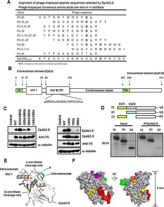
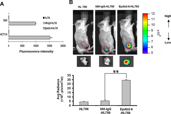
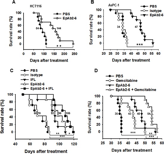
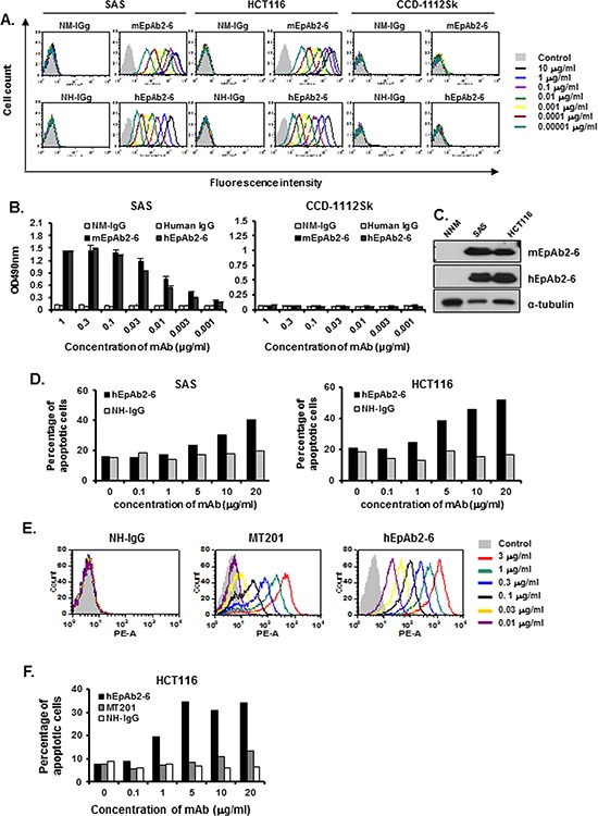
Epithelial cell adhesion molecule (EpCAM) is known to be overexpressed in epithelial cancers associated with enhanced malignant potential, particularly colorectal carcinoma (CRC) and head and neck squamous cell carcinoma (HNSCC). However, it is unknown whether progression of malignance can be directly inhibited by targeting EpCAM. Here, we have generated five novel monoclonal antibodies (mAbs) against EpCAM. One of these anti-EpCAM mAbs, EpAb2-6, was found to induce cancer cell apoptosis in vitro, inhibit tumor growth, and prolong the overall survival of both a pancreatic cancer metastatic mouse model and mice with human colon carcinoma xenografts. EpAb2-6 also increases the therapeutic efficacy of irinotecan, fluorouracil, and leucovorin (IFL) therapy in a colon cancer animal model and gemcitabine therapy in a pancreatic cancer animal model. Furthermore, EpAb2-6, which binds to positions Y95 and D96 of the EGF-II/TY domain of EpCAM, inhibits production of EpICD, thereby decreasing its translocation and subsequent signal activation. Collectively, our results indicate that the novel anti-EpCAM mAb can potentially be used for cancer-targeted therapy.

Keywords: EpCAM; cancer therapy; colorectal carcinoma; targeting imaging; therapeutic antibody.


Conflict of interest statement

CONFLICTS OF INTEREST

The authors have no potential conflicts of interest to declare.

Figures

Figure 1
Figure 1. Characterization of anti-EpCAM mAbs (EpAb1-3, EpAb2-6, EpAb3-5, EpAb4-1, and EpAb5-4)
Binding activities of anti-EpCAM mAbs were measured by Western blotting A. and B. immunofluorescent staining C. and flow cytometry D.
Figure 2
Figure 2. Inhibition of cancer cell growth in vitro by EpAb2-6
HCT116 and SAS cells were transfected with EpCAM shRNA plasmids (shEpCAM). A. Western blot analyses were performed to evaluate EpAb2-6 binding to EpCAM-knockdown HCT116, SAS, and mock cells. B. HCT116, HCT116/shEpCAM, and HCT116 (TP53−/−) cells were treated with EpAb2-6 (0–20 μg/ml) or isotype control (mouse myeloma IgG2a) for 6 h, and cell death was measured by flow cytometry with Annexin V-FITC and PI double staining. Annexin V-FITC was used to determine the percentage of cells within the population that were actively undergoing apoptosis at an early stage (6 hours). Propidium iodide (PI) was used to distinguish between viable and nonviable cells. C. EpCAM knockdown inhibits EpAb2-6, which induces repression of cell viability. HCT116 cells stably expressing control shRNA (shLuc) or shEpCAM were treated with EpAb2-6 (0 – 20 μg/ml) for 48 h. Error bars show mean ± SD (Student's t-test, **p < 0.01). D. Non-attachment assay. HCT116 cells were incubated under Non-attachment conditions with EpAb2-6, which increased cleavage of capase-3 and PARP. E. EpAb2-6 inhibits EpICD cleavage and nuclear localization. Immunofluorescence images of EpICD cellular localization in HCT116 cells treated with EpAb2-6 (40 μg/ml), isotype control (mouse IgG2a, 40 μg/ml), or PBS for 48 hours. (Bar = 10 μm.) F. EpAb2-6 inhibits EpICD production and nuclear translocation. HCT116 cell lines transfected with EpCAM-v5 plasmids were treated with either NM-IgG or EpAb2-6 (40 μg/ml) for 48 hours, and the lysates were subsequently subjected to Western blotting.
Figure 3
Figure 3. Identification of the B cell epitope of EpAb2-6
A. Alignment of phage-displayed peptide sequences selected by EpAb2-6. B. EpCAM mutations with amino acid substitutions in the EGF-I (Q54A/N55A) or EGF-II domain (Q89A/N90A, D92A/G93A, L94A/Y95A, L94A, Y95A, or D96A). C. The indicated EpCAM mutants were expressed in HEK293 cells. Cellular protein extracts were subjected to Western blot analysis using EpAb2-6 and EpAb3-5 antibodies. Substitutions of Y95 and D96 reduced EpAb2-6 binding activity. D. Various EpCAM constructs with different EGF-domains of EpAB2-6 binding sites (FE: full length EpCAM; D1: EGF-I domain deletion; D2: EGF-II/TY domain deletion) are shown. These constructs were transiently transfected into HEK293 cells to evaluate their binding ability with EpAb2-6. Epitopes of anti-EpCAM antibodies are mapped to a structural model of EpEx. E. A ribbon diagram representation of the complete EpEx structure. The epitope of Edrecolomab (mouse Ab) and ING-1 (humanized Ab) is shown in orange. The epitopes of Adecatumumab (MT201; human Ab) and EpAb2-6 are colored green and red, respectively. The cleavage sites of α-secretase (Adam) and β-secretase (BACE1) are colored purple and yellow, respectively. N and C indicate the N and C terminus of EpEx, respectively. F. The molecular surface of EpEx is color coded as described in E.
Figure 4
Figure 4. Tumor-homing ability of anti-EpCAM mAb in human colon cancer xenografts
A. The expression level of EpCAM on cancer cell surfaces was determined by flow cytometry analysis using EpAb2-6-HL750. HL750 and NM-IgG-HL750 were used as controls. B. In vivo imaging of SCID mice bearing HCT116 human colon tumor xenografts was performed after intravenous injection of EpAb2-6-HL750, NM-IgG-HL750, or HL750. NIR fluorescence images were acquired at 48 hours post-injection (top). Red circles indicate the tumor loci. The signal intensity of the tumor area was quantified using IVIS software. Tumor distributions of EpAb2-6-HL750, NM-IgG-HL750, and HL750 at 72 hours post-injection are shown. Signal intensities for the tumor and organs were measured using IVIS software. Error bars show mean ± SD (n = 3) (Student's t-test, **p < 0.01) (below).
Figure 5
Figure 5. Effect of combinatorial treatment with EpAb2-6 and IFL on mice bearing HCT116 tumors
A, D. Mice bearing HCT116-derived tumor xenografts were treated with EpAb2-6, IFL, EpAb2-6 in combination with IFL, or PBS. The sizes of tumors in each group were determined on the indicated days. Error bars show mean ± SD (n = 6) (Student's t-test, *p < 0.05). B, E. Average body weight of each group is shown on the indicated days. Error bars show mean ± SD. C. Tumor weight from (A) was measured at the end of the treatment period. (Student's t-test, *p < 0.05.) F. A Kaplan-Meier survival curve from (D) indicates that mice bearing xenografts treated with EpAb2-6 or EpAb2-6 in combination with IFL had a greater survival rate than those treated with IFL or PBS (n = 6).
Figure 6
Figure 6. EpAb2-6 enhances survival in an animal model of tumor metastasis
A. NOD/SCID mice were intravenously injected with 1 × 10⁶ HCT116 cells, and then were treated with either PBS or EpAb2-6 (n = 10). The survival curves indicate that mice treated with EpAb2-6 exhibited a greater survival rate than those treated with PBS. B. NOD/SCID mice were intravenously injected with 1 × 10⁶ AsPC-1 cells, and were then treated with either PBS, isotype control (Myeloma IgG2a), or EpAb2-6 (n = 10). The survival curves indicate that mice treated with EpAb2-6 exhibited a greater survival rate than those treated with Isotype. C. Mice bearing metastatic HCT116-derived tumors were treated with EpAb2-6, Isotype, IFL, EpAb2-6 in combination with IFL, or PBS. Kaplan-Meier survival curves indicate that mice bearing metastatic cancer cells treated with EpAb2-6 or EpAb2-6 in combination with IFL had a greater survival rate than mice treated with IFL alone or PBS (n = 7) (Log rank test, **p < 0.01, ***p < 0.001). D. Mice bearing metastatic AsPC-1-derived tumors were treated with EpAb2-6, Gemcitabine, EpAb2-6 in combination with Gemcitabine, or PBS. Kaplan-Meier survival curves indicate that mice bearing metastatic cancer cells treated with EpAb2-6 or EpAb2-6 in combination with Gemcitabine had a greater survival rate than mice treated with Gemcitabine alone or PBS (n = 10) (Log rank test, **p < 0.01, ***p < 0.001).
Figure 7
Figure 7. Development of a humanized antibody against EpCAM
The CDRs of EpAb2-6 were grafted onto a human IgG1 backbone to create humanized EpAb2-6 (hEpAb2-6). The binding activity of hEpAb2-6 to human cancer cell lines is shown. Flow cytometry analysis A. and ELISA B. were performed to measure the binding activity of mEpAb2-6 and hEpAb2-6 to SAS, HCT116, and CCD-1112Sk cells. Normal mouse IgG (NM-IgG) and normal human IgG (human IgG) were used as negative controls. C. Western blot analyses of mEpAb2-6 and hEpAb2-6 against NNM, SAS, and HCT116 cells. D. SAS and HCT116 cells were treated with hEpAb2-6 (0–20 μg/ml) for 6 h, and cell death was measured by flow cytometry with Annexin-V FITC and PI double staining. Binding activity of hEpAb2-6 and MT201 to human cancer cell lines. Flow cytometry analysis E. was performed to measure the binding activity of hEpAb2-6 and MT201 to HCT116 cells. Normal human IgG (NH-IgG) was used as a negative control. F. HCT116 cells were treated with hEpAb2-6 and MT201 (0–20 μg/ml) for 6 h, and cell death was measured by flow cytometry with Annexin-V FITC and PI double staining.

References

    1. Balzar M, Prins FA, Bakker HA, Fleuren GJ, Warnaar SO, Litvinov SV. The structural analysis of adhesions mediated by Ep-CAM. Exp Cell Res. 1999;246:108–121. - PubMed
    1. de Boer CJ, van Krieken JH, Janssen-van Rhijn CM, Litvinov SV. Expression of Ep-CAM in normal, regenerating, metaplastic, and neoplastic liver. J Pathol. 1999;188:201–206. - PubMed
    1. Ni J, Cozzi P, Hao J, Beretov J, Chang L, Duan W, Shigdar S, Delprado W, Graham P, Bucci J, Kearsley J, Li Y. Epithelial cell adhesion molecule (EpCAM) is associated with prostate cancer metastasis and chemo/radioresistance via the PI3K/Akt/mTOR signaling pathway. The international journal of biochemistry & cell biology. 2013;45:2736–2748. - PubMed
    1. Gastl G, Spizzo G, Obrist P, Dunser M, Mikuz G. Ep-CAM overexpression in breast cancer as a predictor of survival. Lancet. 2000;356:1981–1982. - PubMed
    1. Patriarca C, Macchi RM, Marschner AK, Mellstedt H. Epithelial cell adhesion molecule expression (CD326) in cancer: a short review. Cancer treatment reviews. 2012;38:68–75. - PubMed

Publication types

MeSH terms