

Review
doi: 10.1021/acs.chemrev.5b00244.
Epub 2015 Aug 28.
Glycosylated Porphyrins, Phthalocyanines, and Other Porphyrinoids for Diagnostics and Therapeutics
Affiliations
- PMID: 26317756
- PMCID: PMC6011754
- DOI: 10.1021/acs.chemrev.5b00244
Item in Clipboard
Review
Glycosylated Porphyrins, Phthalocyanines, and Other Porphyrinoids for Diagnostics and Therapeutics
Chem Rev.
.
Abstract
Conflict of interest statement
The authors declare no competing financial interest.
Figures
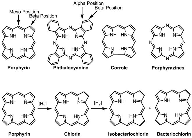
Structures of common porphyrinoid macrocycles (top). Structures of reduced porphyrins, such as chlorin, isobacteriochlorin, and bacteriochlorin (bottom).
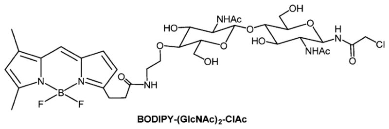
A representative example of a BODIPY conjugated to a simplified chloroacetamidyl chitobiose derivative.
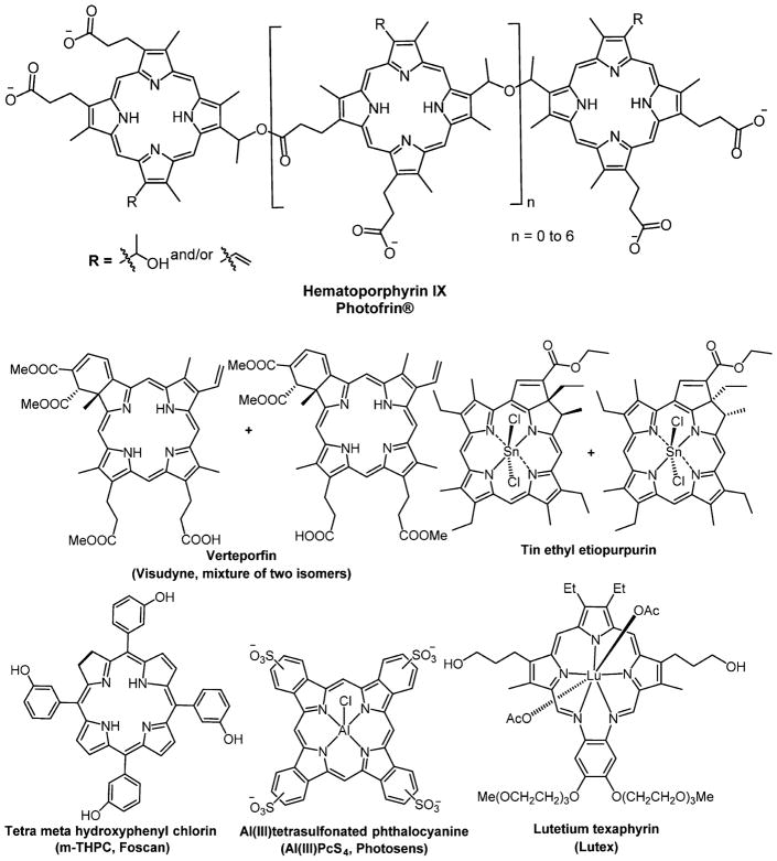
Structures of photosensitizers in clinical or preclinical trials for PDT.
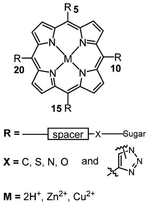
Generalized structure representation of meso-5,10,15,20-substituted porphyrin–carbohydrate conjugates. Spacers vary widely and include phenyl, phenylalkyl, and polyethylene glycol (PEG). Many reports examine the role of the substitution pattern on biochemical properties, for example, the six possible compounds using two different meso substituents.
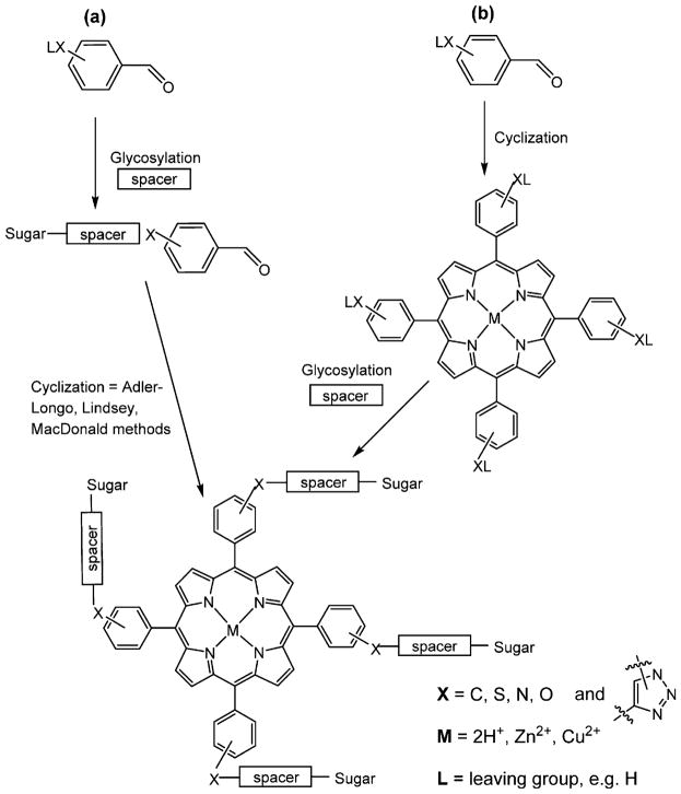
General scheme of the two major synthetic routes toward multiglycosylated porphyrins: (a) cyclization of glycosylated benzaldehydes by the Lindsey, Adler-Longo, or MacDonald methods;,,, (b) glycosylation of porphyrins by reactions with functional groups on the meso-aryl groups., The spacers may or may not be used.
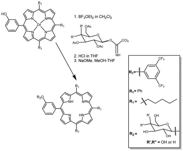
Glycosylated derivatives of hydroxyphenylporphyrin using trichloroacetimidate reagents reported by Aicher et al.
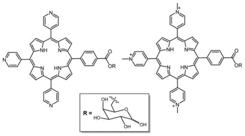
Structure of neutral and cationic tripyridyl porphyrin appended with D-galactose.
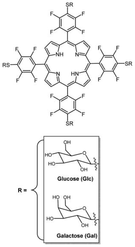
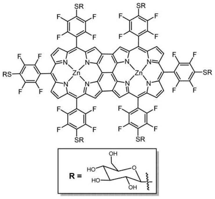
Nearly any primary thiol and unencumbered secondary thiol can substitute for the 4-fluoro group of the commercially available TPPF20 in high yields under mild conditions in this click-type reaction. Here, the tetra glycosyl- and tetra galactosyl- conjugates are shown, PGlc4 and PGal4, respectively, reported by Drain and co-workers.,
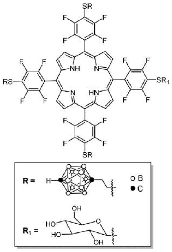
Glucose carboranylporphyrin conjugate reported by Vicente and co-workers made by first appending the thiol carborane and then the thiol glucose.
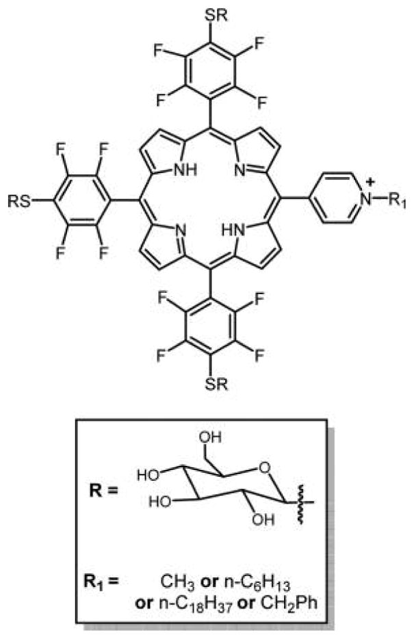
Porphyrin substituted by three glycosyl units and one pyridinium substituent reported by Boyle and co-workers probed lipophilic balance and synergy between glycosylation and cationic moieties. The core porphyrin was made from a mixed aldehyde reaction using the Adler and Longo method, and the compounds separated.
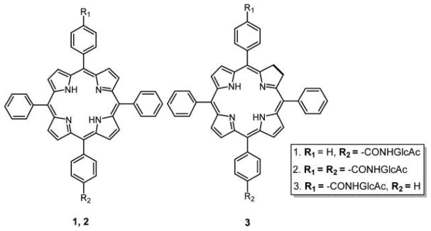
Glycosamide porphyrins and the corresponding chlorins studied reported by DiStasio et al. The mono carboxylic acid porphyrins were synthesized via mixed aldehyde condensation using the Adler and Longo method, and the compounds separated. The carboxylic acid chlorin was obtained by diimide reduction of corresponding porphyrin. Amino sugars were conjugated to carboxylic porphyrin and chlorin using a typical amide coupling reaction. trans-Bis-glucose porphyrin was obtained using the [2+2] Mcdonald condensation method starting with O-acetylated glucosamine benzaldehyde.
meso-C-Glycosylated porphyrins reported by Drasar and co-workers.,
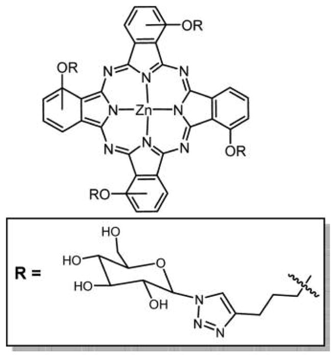
Structure of TPP–galactose conjugate linked via triazole unit.
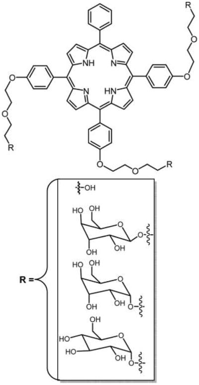
Structures of the parent unconjugated DEG porphyrin and corresponding sugar conjugate; the latter binds to human retinoblastoma cells. The porphyrin core was synthesized via mixed aldehyde condensation using the Adler and Longo method. The DEG with or without sugars was substituted onto the porphyrin core via a Williamson type reaction.
Thioglycosylated meso-porphyrins reported by Krausz and co-workers synthesized via condensation of 1-thioacetylated sugars with monobromotritolyl-porphyrins followed by deprotection of acetate groups using NaOMe.
Glycosylated porphyrin reported by Krausz and co-workers starts by using a mixed aldehyde condensation to yield the monohydroxyphenyltritolylporphyrin.,
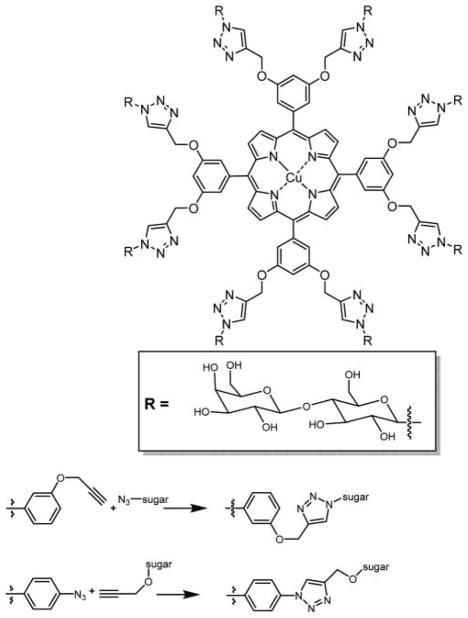
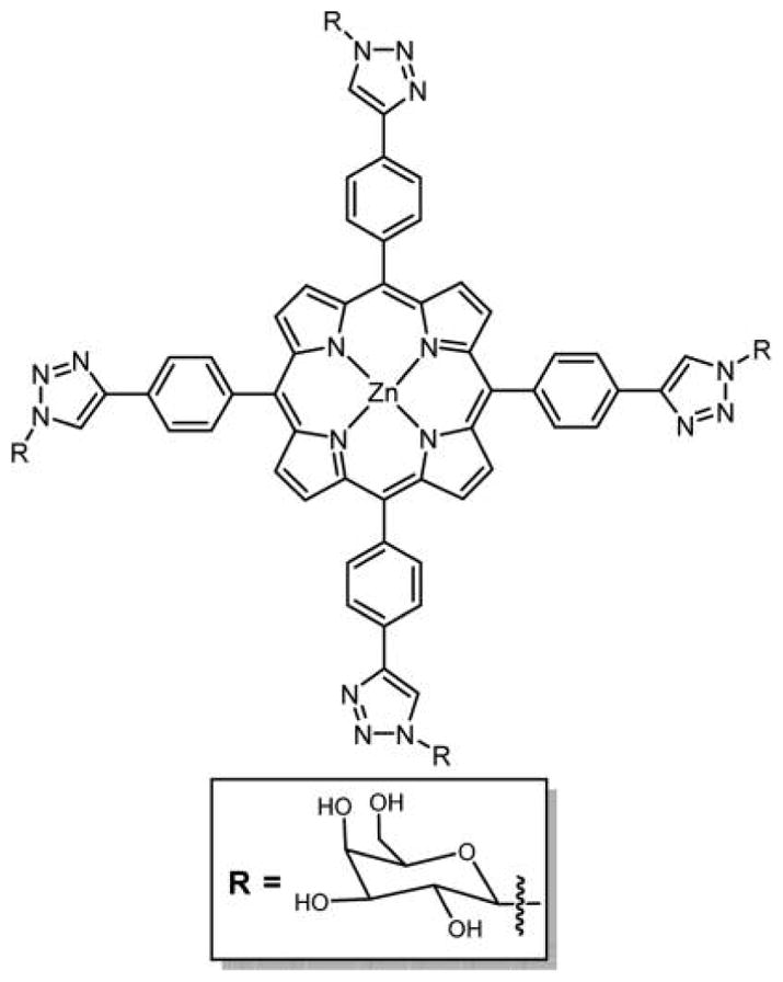
Top: Octa-β-lactoglycosylated porphyrinatocopper (PorCu-Lac8) prepared by click chemistry. Bottom: Two general routes to azide click chemistry to append sugars onto meso-arylporphyrins.,
5,10-Bis-glycoporphyrin synthesized via a microwave heated sequential double click reaction. Core 5,10-bis-azido porphyrin was obtained first by synthesizing 5,10-bis-nitroporphyrin using the Adler and Longo method followed by reduction of nitro groups.
Glycoconjugated dendrimers symmetrically appended to a porphyrin core with 4 and 12 β-D-glucopyranosyl residues reported by Stoddart and co-workers.
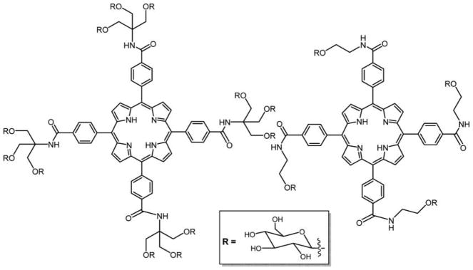
Structures of glycodendrimeric porphyrins reported by Rosilio and co-workers, where the porphyrin core was made by mixed aldehyde condensations using the Adler and Longo method.
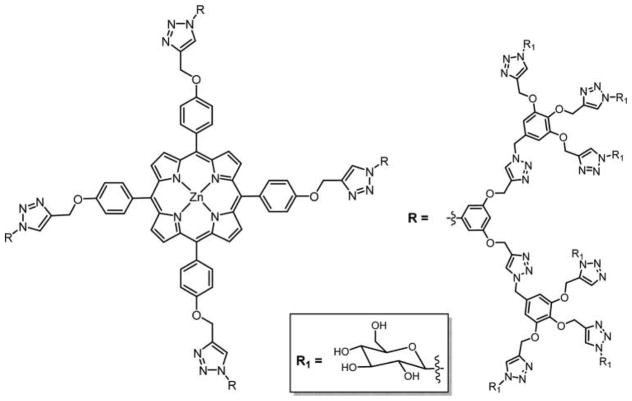
Structure of azide-functionalized glycodendrimers of porphyrin having 24 β-glucopyranose units.
Structure of porphyrin glycodendritic conjugate with eight galactopyranose units.
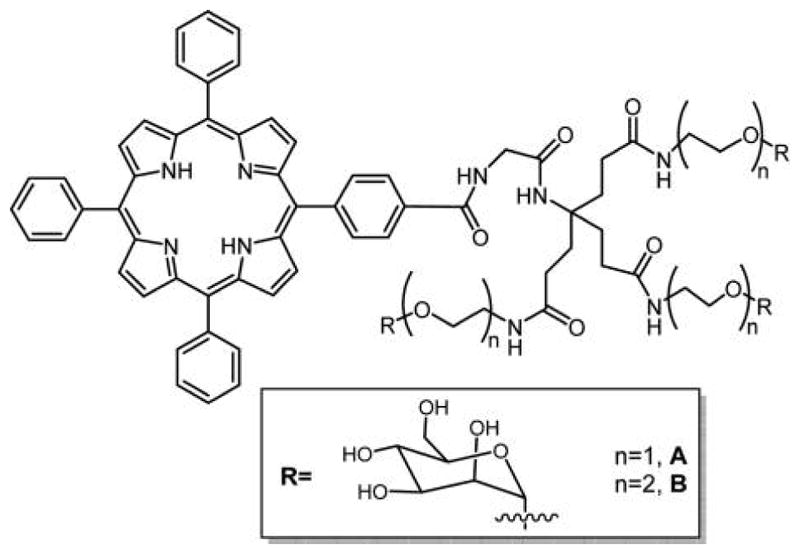
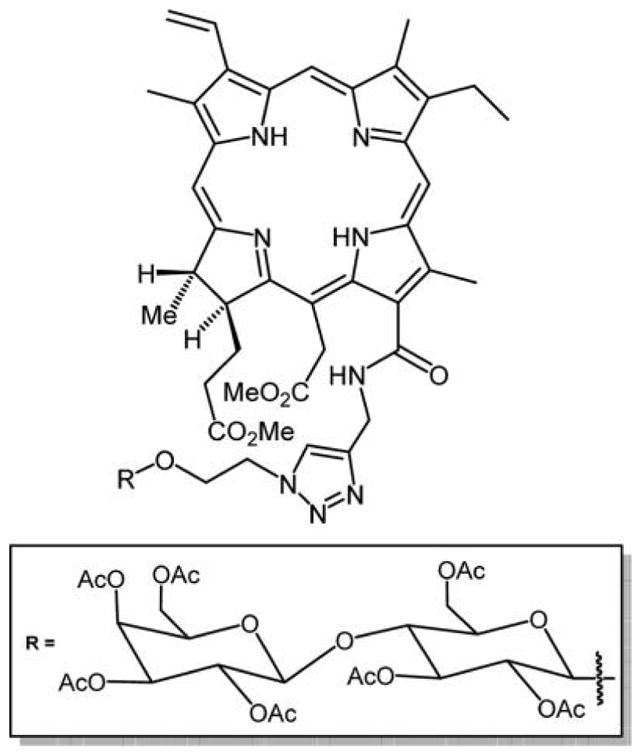
Porphyrin–CD conjugates used as carrier–drug complex for therapy., Other porphyrin–CD conjugates formed using click chemistry and amide linkers are reported, and there are various strategies that self-assemble CD with porphyrins into supramolecular materials.
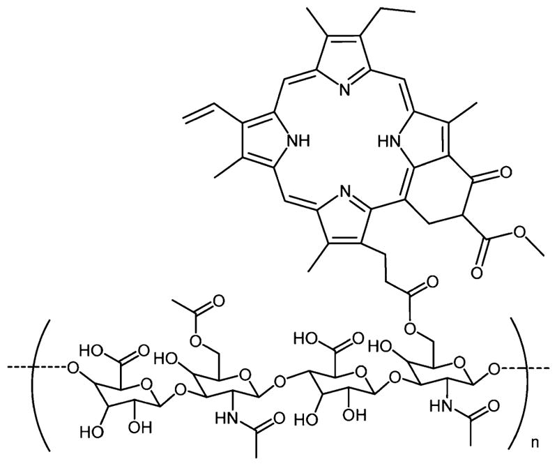
Hyaluronic acid porphyrin conjugate as a PDT agent.
Recently reported β-substituted porphyrin sugar by Cavaleiro and co-workers.
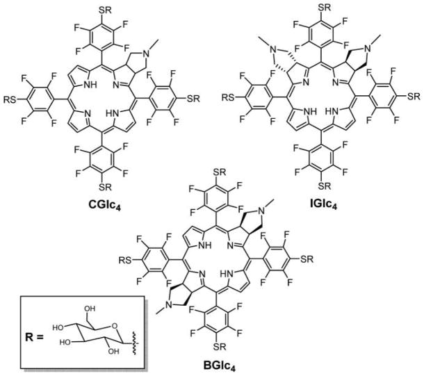
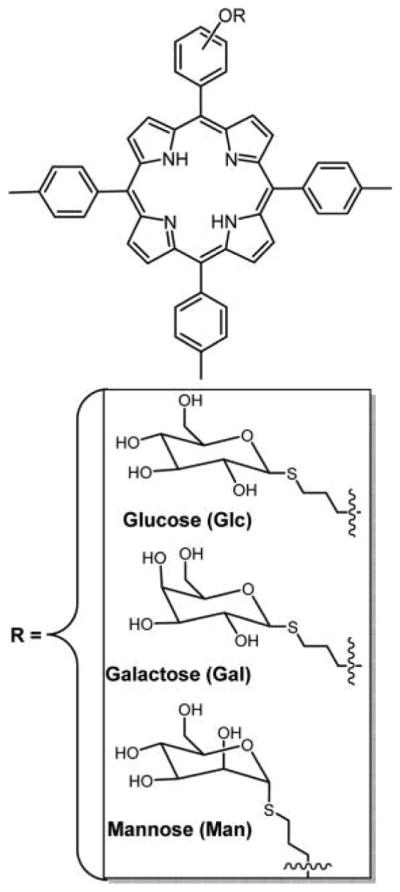
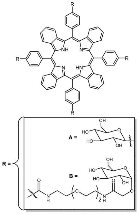
Structures of thioglycosylated chlorin (CGlc4), isobacteriochlorin (IGlc4), and bacteriochlorin (BGlc4) appended with four thioglucose units reported by Singh et al.
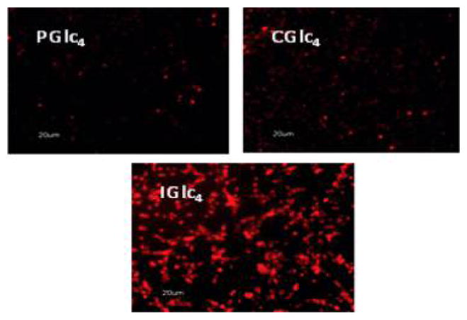
Fluorescence microscopy of K:Molv NIH 3T3 cells treated with 2.5 μM PGlc4, CGlc4, and IGlc4. K:Molv NIH 3T3 cells were incubated for 20 h with porphyrinoids, followed by removal of unbound dye from the cell culture by repeated rinsing with PBS, and the cells were imaged under identical microscope settings and not enhanced; magnification 10×. Reproduced with permission from ref . Copyright 2010 American Chemical Society.
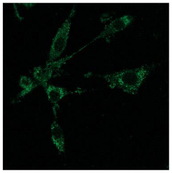
K:Molv NIH 3T3 cells were incubated with 10 μM BGlc4 for 24 h, rinsed three times with PBS buffer, and fixed with 4% paraformaldehyde solution. Confocal microscope excitation at 514 nm, emission monitored with a 710–750 band-pass filter. Under similar conditions using the IGlc4, CGlc4, or PGlc4, no fluorescence images are observed using a 610–650 nm emission band-pass filter. The image is not enhanced; magnification is 60×. Reproduced with permission from ref . Copyright 2010 American Chemical Society.
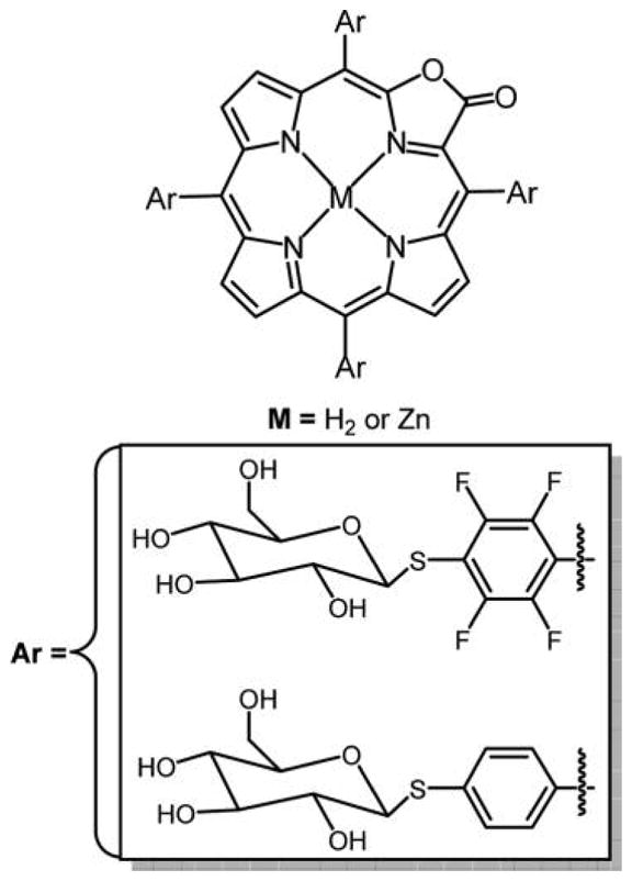
Structure of gluco-conjugate of porpholactone.
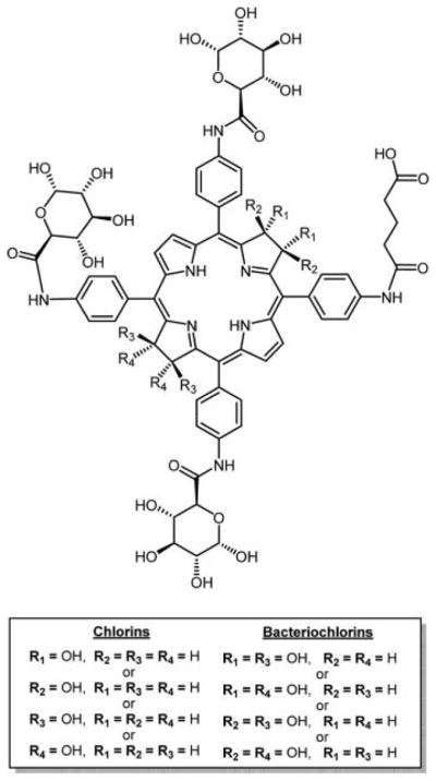
Glycosylated chlorins and bacteriochlorins synthesized from 5,10,15-tris(4-1′,2′,3′,4′-O-acetyl-glucopyranuron-N-phenylamide)-20-[4-(5′methoxy-1′,5′-dioxopentyl) aminophenyl]porphyrin, reported by McCarthy et al., use OsO4 to make the chlorin and bacteriochlorin.
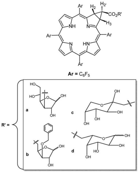
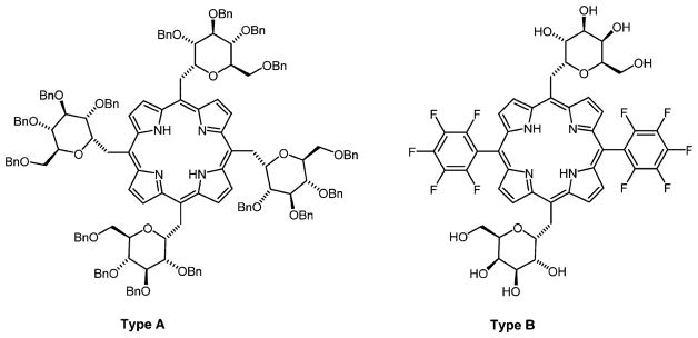
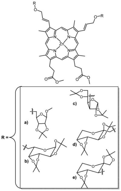
Cavaleiro and co-workers made chlorins by reactions of meso-tetrakis(pentafluorophenyl) porphyrinatozinc(II) with α-diazoacetates derived from diacetonides of the glucofuranose (a), monoacetonide of xylofuranose (b), fructopyranose (c), and galactopyranose (d).
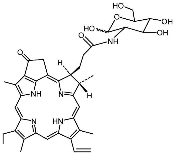
Structure of pyropheophorbide 2-deoxyglucosamide (Pyro-2DG) theragnostic reported by Zhang et al.
Structure of the chlorin e6–carbohydrate conjugate reported by Grin et al.
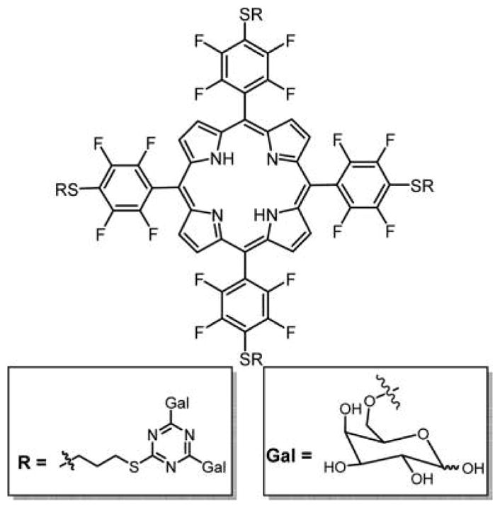
Top: Three thioglucose or three thioxyloses appended to corroles reported by Samaroo et al., made by substitution of the para fluoro group. Note that the para fluoro groups on opposite perfluorophenyls substitute somewhat faster than the central. The bottom pentafluorophenylcorrole-D-galactose conjugate was reported by Röder and co-workers.
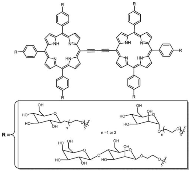
Porphyrin dimers conjugated to carbohydrates were evaluated for potential applications in one-photon and two-photon PDT by Maillard and co-workers.
Structure of conjugated zinc porphyrin oligomer reported by Achelle et al. with good two-photon cross sections.
Triply bridged hexaglycosylated fused porphyrin dye with large two-photon cross section reported by Singh et al.
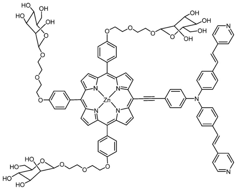
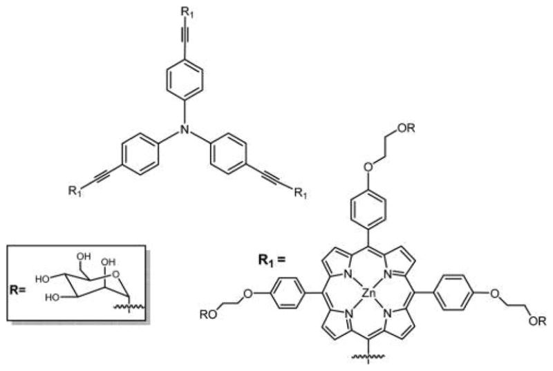
Structure of triphenylamine porphyrin bearing three α-mannose groups attached via DEG tether for two-photon activity reported by Millard and co-workers.
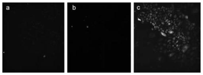
Two-photon microscope images of (a) PGlc4, (b) CGlc4, and (c) IGlc4:BGlc4 (5:1) on Chinese hamster ovary cells excited at 860 nm. Reproduced with permission from ref . Copyright 2014 John Wiley and Sons, Inc.
Structures of the glycosylated tetrabenzoporphyrins reported by Krausz and co-workers.

Structure of tetraphenylbenzoporphyrin (TBP)–galactose conjugate linked via triazole unit. In these and similar compounds, the rotational barriers between the phenyl and triazole units are likely low enough that interconversion between atropisomers is facile at room temperature, such that they cannot be isolated.
Galactose-containing Si(IV)Pc reported by Lee et al.
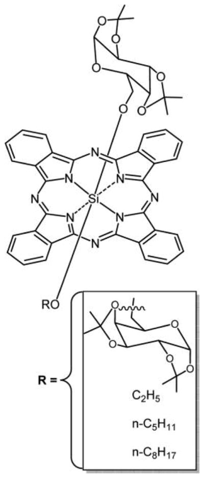
Structure of glycosylated Si(IV)Pc from a mixed condensation reaction in which the D-glucofuranose unit binds axially to the Si(IV) center through the tetraethylene glycol spacer.
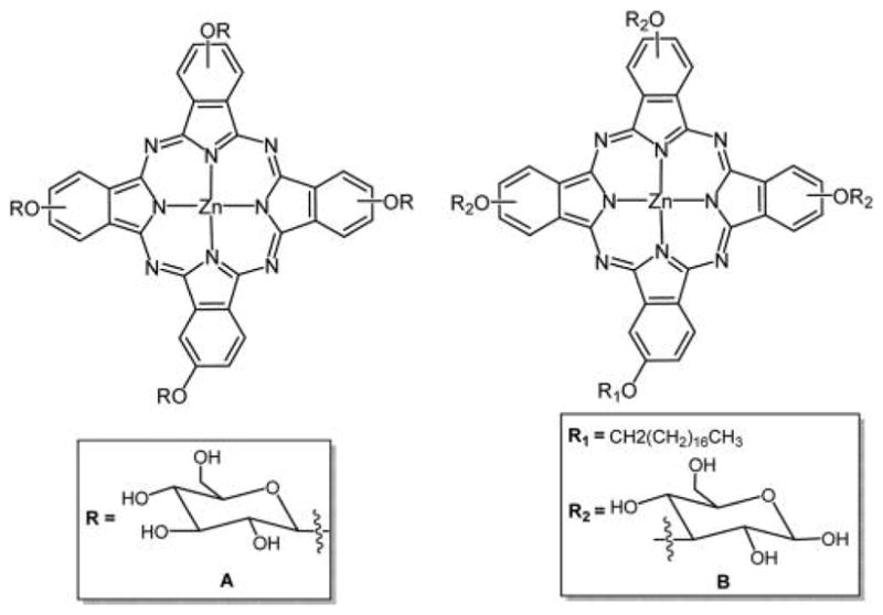
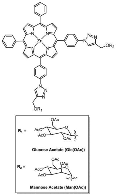
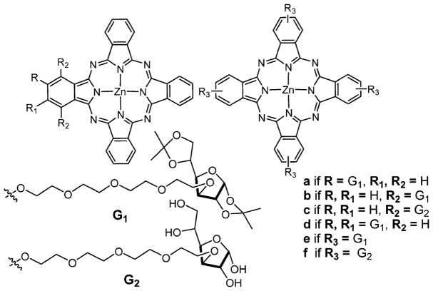
(A) Asymmetric anomeric glycoconjugated Zn(II)Pc reported by Iqbal et al. (B) Asymmetric Zn(II)Pc bearing octadecyloxy and glucosyl groups by Zhang et al. The way of representing the peripheral-OR groups indicates that a mixture of positional isomers was used, including α or β.
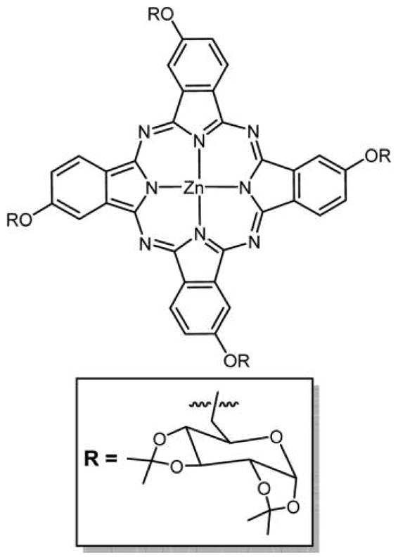
Structure of isopropylidene-protected tetra-β-glycosylated Zn(II)Pc (one isomer shown).
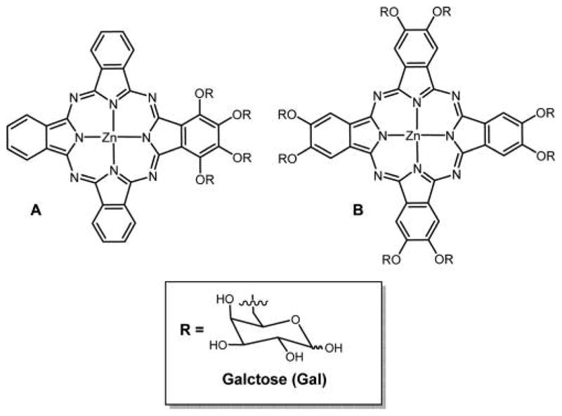
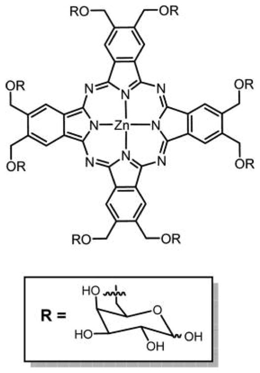
Structure of pure glycophthalocyanine appended with (A) four galactose units from a mixed condensation reaction, and (B) eight galactose units made from the phthalonitrile in 21% overall yield.
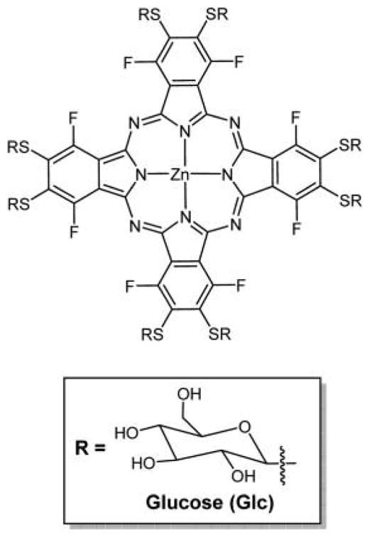
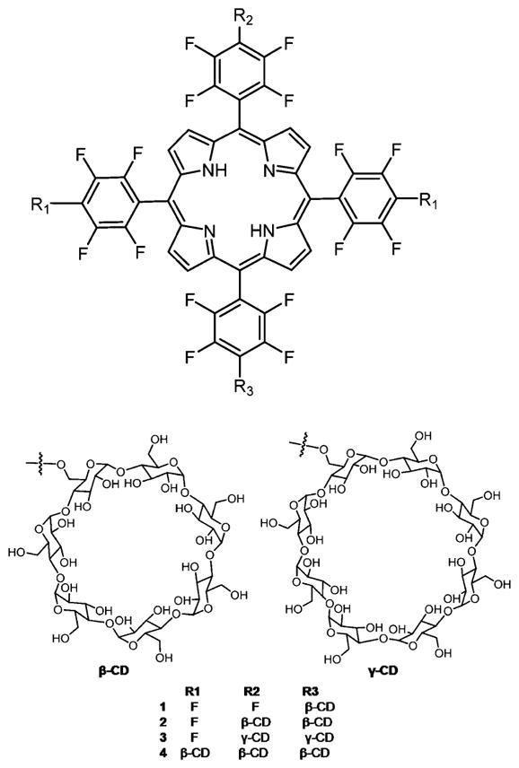
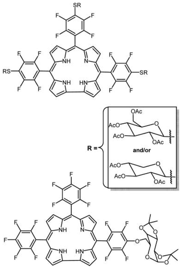
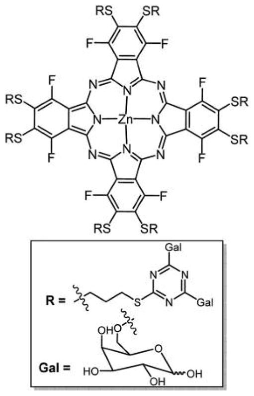
Symmetrical octa-substituted Zn(II)Pc’s with thioglucose units derived from a core perfluorophthalocyanine platform made in ca. 70% yield in two steps. Similar conjugates are also reported using different linkers, for example, Figure 46.,,
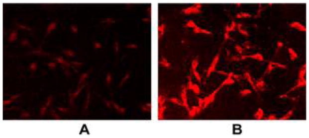
MDA-MB-231 cells were incubated with 50 nM ZnPcGlc8 for 24 h, rinsed three times with PBS buffer to remove unbound dye, and fixed with 4% paraformaldehyde solution. Fluorescence images were captured by exciting at 540–580 nm, magnification 20× under identical conditions. (A) Just after preparation of the fixed cells slide, and (B) 4 days after later. The contrast of each was enhanced by 40% for publication. Reproduced with permission from ref . Copyright 2011 Elsevier, Inc.
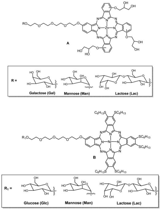
Structures of glycosylated glycerol-Zn-phthalocyanine (A) with an intervening tetraethylene glycol spacer, and glycosylated thiol-hexane-Ni-phthalocyanine (B) with an intervening tetraethylene glycol spacer; the core Pc was made by a mixed condensation reaction. Ni(II)Pc generally have unfavorable photophysical properties for PDT because of a low-lying d,d state.
Mono-, di-, and tetra-O-glycosyl-substituted Pc containing tetraethylene glycol spacer synthesized via condensation of glycosyl phthalonitrile reported by Liu et al.
Pc’s bearing eight D-galactose units on the periphery synthesized via condensation of 4,5-(di-D-galactose) phthalonitrile.
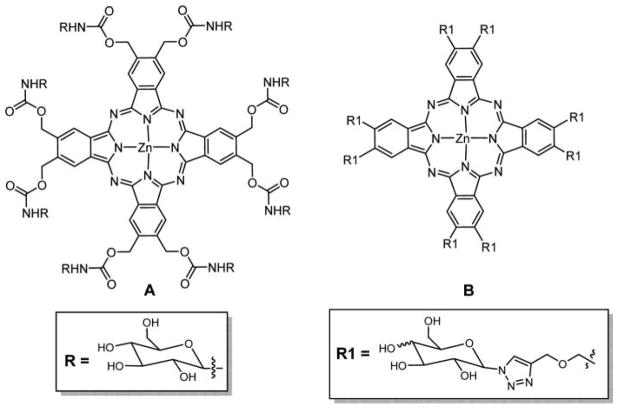
(A) Structure of octacarbamoyl glycosylated-substituted Zn(II)Pc made from the isocyanate and the hydroxymethylphthalocyanine, and (B) click chemistry to prepare the octaglycosylated Pc starting with the Zn(II)Pc-octa-β-CH2OCH2C≡CH.
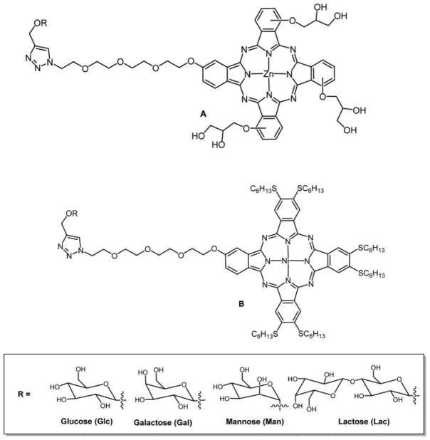
Structures of glycosylated glycerol-Zn-phthalocyanine (A) with an intervening triethylene glycol spacer and glycosylated thiol-hexane-Ni-phthalocyanine, and (B) with an intervening triethylene glycol spacer prepared via 1,3-dipolar cycloaddition reported by Lafont and coworkers, wherein the core Pc was made from a mixed condensation reaction or from a core hydroxyl Pc.
Structure of tetra glycosyl-substituted Zn(II)Pc on the α position. The core Pc was obtained by cyclotetramerization of 3-pent-4-ynyloxy phthalonitrile.
Structure of Zn(II)Pc glycodendritic conjugate with 16 galactopyranose units.
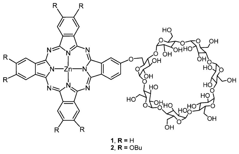
A Zn(II)Pc functionalized with β-CD from a mixed condensation reaction.
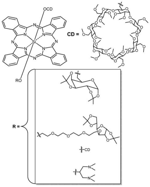
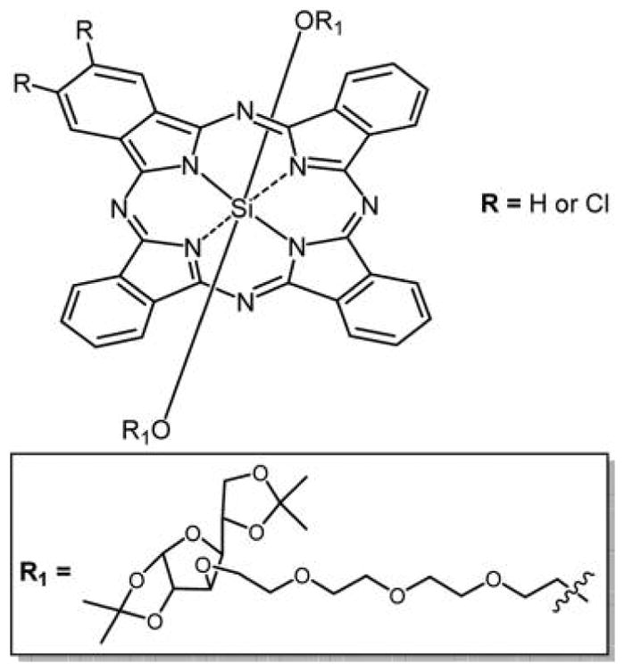
Structure of a series of symmetrical and unsymmetrical Si(IV)Pc with a permethylated β-CD unit and a sugar as axial substituents.
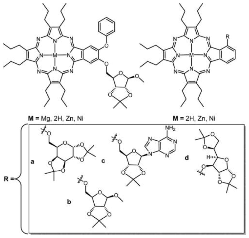
Structure of Pz’s substituted with (a) galactopyranose and (b–d) ribose derivatives.,
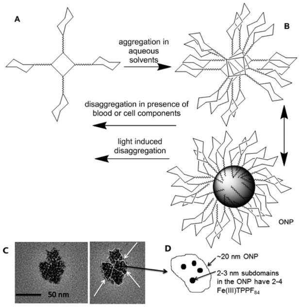
Hydrophobic core of porphyrinoid dyes (A) often drives aggregation in aqueous media into small π-stacked aggregates (B), which can further aggregate into larger organic nanoparticles (ONP). The nanoaggregates can be driven to disassemble by interaction with cellular components such as lipid head groups and protein and/or photothermal processes. While the free bases do not have sufficient contrast in transmission electron microscope studies, the Fe(III) complexes of a fluorous porphyrin do, and reveal the presence of small domains within a larger ONP (C and D). Reproduced with permission from ref . Copyright 2012 John Wiley and Sons, Inc. If <ca. 50 nm, the aggregate can be taken up by the cell, for example, by endocytosis, where it can disaggregate because of interactions with cellular components, photothermally by internal conversion, and PDT damage to the endosome. Also, the loosely bound subdomains may be induced to break out of the ONP by the same processes and be taken up by the cells. Polysaccharide possessing in the cell will also contribute to the dye distributions, and is a key factor in why nonhydrolyzable bioconjugates can be more effective therapeutics.
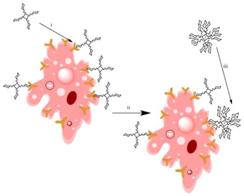
(i) At low concentrations, the porphyrinoid dyes are solvated and not aggregated. (ii) Upon binding to the target cell, the local concentration increases and the dyes then aggregate. (iii) Nanoaggregates in aqueous media have sugar-coated shells around the dye core, which then can bind to cell receptors. Either or both processes can be present depending on the specific dye and cell types and media.
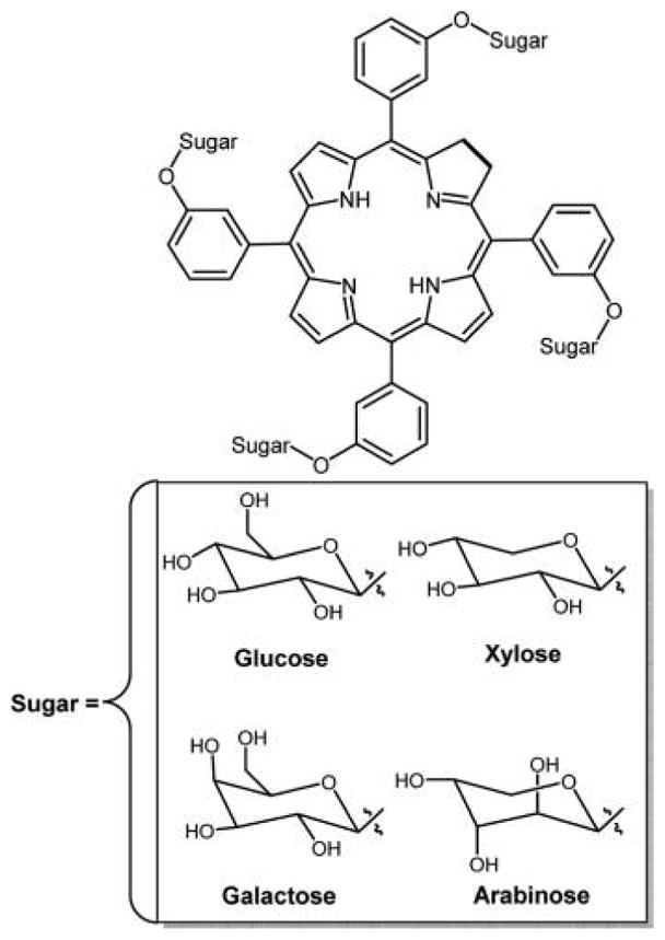
Aggregation properties were correlated with the 5- and 6-carbon sugars, where the former is reported to have a propensity to form H-aggregates and the latter J-aggregates.
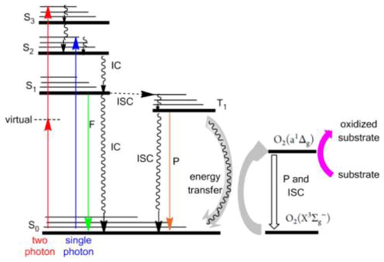
aS is the singlet manifold and T is the triplet manifold, IC is internal conversion by heat loss, F is fluorescence, P is phosphorescence, ISC is intersystem crossing from one manifold to another, and collision of the dye in the triple state with ground-state triplet oxygen photosensitizes the formation of singlet oxygen. Singlet oxygen and hydroxyl radicals react with cell constituents such as double bonds in lipids, flavins, heterocycles, sugar–phosphate backbones of nucleic acids, as well as systems to mitigate oxidative stress like super oxide dismutase.
References
-
- Ogilby PR. Singlet oxygen: there is indeed something new under the sun. Chem Soc Rev. 2010;39:3181–3209. - PubMed
-
- DeRosa MC, Crutchley RJ. Photosensitized singlet oxygen and its applications. Coord Chem Rev. 2002;233–234:351–371.
-
- Macdonald IJ, Dougherty TJ. Basic principles of photodynamic therapy. J Porphyrins Phthalocyanines. 2001;5:105–129.
-
- Henderson BW, Dougherty TJ. How Does Photodynamic Therapy Work? Photochem Photobiol. 1992;55:145–157. - PubMed
Publication types
MeSH terms
Substances
Grants and funding
LinkOut - more resources
Full Text Sources
Other Literature Sources
