. 2015 Aug 28;9(8):e0004031.
doi: 10.1371/journal.pntd.0004031. eCollection 2015.

Bile Salts Modulate the Mucin-Activated Type VI Secretion System of Pandemic Vibrio cholerae

Affiliations

Bile Salts Modulate the Mucin-Activated Type VI Secretion System of Pandemic Vibrio cholerae

Verena Bachmann et al. PLoS Negl Trop Dis. .

Abstract

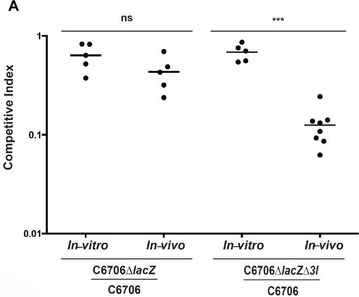
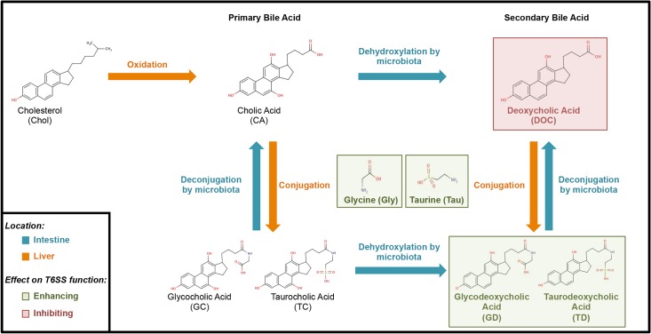
The causative agent of cholera, Vibrio cholerae, regulates its diverse virulence factors to thrive in the human small intestine and environmental reservoirs. Among this pathogen's arsenal of virulence factors is the tightly regulated type VI secretion system (T6SS). This system acts as an inverted bacteriophage to inject toxins into competing bacteria and eukaryotic phagocytes. V. cholerae strains responsible for the current 7th pandemic activate their T6SS within the host. We established that T6SS-mediated competition occurs upon T6SS activation in the infant mouse, and that this system is functional under anaerobic conditions. When investigating the intestinal host factors mucins (a glycoprotein component of mucus) and bile for potential regulatory roles in controlling the T6SS, we discovered that once mucins activate the T6SS, bile acids can further modulate T6SS activity. Microbiota modify bile acids to inhibit T6SS-mediated killing of commensal bacteria. This interplay is a novel interaction between commensal bacteria, host factors, and the V. cholerae T6SS, showing an active host role in infection.


Conflict of interest statement

The authors have declared that no competing interests exist.

Figures

Fig 1
Fig 1. The T6SS of V. cholerae is functional in vivo.
The T6SS of V. cholerae O1 strain C6706 is active in the infant mouse model of infection. V. cholerae C6706, C6706ΔlacZ, and C6706ΔlacZΔtsiV1-3 were mixed in pairwise combinations and administered to the infant mouse as mixtures via oral gavage. As an in-vitro control, 2 mL LB were inoculated with the same bacterial mixtures and grown overnight at 37°C. After a 16-h incubation, the mice were sacrificed and their small intestines were harvested and plated on X-gal plates to count surviving bacteria. The competitive index of the two competing strains is shown on the y-axis. Horizontal bars represent the arithmetic mean of one experiment performed with a minimum of 5 mice in each group. Stars indicate statistical significance (unpaired two-tailed Student’s t-test), with *** p < 0.0005, ns = not significant.
Fig 2
Fig 2. V. cholerae O1 strains have an activated T6SS in the presence of mucins.
(A) Survival assay of V. cholerae C6706 mutants on mucins. 1 × 10⁸ V. cholerae C6706, C6706ΔtsiV1–3 (lacking all three immunity genes), or C6706ΔtsiV1–3ΔvasK (lacking all three immunity genes and the T6SS gene vasK) were loaded separately on columns containing 3% mucins or 3% gelatin. After incubation for 2 h at 37°C, eluents were collected, and serial dilutions were plated on selective LB agar plates. Surviving numbers of V. cholerae bacteria were plotted as CFUs. Bars represent mean values ± SD of two independent experiments done in duplicate. (B) Intraspecies killing by V. cholerae C6706 on mucin columns. Killing assays were performed by mixing predator C6706 and prey C6706ΔtsiV1–3ΔvasK at a ratio of 10:1 and loading them together on gelatin or mucin columns. After incubation for 2 h at 37°C, cells were collected and serial dilutions were plated on LB agar plates. Bars show mean values ± SD of two independent experiments done in duplicate. (C) Intraspecies killing by V. cholerae N16961 on mucin columns. Killing assays were performed by mixing predator N16961 and prey C6706ΔtsiV1–3ΔvasK at a ratio of 10:1 and loading them together on either gelatin or mucin columns. After incubation for 2 h at 37°C, eluents were collected and serial dilutions were plated on LB agar plates. Bars show mean values ± SD of two independent experiments done in duplicate.
Fig 3
Fig 3. Bile acid metabolism.
Flow diagram showing the metabolism and fate of bile acids. In the liver (orange arrows), cholesterol is enzymatically converted to primary bile acids such as cholic acid. After conjugation of taurine or glycine to cholic acid by hepatocytes, the resulting glycocholic acid or taurocholic acid are stored in the gall bladder until released into the small intestine in response to food ingestion. In the small intestine (blue arrows), several commensals deconjugate and/or dehydroxylate bile acids, producing unconjugated deoxycholic acid and cholic acid. Alternatively, glycholic acid or taurocholic acid can be dehydroxylated to the conjugated secondary bile acids glycodeoxycholic acid or taurodeoxycholic acid. In a process called enterohepatic circulation, unconjugated bile acids are excreted with the feces or reabsorbed from the ileum back into the liver. Depending on the types of microbiota and nutrition ingested, bile acid composition may vary throughout the intestines. Therefore, the inhibitory effect identified in this study (red) of deoxycholic acid and the enhancing effect (green) of glycodeoxycholic acid or taurodeoxycholic acid on the T6SS of V. cholerae may vary depending on the microbiota present.
Fig 4
Fig 4. Deoxycholate diminishes T6SS function.
(A) Bile acids affect a mucin-activated T6SS of V. cholerae C6706. 1 × 10⁸ V. cholerae C6706 or C6706ΔtsiV1–3 were loaded separately on columns containing 3% mucins. After incubation for 1 h at 37°C, either cholic acid (CA), glycocholic acid (GC), taurocholic acid (TC), deoxycholic acid (DOC), taurodeoxycholic acid (TD), glycodeoxycholic acid (GD), glycine (Gly), or taurine (Tau) were added to the mucin columns at a final concentration of 1.2 mM each. After incubation for 2 h at 37°C, cells were collected, serial dilutions were plated on LB agar plates, and surviving numbers of C6706 or C6706ΔtsiV1–3 were plotted. Bars show mean values ± SD of two independent experiments done in duplicate. (B) Individual bile acids affect the T6SS of V. cholerae V52. Predator V. cholerae V52 or V52ΔvasK were mixed at a 10:1 ratio with prey E. coli MG1655 and spotted on LB agar plates supplemented with 1.2 mM cholic acid (CA), glycocholic acid (GC), taurocholic acid (TC), deoxycholic acid (DOC), glycodeoxycholic acid (GD), taurodeoxycholic acid (TD), glycine (Gly), or taurine (Tau) for 4 h at 37°C. CFUs were counted after incubation of serial dilutions of eluent on LB agar plates overnight. The killing index was calculated by the ratio of surviving prey in the presence of V52 versus attenuated V52. The graph gives mean values ± SD of two experiments done in duplicate. A Student’s t-test was performed for significance, with *p < 0.05, ** p < 0.005, *** p < 0.0005, **** p < 0.0001.
Fig 5
Fig 5. Deoxycholic acid regulates tube formation.
(A) Bile salts do not affect the transcription of hcp in V52. V52 was incubated for two hours with 1.2 mM deoxycholic acid, glycine, taurine and combinations of the three. RNA was then isolated, converted to cDNA, quantified using qPCR, and compared to 16S rRNA gene. Experiments were performed in triplicates and normalized to the LB control. (B) Deoxycholic acid does not affect the transcription of tseL, vasX, vgrG3, vasH in V52. V52 was incubated with 1.2 mM deoxycholic acid for two hours. RNA was isolated, converted to cDNA and quantified using qPCR. Experiments were performed in triplicates and normalized to the LB control. (C) Bile salts do not affect Hcp-2 levels in V52. V52 was incubated with 1.2 mM deoxycholic acid, glycine, taurine and combinations of the three for two hours. Bacteria were harvested and western blot analysis was performed using antibodies for Hcp-2 and DnaK. (D) Deoxycholic acid affects the ability of the T6SS to form tubes. V. cholerae 2740–80 with a sfGFP labeled vipA was incubated with 1.2 mM cholate (negative control), deoxycholic acid or taurine for thirty minutes. After this incubation, the cells were imaged for ten minutes using the Super Resolution OMX microscope. Three frames were chosen and the number of extended T6SS tubes were counted. A Student’s t-test was performed for significance, with ** p < 0.005; ns is no significance.
Fig 6
Fig 6. Commensal gut bacteria influence T6SS function.
(A) Metabolism of bile acids by B. bifidum. TLC was performed with indicated bile acids in the presence or absence (control) of B. bifidum: cholic acid (CA), deoxycholic acid (DOC), glycocholic acid (GC), taurocholic acid (TC), glycodeoxycholic acid (GD), or taurodeoxycholic acid (TD), (B) Metabolism of bile acids by B. adolescentis. TLC was performed with indicated bile acids in the presence or absence (control) of B. adolescentis. (C) Rf-values for TLC experiments. (D) B. bifidum metabolizes bile acids that lead to T6SS inhibition. B. bifidum was incubated under anaerobic conditions for 2 days on LB agar plates supplemented with 1.2 mM of one of the indicated bile acids. ‘Mock’ indicates the plates with no bile acids added. After removal of the anaerobes, V. cholerae V52 or V52ΔvasK were mixed at a 10:1 ratio with E. coli MG1655 and spotted on either on top of the removed commensal spot or 2 cm away from the commensal spot. Surviving E. coli bacteria were enumerated after 4 h-incubation at 37°C. Bars show mean values ± SD of two independent experiments done in triplicate. (E) B. adolescentis can metabolize GD and TD to inhibit the T6SS. B. adolescentis was incubated under anaerobic conditions for 2 days on LB agar plates supplemented with 1.2 mM of one of the indicated bile acids. ‘Mock’ indicates the plates with no bile acids added. After removal of the anaerobes, V. cholerae V52 or V52ΔvasK were mixed at a 10:1 ratio with E. coli MG1655 and spotted on top of the removed commensal spot or 2 cm away from the commensal spot. Surviving E. coli bacteria were enumerated after 4 h incubation at 37°C. Bars show mean values ± SD of two independent experiments done in triplicate. A Student’s t-test was performed for significance, with *p < 0.05, ** p < 0.005, *** p < 0.0005, ns = not significant.

Similar articles

Cited by

References

    1. MacIntyre DL, Miyata ST, Kitaoka M, Pukatzki S (2010) The Vibrio cholerae type VI secretion system displays antimicrobial properties. Proc Natl Acad Sci U S A 107: 19520–19524. 10.1073/pnas.1012931107 - DOI - PMC - PubMed
    1. Pukatzki S, Ma AT, Sturtevant D, Krastins B, Sarracino D, et al. (2006) Identification of a conserved bacterial protein secretion system in Vibrio cholerae using the Dictyostelium host model system. Proc Natl Acad Sci U S A 103: 1528–1533. - PMC - PubMed
    1. Butler SM, Camilli A (2005) Going against the grain: chemotaxis and infection in Vibrio cholerae . Nat Rev Microbiol 3: 611–620. - PMC - PubMed
    1. Tacket CO, Losonsky G, Nataro JP, Cryz SJ, Edelman R, et al. (1993) Safety and immunogenicity of live oral cholera vaccine candidate CVD 110, a delta ctxA delta zot delta ace derivative of El Tor Ogawa Vibrio cholerae. J Infect Dis 168: 1536–1540. - PubMed
    1. Miyata ST, Kitaoka M, Brooks TM, McAuley SB, Pukatzki S (2011) Vibrio cholerae requires the type VI secretion system virulence factor VasX to kill Dictyostelium discoideum . Infect Immun 79: 2941–2949. 10.1128/IAI.01266-10 - DOI - PMC - PubMed

Publication types

MeSH terms