. 2015 Aug 28;10(8):e0136994.
doi: 10.1371/journal.pone.0136994. eCollection 2015.

Molecular Markers of Tubulointerstitial Fibrosis and Tubular Cell Damage in Patients with Chronic Kidney Disease

Affiliations

Molecular Markers of Tubulointerstitial Fibrosis and Tubular Cell Damage in Patients with Chronic Kidney Disease

Shunsaku Nakagawa et al. PLoS One. .

Abstract

In chronic kidney disease (CKD), progressive nephron loss causes glomerular sclerosis, as well as tubulointerstitial fibrosis and progressive tubular injury. In this study, we aimed to identify molecular changes that reflected the histopathological progression of renal tubulointerstitial fibrosis and tubular cell damage. A discovery set of renal biopsies were obtained from 48 patients with histopathologically confirmed CKD, and gene expression profiles were determined by microarray analysis. The results indicated that hepatitis A virus cellular receptor 1 (also known as Kidney Injury Molecule-1, KIM-1), lipocalin 2 (also known as neutrophil gelatinase-associated lipocalin, NGAL), SRY-box 9, WAP four-disulfide core domain 2, and NK6 homeobox 2 were differentially expressed in CKD. Their expression levels correlated with the extent of tubulointerstitial fibrosis and tubular cell injury, determined by histopathological examination. The expression of these 5 genes was also increased as kidney damage progressed in a rodent unilateral ureteral obstruction model of CKD. We calculated a molecular score using the microarray gene expression profiles of the biopsy specimens. The composite area under the receiver operating characteristics curve plotted using this molecular score showed a high accuracy for diagnosing tubulointerstitial fibrosis and tubular cell damage. The robust sensitivity of this score was confirmed in a validation set of 5 individuals with CKD. These findings identified novel molecular markers with the potential to contribute to the detection of tubular cell damage and tubulointerstitial fibrosis in the kidney.


Conflict of interest statement

Competing Interests: The authors have declared that no competing interests exist.

Figures

Fig 1
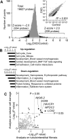
Fig 1. The Transcriptional Profile Related to Chronic Kidney Disease (CKD).
(A) Distribution of differences in gene expression between control kidney RNA (Control) and RNA extracted from 48 CKD biopsies. Microarray analysis was performed and genes down-regulated (z-score < -2.0, green) or up-regulated (z-score > 2.0, blue) in CKD are indicated. (B) Biological functions of the genes showing differential expression in CKD samples were classified according to their Gene Ontology and P values were calculated with MetaCore software. (C) Relationship between P values from Kruskal-Wallis test on tubulointerstitial fibrosis and tubular cell damage. Each symbol represents one gene. Gray or black circles indicate genes with any P values < 0.05; open circles represent genes with both P values > 0.05. HAVCR1, hepatitis A virus cellular receptor 1; LCN2, lipocalin 2; SOX9, SRY-box 9; WFDC2, WAP four-disulfide core domain 2; NKX6-2, NK6 homeobox 2.
Fig 2
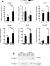
Fig 2. Mouse Kidney Gene Expression Profiles following Unilateral Ureteral Obstruction (UUO).
(A) Real-time PCR analysis was used to determine the relative mRNA levels of alpha smooth muscle actin (Acta2), hepatitis A virus cellular receptor 1 (Havcr1), lipocalin 2 (Lcn2), SRY-box 9 (Sox9), WAP four-disulfide core domain 2 (Wfdc2), and NK6 homeobox 2 (Nkx6-2) at the indicated time-points. Glyceraldehyde-3-phosphate dehydrogenase (GAPDH) mRNA was used as an internal control. Data are expressed as the mean ± standard deviation of 4–6 mice. *P < 0.05; **P < 0.01; one-way analysis of variance. (B) Western blot analysis was used to determine the leakage of Ngal (Lcn2) and Kim-1 (Havcr1) in mouse urine.
Fig 3
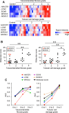
Fig 3. Analysis of the Molecular Score Performance.
(A) Relative signal intensity heatmaps for hepatitis A virus cellular receptor 1 (HAVCR1), lipocalin 2 (LCN2), SRY-box 9 (SOX9), WAP four-disulfide core domain 2 (WFDC2), and NK6 homeobox 2 (NKX6-2) in relation to renal histopathology. (B) The distribution of molecular score based on histological grades. (C) The area under the receiver operating characteristics curve (AUC) for the molecular score of each biopsy, plotted against the grade of tubulointerstitial fibrosis and tubular cell damage.
Fig 4
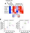
Fig 4. Validation Set Study.
Microarray analysis was performed on 5 biopsies from CKD patients in the validation set. (A) Heatmap of the relative signal intensities of hepatitis A virus cellular receptor 1 (HAVCR1), lipocalin 2 (LCN2), SRY-box 9 (SOX9), WAP four-disulfide core domain 2 (WFDC2), and NK6 homeobox 2 (NKX6-2). (B) Comparison of the molecular score with the grade of tubulointerstitial fibrosis and tubular cell damage. Dotted lines represent the threshold values corresponding to histopathology grade 3 (Threshold I) or grade 4 (Threshold II).

Similar articles

Cited by

References

    1. Nath KA. Tubulointerstitial changes as a major determinant in the progression of renal damage. Am J Kidney Dis. 1992;20(1):1–17. . - PubMed
    1. Remuzzi G, Bertani T. Pathophysiology of progressive nephropathies. N Engl J Med. 1998;339(20):1448–56. 10.1056/NEJM199811123392007 . - DOI - PubMed
    1. Nishihara K, Masuda S, Nakagawa S, Yonezawa A, Ichimura T, Bonventre JV, et al. Impact of Cyclin B2 and Cell division cycle 2 on tubular hyperplasia in progressive chronic renal failure rats. Am J Physiol Renal Physiol. 2010;298(4):F923–34. 10.1152/ajprenal.00567.2009 . - DOI - PMC - PubMed
    1. Mishra J, Ma Q, Prada A, Mitsnefes M, Zahedi K, Yang J, et al. Identification of neutrophil gelatinase-associated lipocalin as a novel early urinary biomarker for ischemic renal injury. J Am Soc Nephrol. 2003;14(10):2534–43. . - PubMed
    1. Schwab K, Witte DP, Aronow BJ, Devarajan P, Potter SS, Patterson LT. Microarray analysis of focal segmental glomerulosclerosis. Am J Nephrol. 2004;24(4):438–47. 10.1159/000080188 . - DOI - PubMed

Publication types

MeSH terms