. 2015 Aug 28;10(8):e0136883.
doi: 10.1371/journal.pone.0136883. eCollection 2015.

Modulation of Innate Immune Signalling by Lipid-Mediated MAVS Transmembrane Domain Oligomerization

Affiliations

Modulation of Innate Immune Signalling by Lipid-Mediated MAVS Transmembrane Domain Oligomerization

Luis Nobre et al. PLoS One. .

Abstract

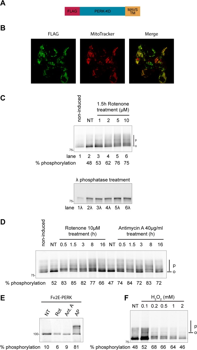
RIG-I-like receptors detect viral RNA in infected cells and promote oligomerization of the outer mitochondrial membrane protein MAVS to induce innate immunity to viral infection through type I interferon production. Mitochondrial reactive oxygen species (mROS) have been shown to enhance anti-viral MAVS signalling, but the mechanisms have remained obscure. Using a biochemical oligomerization-reporter fused to the transmembrane domain of MAVS, we found that mROS inducers promoted lipid-dependent MAVS transmembrane domain oligomerization in the plane of the outer mitochondrial membrane. These events were mirrored by Sendai virus infection, which similarly induced lipid peroxidation and promoted lipid-dependent MAVS transmembrane domain oligomerization. Our observations point to a role for mROS-induced changes in lipid bilayer properties in modulating antiviral innate signalling by favouring the oligomerization of MAVS transmembrane domain in the outer-mitochondrial membrane.


Conflict of interest statement

Competing Interests: The authors have declared that no competing interests exist.

Figures

Fig 1
Fig 1. Rotenone potentiates RLR signalling.
HEK 293T cells were treated with 1 μM rotenone alone or in combination with 0.1 μg/ml or 1 μg/ml of the intracellularly delivered RLR ligand poly(I:C)/Lyovec (poly(I:C)-LV) for 16h. (A) IFNβ mRNA levels and (B) CXCL10 mRNA levels were quantified by qRT-PCR and normalized to the level of β actin mRNA. Shown are the mean ± standard error of the mean (sem) of three independent experiments. *, p ≤ 0.05
Fig 2
Fig 2. Increased MAVS-TM domain oligomerization in cells with elevated reactive oxygen species.
(A) Schema of Flag_PERK-KD_MAVS-TM, a N-terminally Flag-tagged fusion protein between the kinase domain of PERK and the TM domain of MAVS. The construct encoding this protein was introduced into Flp-In T-Rex-293 cells by recombination (B) Intracellular localization of Flag_PERK-KD_MAVS-TM following doxycycline induction in Flp-In T-Rex-293 cells. Cells were stained for the Flag tag and the mitochondrial specific dye MitoTracker. Images were acquired sequentially on a confocal microscope. The merge panel shows an overlap of Flag and Mitotracker signals. (C) Immunoblot of Flag_PERK-KD_MAVS-TM immunopurified from Flp-In T-Rex-293 cells. Cells were either left untreated (non-induced) or exposed to doxycycline to induce the expression of Flag_PERK-KD_MAVS-TM. In addition, cells were treated for 90min with the indicated doses of rotenone. The position of hypophosphorylated (0) and phosphorylated (P) Flag_PERK-KD_MAVS-TM is indicated in the top panel. The bottom panel is an immunoblot of the same samples treated in vitro with λ-Phosphatase. (D) Immunoblot of Flag_PERK-KD_MAVS-TM immunopurified from Flp-In T-Rex-293 cells treated with 10 μM rotenone or 40 μg/ml antimycin A for the indicated time. (E) Immunoblot of Fv2E-PERK, immunopurified from cells treated with 10 μM rotenone (Rot) or 40 μg/ml antimycin A (Ant A), or with the Fv2E-PERK dimerizing ligand AP20187 (AP) at 10 nM for 90 min. (F) Immunoblot of Flag_PERK-KD_MAVS-TM immunopurified from Flp-In T-Rex-293 cells exposed for 2 hours to the indicated concentrations of hydrogen peroxide. The percentage of PERK-KD phosphorylation is indicated under each sample.
Fig 3
Fig 3. Sendai virus infection promotes lipid-dependent MAVS-TM domain oligomerization and increases lipid peroxidation.
(A) Immunoblot of Flag_PERK-KD_MAVS-TM immunopurified from Flp-In T-Rex-293 cells infected with 100 HA/ml of Sendai virus strain Cantell for the indicated time. (B) Quantification of the level of Flag_PERK-KD_MAVS-TM phosphorylation in Sendai virus-infected cells. Shown are mean ± sem values of three independent experiments. *, p ≤ 0.05; **, p ≤ 0.01 (C) Immunoblot of Fv2E-PERK immunopurified from cells treated with 10 μM rotenone (Rot) for 90 min, or infected with 100 HA/ml Sendai virus for 16h (SeV), or treated with the Fv2E-PERK dimerizing ligand AP20187 (AP) at 10 nM for 90 min. The percentage of Fv2E-PERK phosphorylation is indicated under each sample. (D) Detection of lipid peroxidation end products by the Thiobarbituric Acid Reactive Substances (TBARS) assay. Pure malondialdehyde was used as a reference in the assay. TBARS were quantified from Flp-In T-Rex-293 cells treated for 90min with 40 μg/ml Antimycin A, 10 μM Rotenone, 1 mM hydrogen peroxide, or infected with 100 HA/ml Sendai virus for 16 hours. Shown is a representative experiment reproduced three times. Data are mean ± sem values of two technical replicates, each analysed in triplicate. *, p ≤ 0.05; **, p ≤ 0.01
Fig 4
Fig 4. Rotenone and Sendai virus infection trigger oligomerization of the TM domain of the unrelated outer mitochondrial membrane protein OMP25.
(A) Schema of Flag_PERK-KD_OMP25-TM, a N-terminally Flag-tagged fusion protein between the kinase domain of PERK and the TM domain of OMP25, an outer mitochondrial membrane protein. The construct encoding this protein was introduced into Flp-In T-Rex-293 cells by recombination (B) Intracellular localization of Flag_PERK-KD_OMP25-TM following doxycycline induction in Flp-In T-Rex-293 cells. Cells were stained for the Flag tag and the mitochondrial specific dye MitoTracker. Images were acquired sequentially on a confocal microscope. The merge panel shows an overlap of Flag and Mitotracker signals. (C) Immunoblot of Flag_PERK-KD fused to the TM domain of MAVS (MAVS-TM) or to the TM domain of OMP25 (OMP25-TM). Cells were treated with 10 μM rotenone (Rot) for 90 min, or infected with 100 HA/ml Sendai virus for 16h (SeV). The percentage of PERK-KD phosphorylation is indicated under each sample.
Fig 5
Fig 5. Proposed model for mROS effects on MAVS signalling.
MAVS is predominantly monomeric on the outer mitochondrial membrane of cells with low levels of mROS (top panel). Increased mROS production promotes lipid-dependent oligomerization of MAVS TM domain (lower panel). Upon viral infection, RLRs bind to viral RNA and expose their CARD domain, which is now available to interact with the CARD domain of MAVS. RLR induced MAVS CARD domains homo-oligomerization is required for MAVS signalling. Lipid-dependent TM domain oligomerization could prime MAVS for RLR-induced MAVS CARD domains homooligomerization, therefore leading to increased MAVS signalling.

Similar articles

Cited by

References

    1. Pandey S, Kawai T, Akira S. Microbial Sensing by Toll-Like Receptors and Intracellular Nucleic Acid Sensors. Cold Spring Harb Perspect Biol. 2015;7: a016246 10.1101/cshperspect.a016246 - DOI - PMC - PubMed
    1. Wu B, Peisley A, Tetrault D, Li Z, Egelman EH, Magor KE, et al. Molecular imprinting as a signal-activation mechanism of the viral RNA sensor RIG-I. Mol Cell. 2014;55: 511–523. 10.1016/j.molcel.2014.06.010 - DOI - PMC - PubMed
    1. Hou F, Sun L, Zheng H, Skaug B, Jiang Q-X, Chen ZJ. MAVS Forms Functional Prion-like Aggregates to Activate and Propagate Antiviral Innate Immune Response. Cell. 2011;146: 448–461. 10.1016/j.cell.2011.06.041 - DOI - PMC - PubMed
    1. Dixit E, Boulant S, Zhang Y, Lee ASY, Odendall C, Shum B, et al. Peroxisomes are signaling platforms for antiviral innate immunity. Cell. 2010;141: 668–681. 10.1016/j.cell.2010.04.018 - DOI - PMC - PubMed
    1. Horner SM, Liu HM, Park HS, Briley J, Gale M. Mitochondrial-associated endoplasmic reticulum membranes (MAM) form innate immune synapses and are targeted by hepatitis C virus. Proc Natl Acad Sci. 2011;108: 14590–14595. 10.1073/pnas.1110133108 - DOI - PMC - PubMed

Publication types

MeSH terms

Substances