

Review
doi: 10.1021/cr4006318.
Epub 2015 Aug 28.
Role of Marine Natural Products in the Genesis of Antiviral Agents
Affiliations
- PMID: 26317854
- PMCID: PMC4883660
- DOI: 10.1021/cr4006318
Item in Clipboard
Review
Role of Marine Natural Products in the Genesis of Antiviral Agents
Chem Rev.
.
Abstract
Conflict of interest statement
The authors declare no competing financial interest.
Figures
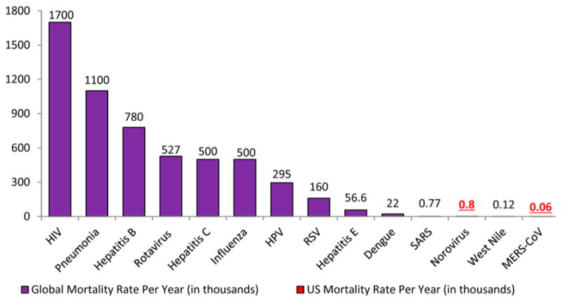
Mortality versus viral diseases.–
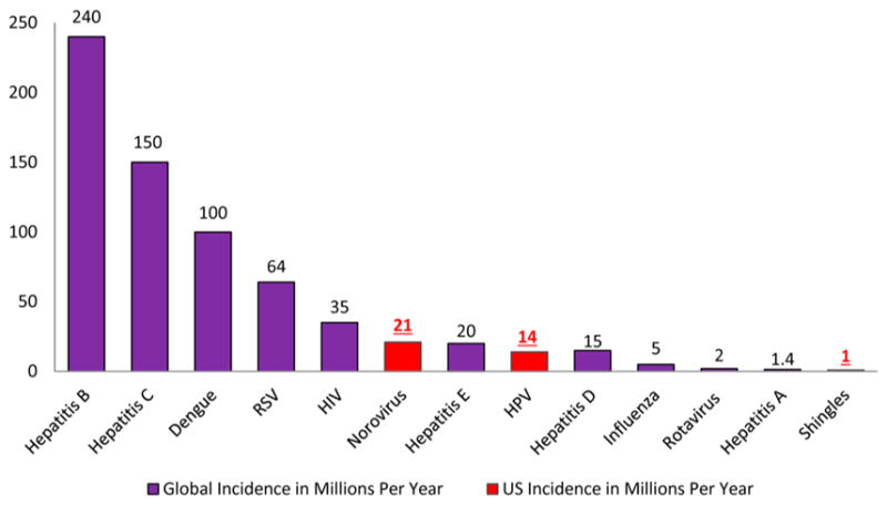
Incidence rates versus viral diseases.–
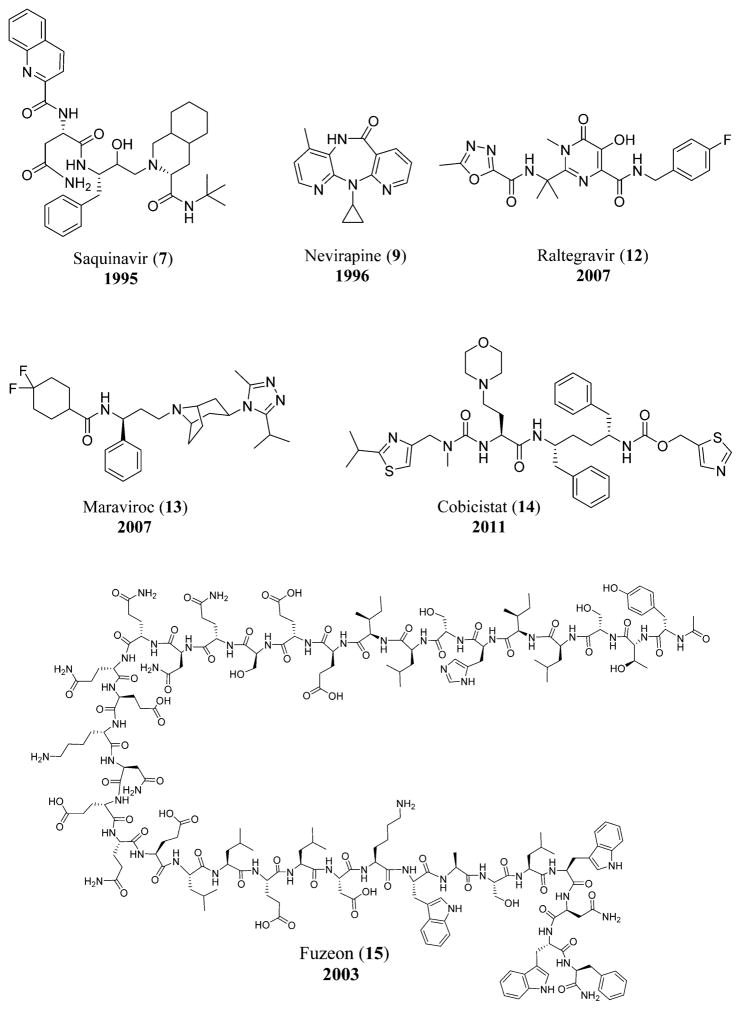
Anti-HIV-1 drugs referenced in the history. Compounds include saquinavir (7), nevirapine (9), raltegravir (12), maraviroc (13), cobicistat (14), and fuzeon (15).
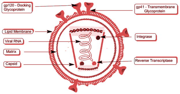
Structure of HIV.
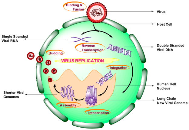
Schematic representation of the virus replication cycle.
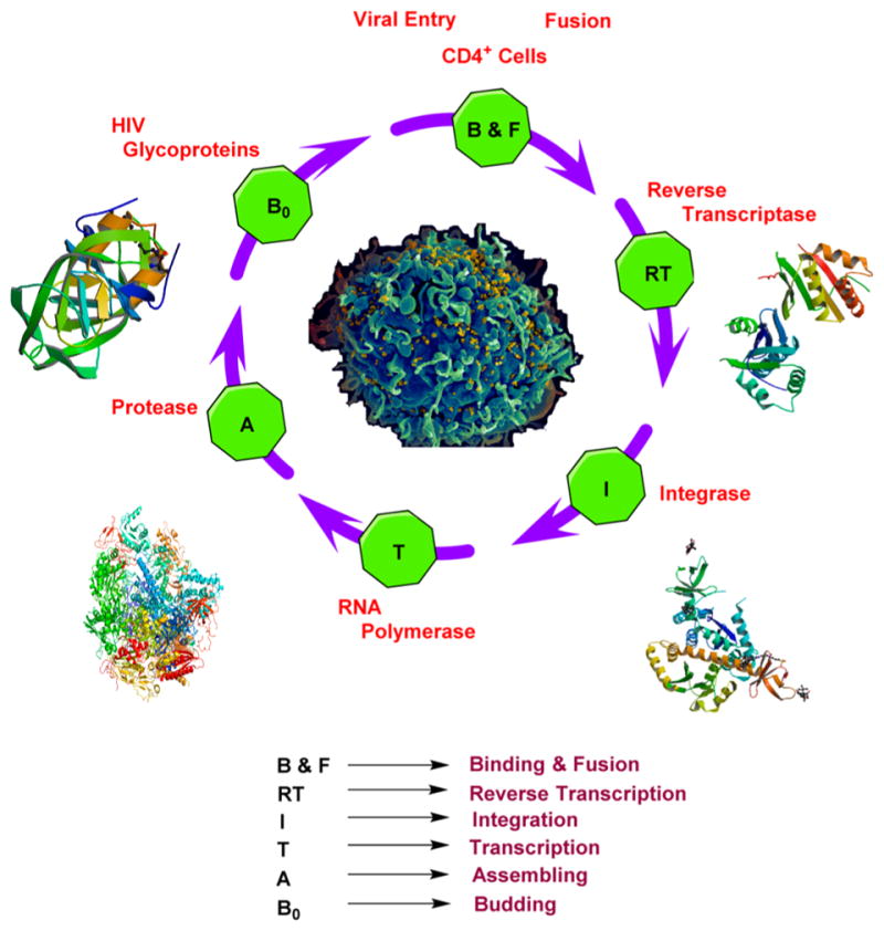
Crystal structures of HIV reverse transcriptase, integrase, RNA polymerase, and protease and scanning electron micrograph of HIV. Courtesy: National Institute of Allergy and Infectious Diseases, http://www.nih.gov/science/hiv/ .
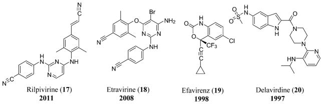
NNRTIs used for the treatment of HIV that include rilpivirine (17), etravirine (18), efavirenz (19), and delavirdine (20).
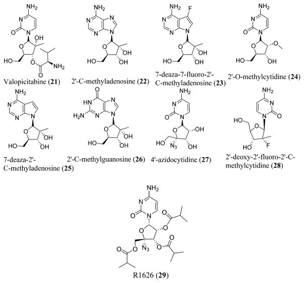
Various NRRIs possessing anti-HCV activity. NRRIs include valopicitabine (21), 2′-C-methyladenosine (22), 7-deaza-7-fluoro-2′-C-methyladenosine (23), 2′-O-methylcytidine (24), 7-deaza-2′-C-methyladenosine (25), 2′-C-methylguanosine (26), 4′-azidocytidine (27), 2′-deoxy-2′-fluoro-2′-C-methylcytidine (28), and R1626 (29).
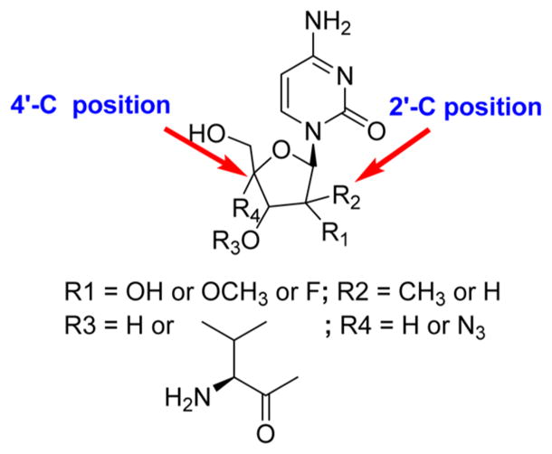
Structure–activity relationship (SAR) of anti-HCV NRRIs.
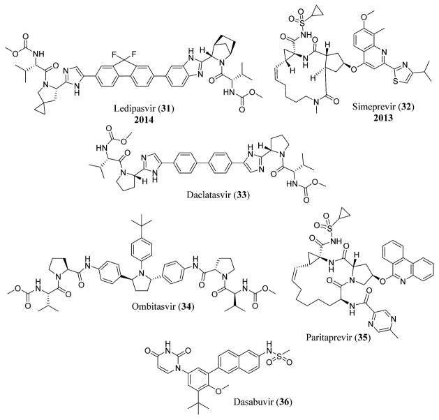
Structures of ledipasvir (31), simeprevir (32), daclatasvir (33), ombitasvir (34), paritaprevir (35), and dasabuvir (36).
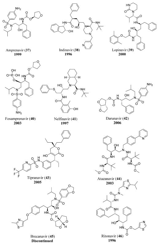
HIV protease inhibitors including amprenavir (37), indinavir (38), lopinavir (39), fosamprenavir (40), nelfinavir (41), darunavir (42), tipranavir (43), atazanavir (44), brecanavir (45), and ritonavir (46).
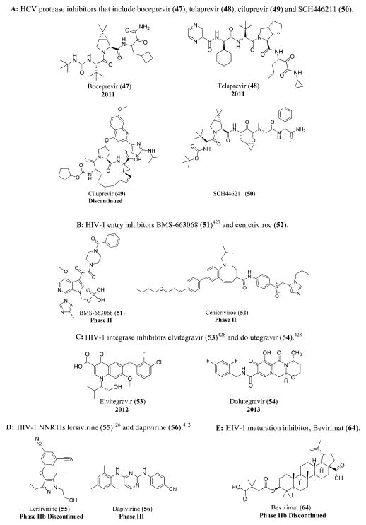
(A) HCV protease inhibitors that include boceprevir (47), telaprevir (48), ciluprevir (49), and SCH446211 (50). (B) HIV-1 entry inhibitors BMS-663068 (51) and cenicriviroc (52). (C) HIV-1 integrase inhibitors elvitegravir (53) and dolutegravir (54). (D) HIV-1 NNRTIs lersivirine (55) and dapivirine (56). (E) HIV-1 maturation inhibitor, Bevirimat (64).
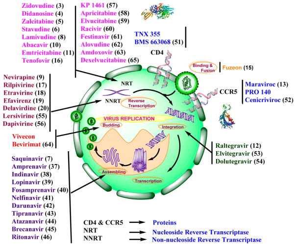
Schematic representation of all the anti-HIV-1 drugs that act at various stages of viral replication cycle including the crystal structures of CD4 and CCR5.
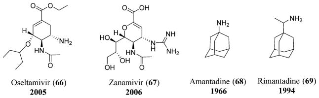
Neuraminidase inhibitors: oseltamivir (66) and zanamivir (67); adamantane drugs: amantadine (68) and rimantadine (69).
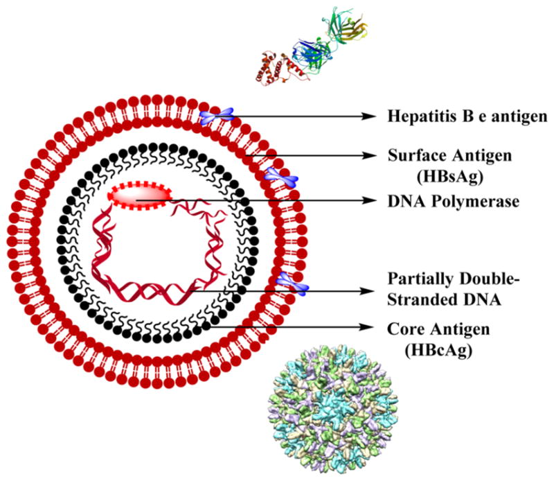
Structure of hepatitis B virus with crystal structure of HBV e antigen and cryo-electron microscopy (CryoEM) structure of HBV core antigen.
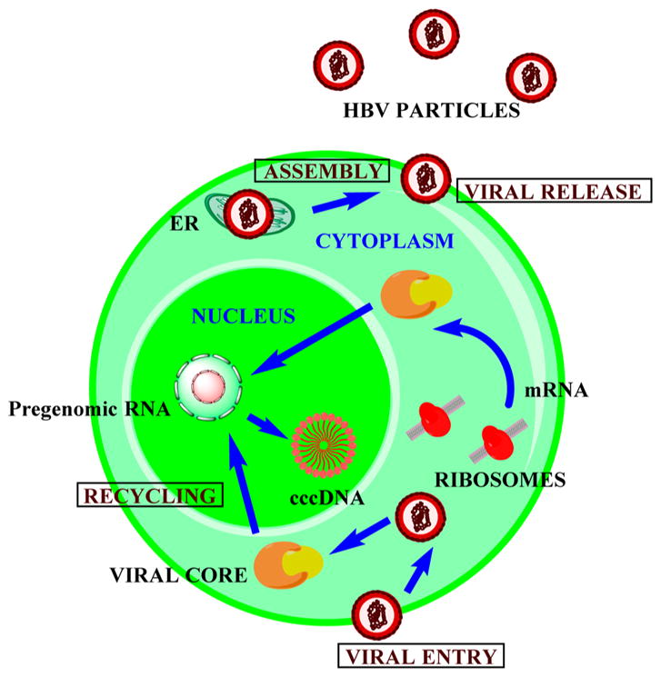
Viral replication of Hepatitis B virus.
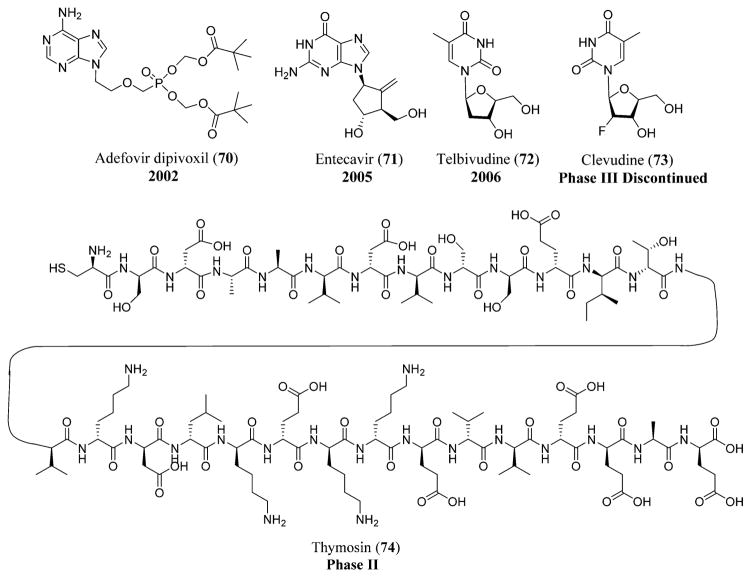
HBV drugs: adefovir dipivoxil (70), entecavir (71), telbivudine (72), clevudine (73), and thymosin (74).
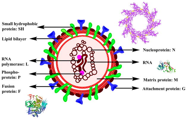
Structure of RSV along with crystal structures of nucleoprotein, matrix protein, and fusion protein.
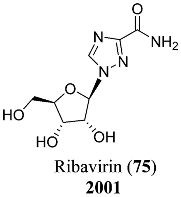
Chemical structure of ribavirin (75).
Structure of dengue virus along with the crystal structure of the envelope protein (E)
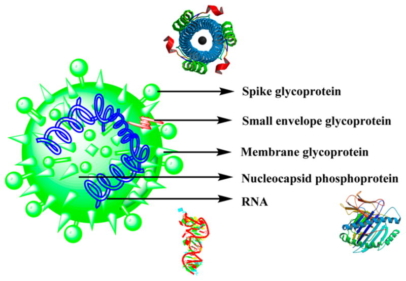
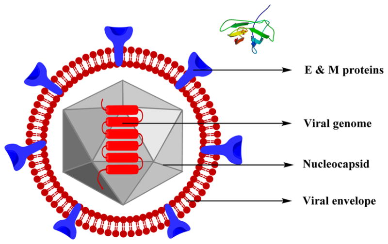
Structure of the SARS virus along with the crystal structures of spike glycoprotein, nucleocapsid phosphoprotein, and RNA.
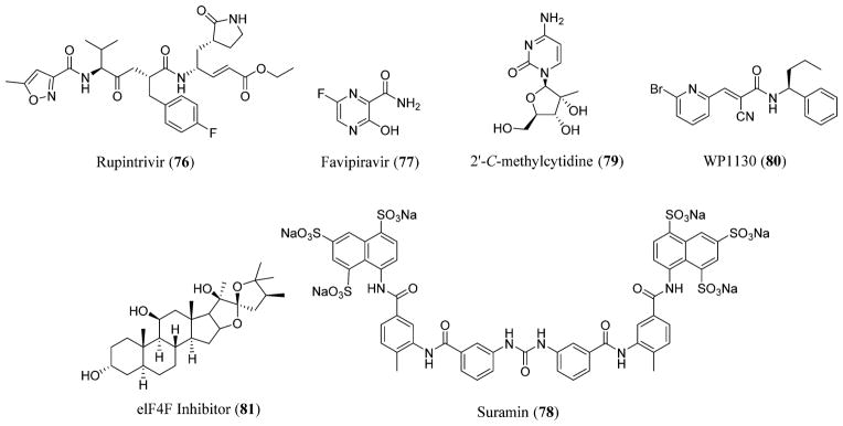
Norovirus inhibitors: rupintrivir (76), favipiravir (77), suramin (78), 2′-C-methylcytidine (79), deubiquitinase [WP1130] (80), and elF4F inhibitors (81).
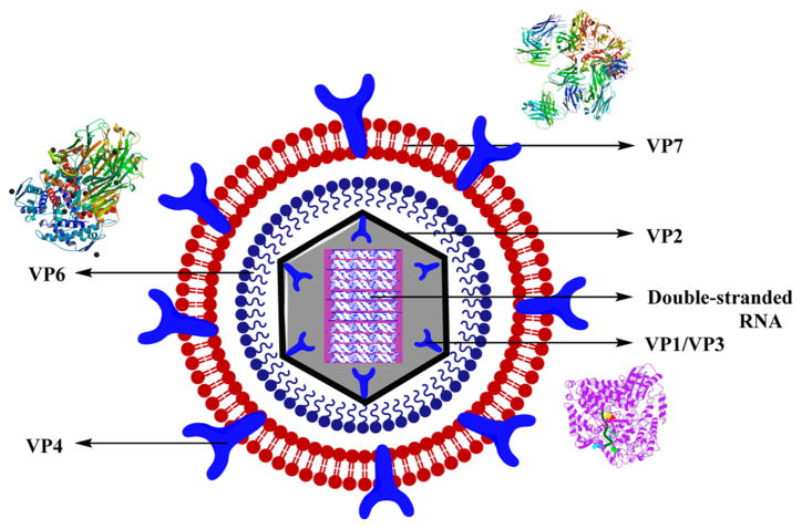
Structure of rotavirus along with the crystal structures of VP7, VP1, and VP6.
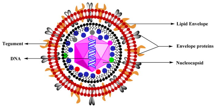
Structure of varicella-zoster virus (VZV).
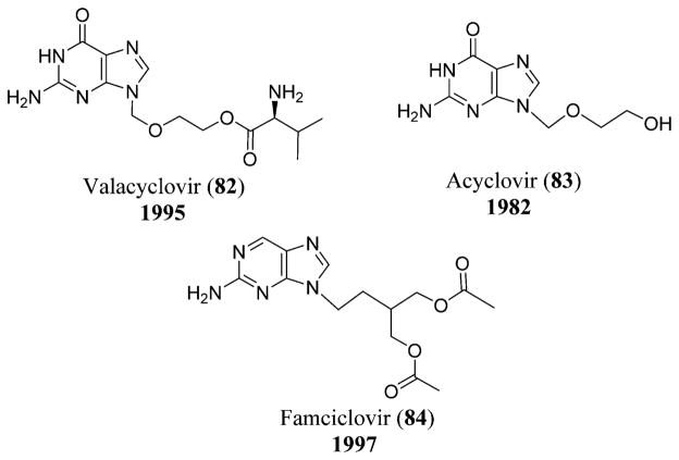
Chemical structures of valacyclovir (82), acyclovir (83), and famciclovir (84).
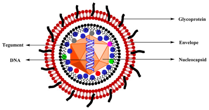
Structure of HSV.
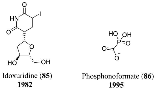
Chemical structures of idoxuridine (85) and phosphonoformate (86).
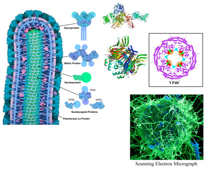
Structure of Ebola virus. Reprinted with permission from ref . Copyright 1989 American Chemical Society. Image from the RCSB PDB October 2014 Molecule of the Month featured by David Goodsell (DOI: 10.2210/rcsb_pdb/mom_2014_10)], crystal structures of glycoprotein, matrix proteins,, and electron scanning microscopic image (Courtesy: National Institute of Allergy and Infectious Diseases, http://www.niaid.nih.gov/news/newsreleases/2014/Pages/EbolaDisparities.aspx ).
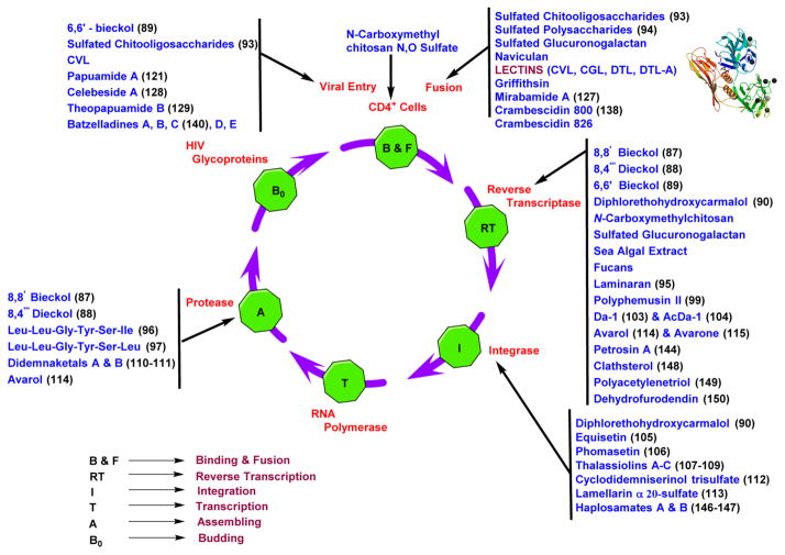
Schematic representation of the marine metabolites that are active at various stages of the viral replication cycle including the crystal structure of lectin.
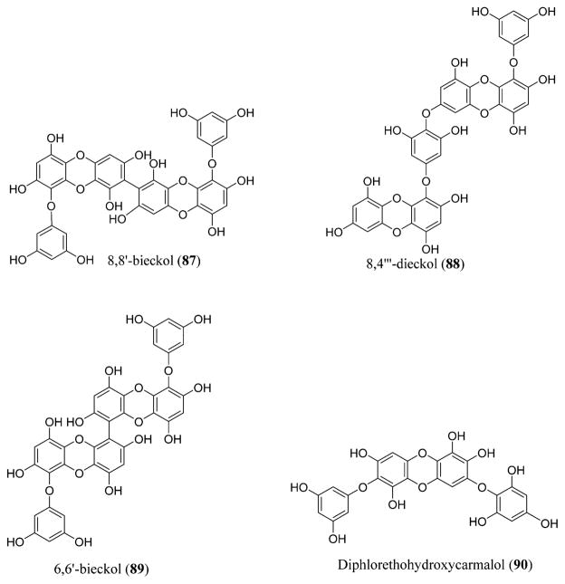
Phlorotannins 8,8′-bieckol (87), 8,4‴-dieckol (88), and 6,6′-bieckol (89) isolated from a brown algae, Ecklonia cava, and diphlorethohydroxycarmalol (90) isolated from Ishige okamurae.
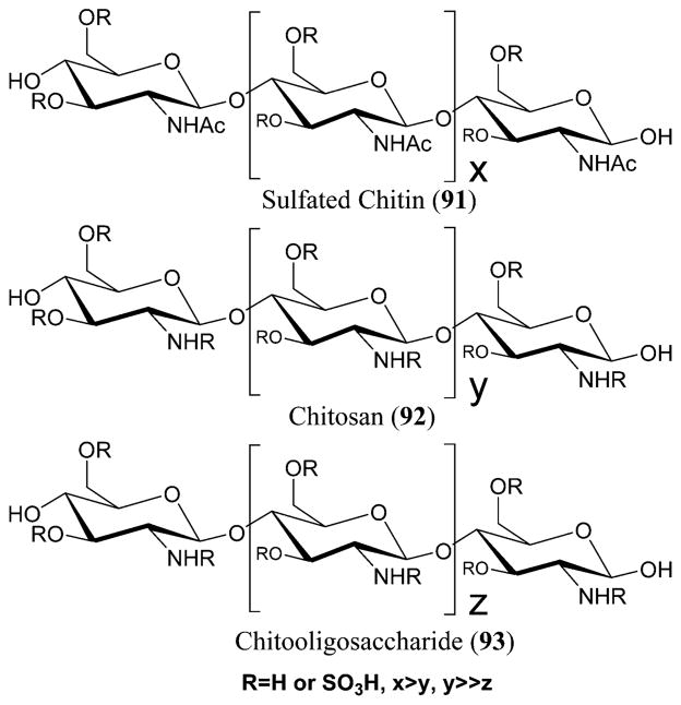
Sulfated chitin (91), chitosan (92), and chitooligosaccharide (93) derivatives.
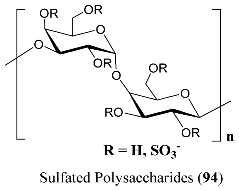
Sulfated polysaccharides (94) isolated from red seaweeds.
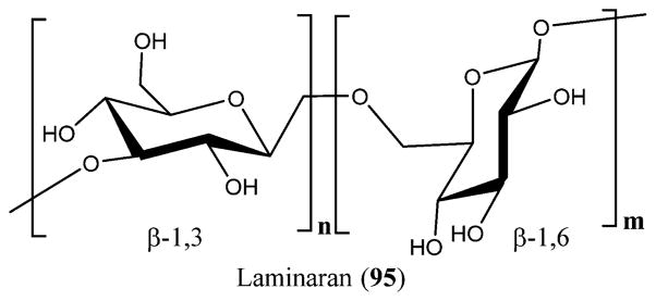
Structure of laminaran (95).
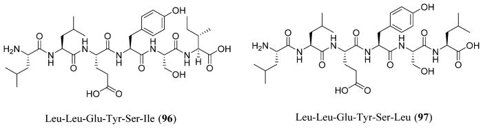
Structures of Leu-Leu-Glu-Tyr-Ser-Ile (96) and Leu-Leu-Glu-Tyr-Ser-Leu (97).
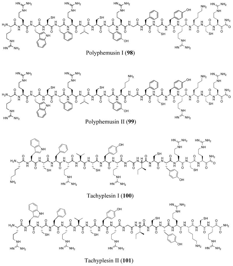
Structures of polyphemusin I (98)–II (99) and tachyplesins I (100)–II (101).
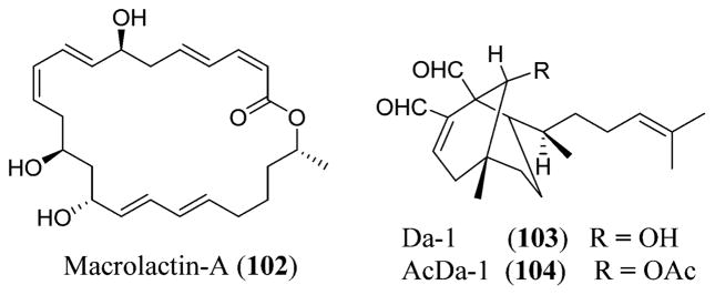
Structures of macrolactin-A (102), Da-1 (103), and AcDa-1 (104).
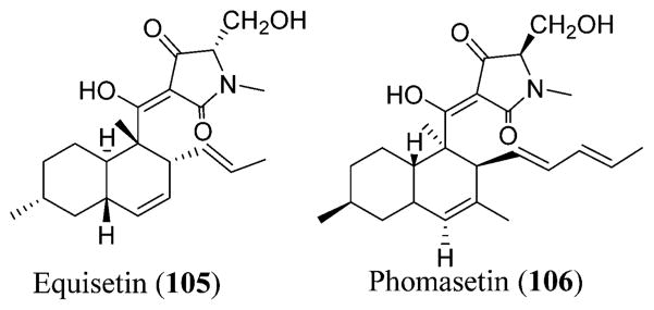
Structures of equisetin (105) and phomasetin (106).
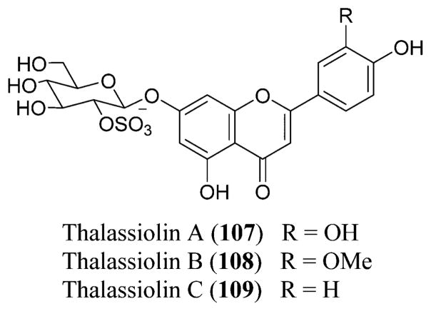
Structures of thalassiolins A (107), B (108), and C (109).
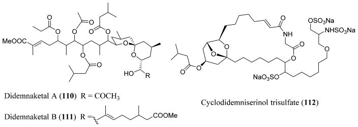
Structures of didemnaketals A (110) and B (111) and cyclodidemniserinol trisulfate (112).
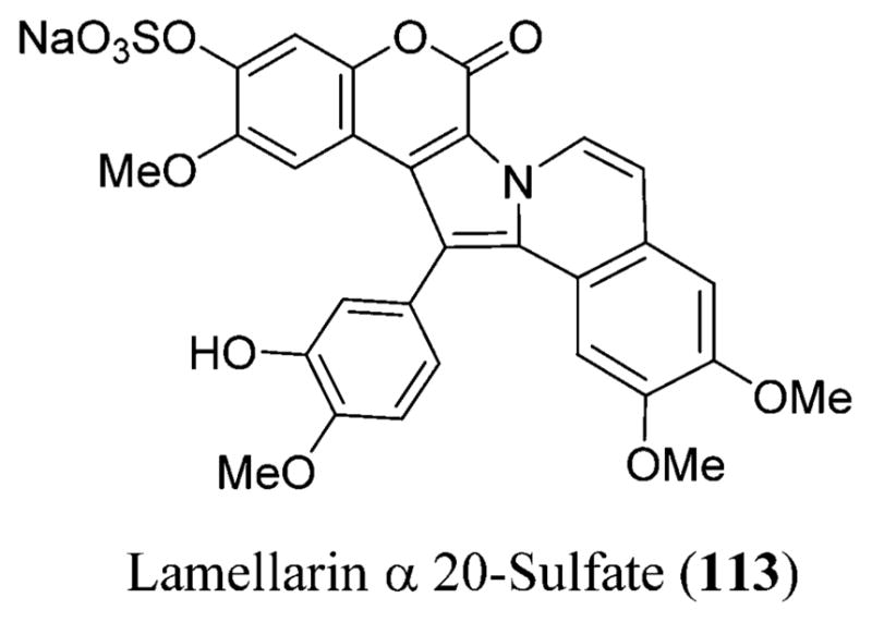
Structure of lamellarin α 20-sulfate (113).
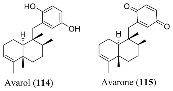
Avarol (114) and avarone (115) possessing anti-HIV-1 activity.
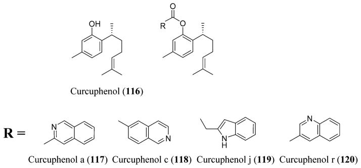
Curcuphenol (116) and its analogues a (117), c (118), j (119), and r (120) isolated and modified from the sponges Didiscus oxeata, Didiscus flavus, Myrmekioderma styx, and Epipolasis sp.
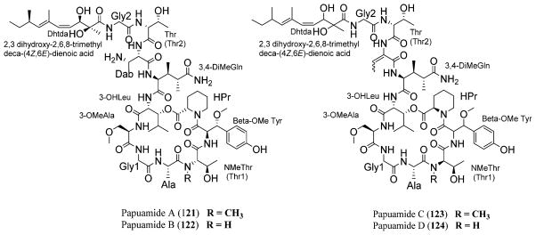
Papuamides A (121), B (122), C (123), and D (124) isolated from the marine sponges Theonella swinhoei and T. mirabilis.
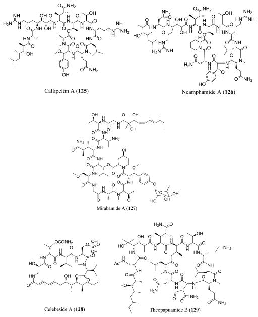
Callipeltin A (125) isolated from Callipelta, neamphamide A (126) isolated from Neamphius huxleyi, mirabamide A (127), celebeside A (128), and theopapuamide B (129) isolated from Siliquariaspongia mirabilis.
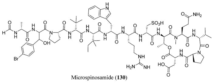
Microspinosamide (130) isolated from the Indonesian sponge Sidonops microspinosa.
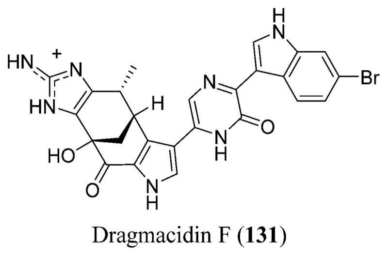
Dragmacidin F (131) isolated from the marine sponge belonging to the Halicortex genus from the southern coast of Ustica Island in Italy.
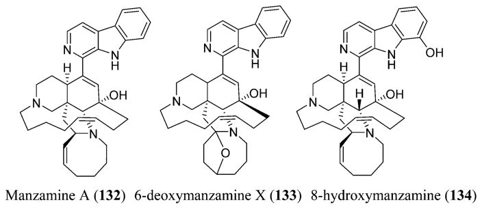
Manzamine A (132), 6-deoxymanzamine X (133), and 8-hydroxymanzamine (134) isolated from Haliclona sp.
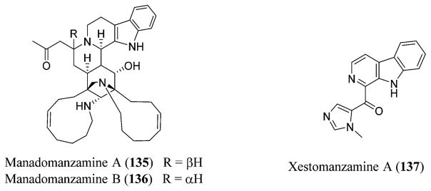
Manadomanzamines A (135) and B (136) and xestomanzamine A (137) isolated from the Indonesian sponge Acanthostrongylophora sp.
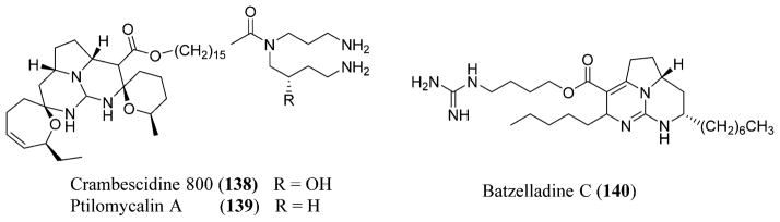
Ptilomycalin A (139), isolated from the sponge Monanchora unguifera, and crambescidine 800 (138) and batzelladine C (140) isolated from the Batzella and Monanchora genera.
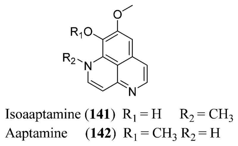
Isoaaptamine (141) and aaptamine (142) isolated from the sponge Aaptos aaptos.
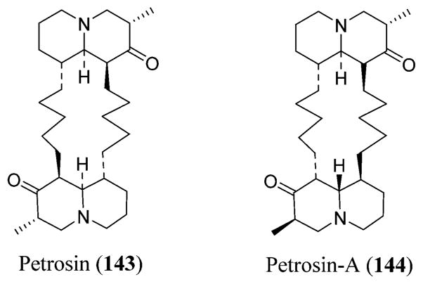
Structures of petrosin (143) and petrosin A (144).
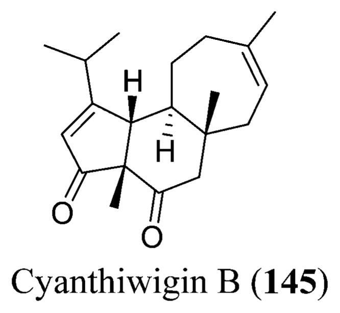
Cyanthiwigin B (145) isolated from the Jamaican sponge Epipolasis reiswigi.
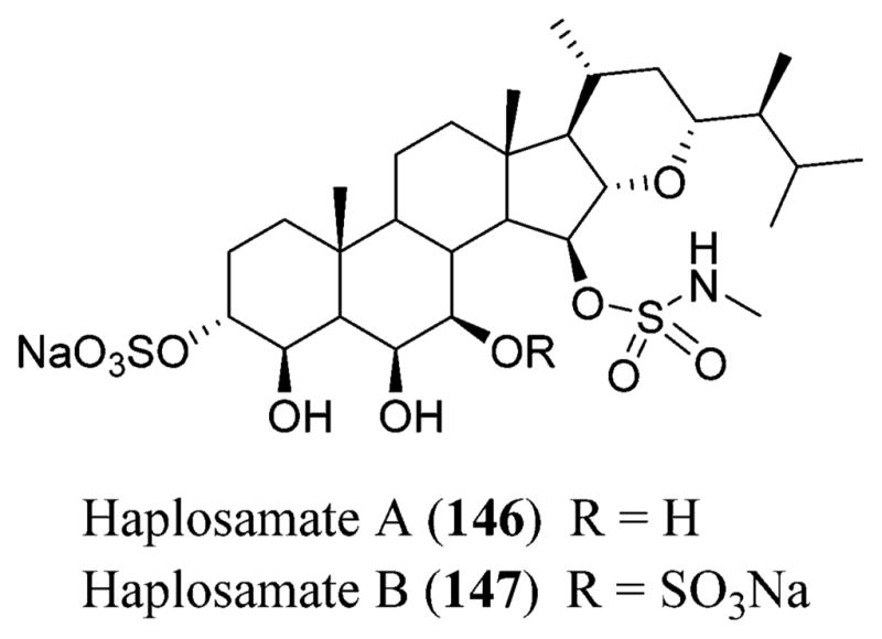
Structures of haplosamates A (146) and B (147).
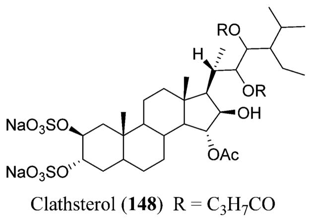
Structure of clathsterol (148).
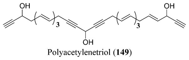
Structure of polyacetylenetriol (149).
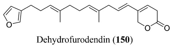
Structure of dehydrofurodendin (150).
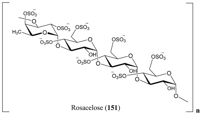
Structure of the repeating unit of rosacelose (151).
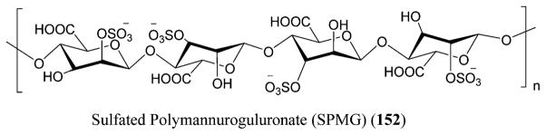
Structure of sulfated polymannuroguluronate (SPMG) (152).
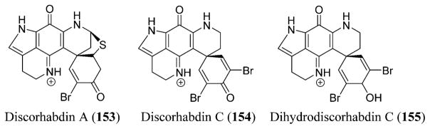
Structures of discorhabdins A (153) and C (154) and dihydrodiscorhabdin C (155).
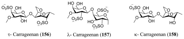
Structures of ι- (156), λ- (157), and κ-carrageenans (158).
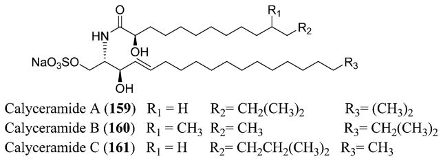
Structures of calyceramides A (159), B (160), and C (161).
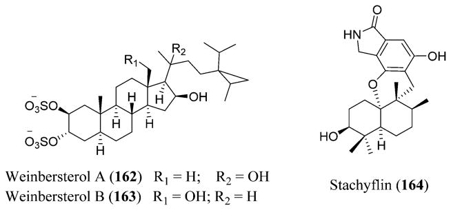
Structures of weinbersterols A (162) and B (163) and stachyflin (164).
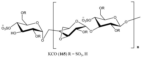
Structure of KCO (165).
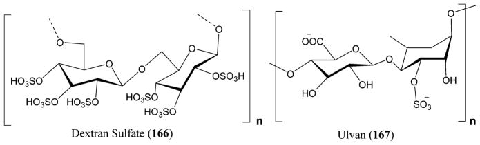
Structures of dextran sulfate (166) and ulvan (167).
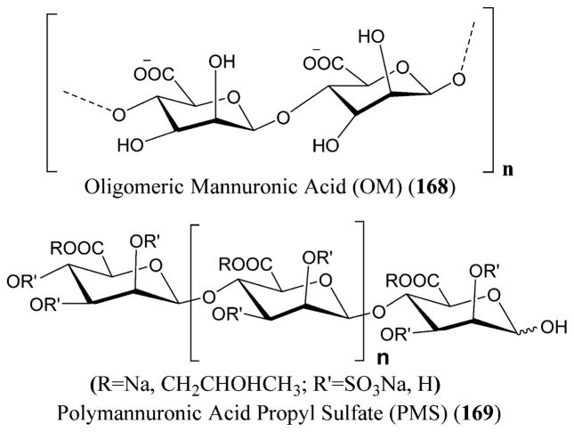
Structures of oligomeric mannuronic acid (OM) (168) and polymannuronic acid propyl sulfate (PMS) (169).
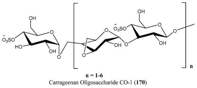
Structure of the carrageenan oligosaccharide CO-1 (170).
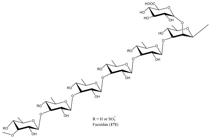
Structure of fucoidan (171).
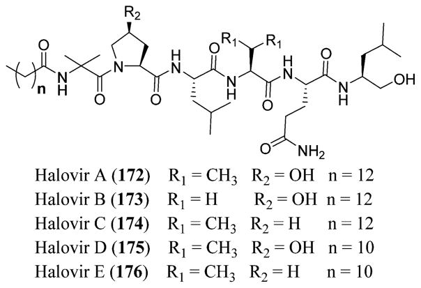
Structures of halovirs A (172), B (173), C (174), D (175), and E (176).
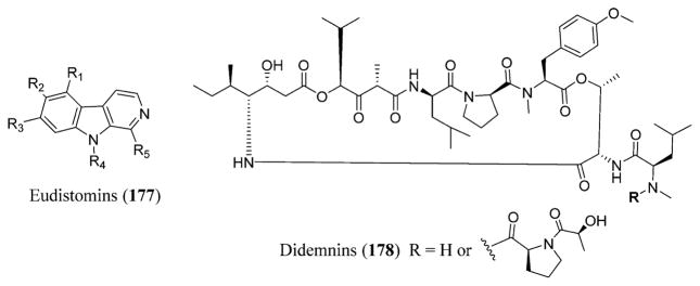
Structures of eudistomins (177) and didemnins (178).
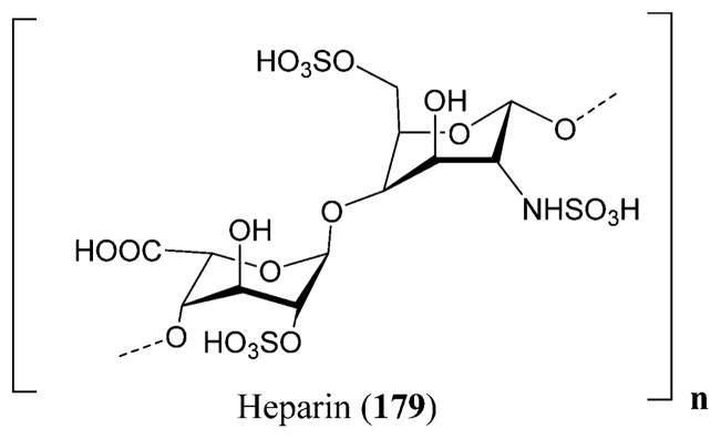
Structure of heparin (179).
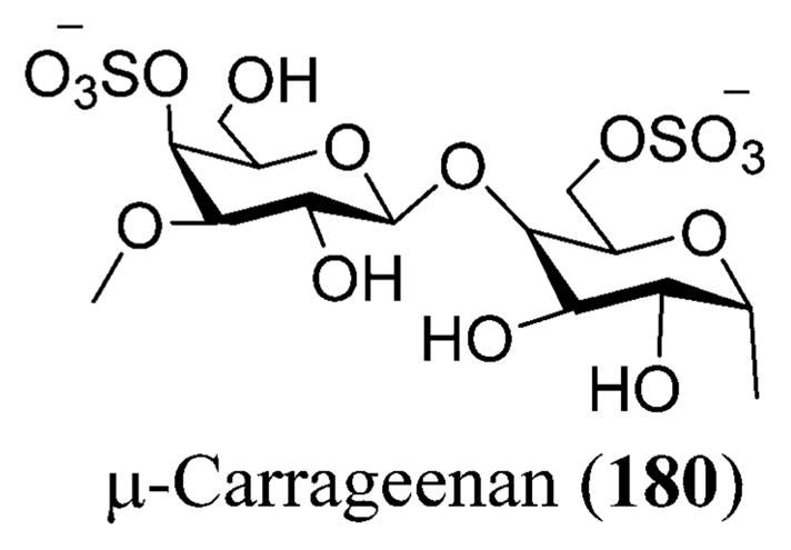
Structure of μ-carrageenan (180).
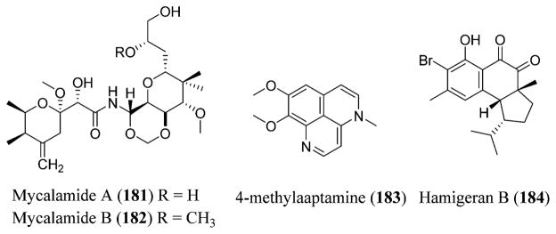
Structures of mycalamide A (181), mycalamide B (182), 4-methylaaptamine (183), and hamigeran B (184).
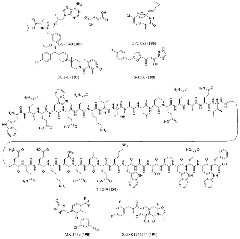
HIV drugs in late stage development: GS 7340 (185), DPC 083 (186), SCH-C (187), S-1360 (188), T-1249 (189), MK-1439 (190), and S/GSK1265744 (191).

Novel HCV drugs: mericitabine (192), danoprevir (193), setrobuvir (194), BI 201335 (195), BI 207127 (196), MK-8742 (197), MK-5172 (198), MK-7009 or vaniprevir (199), and GSK 2336805 (200).
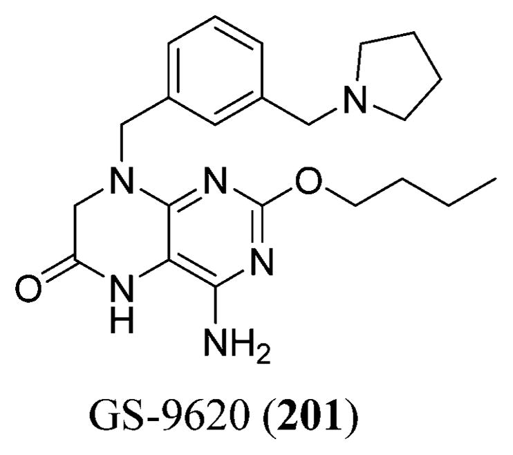
Structure of GS-9620 (201).
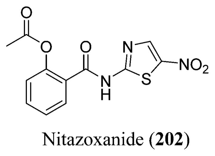
Structure of nitazoxanide (202).
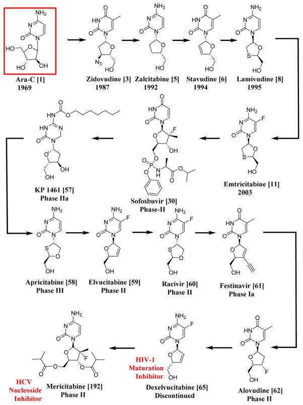
Systematic Representation of the Chronological Order of HIV-1 and HCV Drugs Having Similar Structures to Ara-C, the First Anticancer Lead from a Sponge; All the Drugs and Compounds Mentioned in This Scheme Are NRTIs except for Dexelvucitabine (65), Which Is an HIV-1 Maturation Inhibitor, and Mericitabine (192), Which Is an HCV Nucleoside Inhibitor
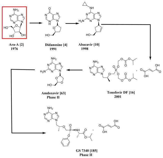
Systematic Representation of the Chronological Order of HIV-1 and HCV Drugs Having Similar Nuclei to Ara-A, an Antiviral Agent; All the Drugs and Compounds Mentioned in This Scheme Are NRTIs
References
-
- MedicineNet.com. [accessed November 25, 2011];Definition of Agent, Anti-Infective. http://www.medicinenet.com/script/main/art.asp?articlekey=2178.
-
- World Health Organization. [accessed May 15, 2014];Media Centre: Fact Sheets. http://www.who.int/mediacentre/factsheets/en/#A.
-
- National Institute of Allergy and Infectious Diseases. [accessed May 15, 2014];Health & Research Topics. http://www.niaid.nih.gov/topics/Pages/default.aspx.
-
- Centers for Disease Control and Prevention. [accessed May 15, 2014];Morbidity and Mortality Weekly Report (MMWR) http://www.cdc.gov/mmwr/indss_2014.html.
Publication types
MeSH terms
Substances
Grants and funding
LinkOut - more resources
Full Text Sources
Other Literature Sources
