. 2015 Aug 28;10(8):e0136533.
doi: 10.1371/journal.pone.0136533. eCollection 2015.

Structural Analysis of the Regulatory Domain of ExsA, a Key Transcriptional Regulator of the Type Three Secretion System in Pseudomonas aeruginosa

Affiliations

Structural Analysis of the Regulatory Domain of ExsA, a Key Transcriptional Regulator of the Type Three Secretion System in Pseudomonas aeruginosa

Manisha Shrestha et al. PLoS One. .

Abstract

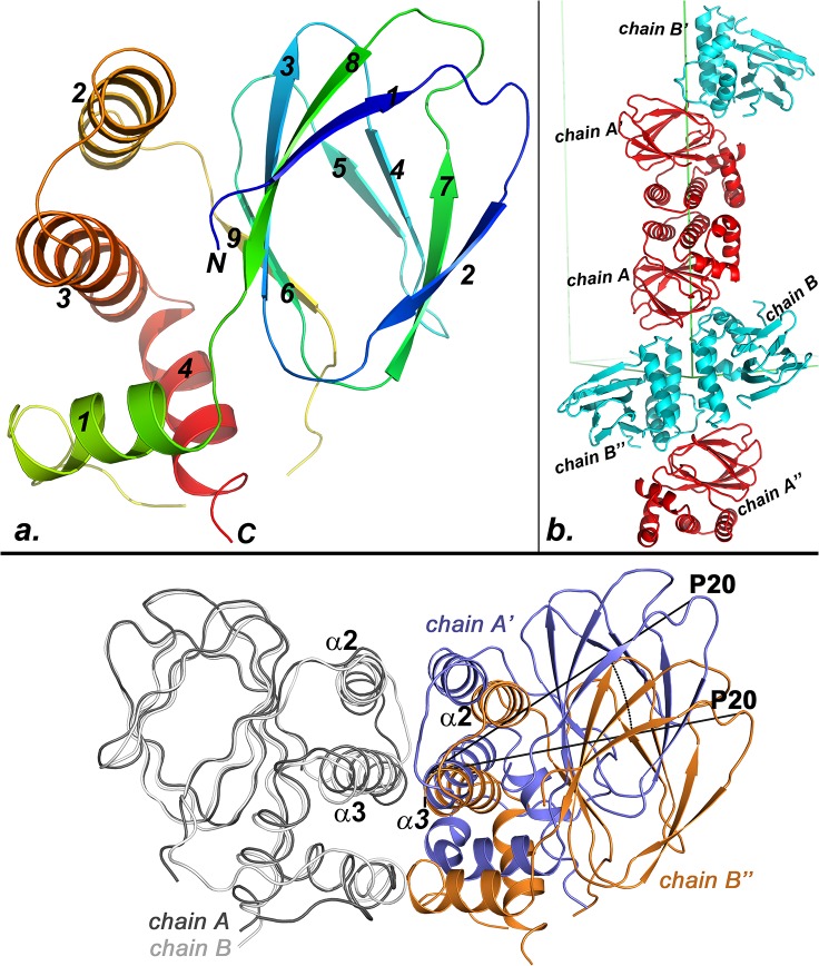
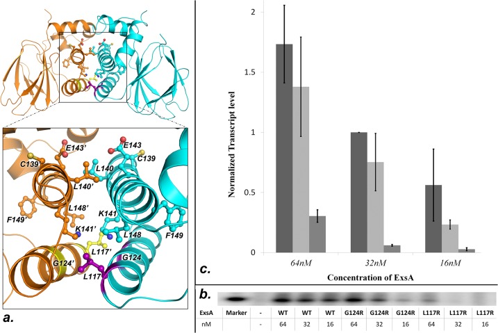
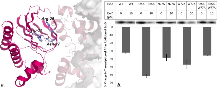
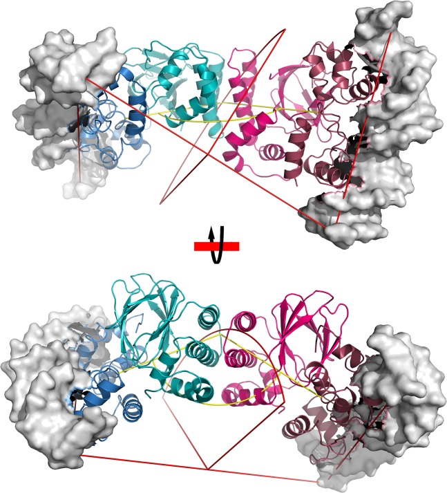
Pseudomonas aeruginosa employs a type three secretion system to facilitate infections in mammalian hosts. The operons encoding genes of structural components of the secretion machinery and associated virulence factors are all under the control of the AraC-type transcriptional activator protein, ExsA. ExsA belongs to a unique subfamily of AraC-proteins that is regulated through protein-protein contacts rather than small molecule ligands. Prior to infection, ExsA is inhibited through a direct interaction with the anti-activator ExsD. To activate ExsA upon host cell contact this interaction is disrupted by the anti-antiactivator protein ExsC. Here we report the crystal structure of the regulatory domain of ExsA, which is known to mediate ExsA dimerization as well as ExsD binding. The crystal structure suggests two models for the ExsA dimer. Both models confirmed the previously shown involvement of helix α-3 in ExsA dimerization but one also suggest a role for helix α-2. These structural data are supported by the observation that a mutation in α-2 greatly diminished the ability of ExsA to activate transcription in vitro. Additional in vitro transcription studies revealed that a conserved pocket, used by AraC and the related ToxT protein for the binding of small molecule regulators, although present in ExsA is not involved in binding of ExsD.


Conflict of interest statement

Competing Interests: The authors have declared that no competing interests exist.

Figures

Fig 1
Fig 1. Crystal structure of the ExsA-NTD.
(A) Model of a monomer encompassing amino acids 2–166 which produced clearly defined electron density. Blue to red rainbow coloring traces the backbone from the N to the C-terminus. Secondary structure elements are numbered. (B) Packing contacts in the crystal suggest the possible structure of the biological dimer. Chains A and B constitute the asymmetric unit of the crystal. Application of two crystallographic two-fold axes produces two additional pairs of chains labeled with a prime and a double-prime, respectively. Contacts between either chains A and A’ or between chains B and B” are proposed to mediate ExsA dimerization in vivo. (C) Shown in gray are the overlaid backbones traces of chains A and B. Also displayed are the symmetry-related molecules A’ and B” to highlight similarities and differences between the two possible quaternary structures. The B” molecule is rotated by approximately 23° around helix α-3. The rotation is visualized by marking the angle between the P20 residues of A’ and B” in the figure.
Fig 2
Fig 2. Mapping of the ExsA dimer interface.
(A) The shown A/A’ ExsA-NTD interface suggests involvement of helix α-2 in ExsA dimerization. Previously identified interface residues are indicated in the same color as the protein backbone. G124 and L117 are colored violet and yellow in the respective molecules. (B) Shown is a sample gel of measurements testing the impact of the L117R and G124R mutations on the ability of ExsA to activate transcription in vitro. Three concentrations of each protein were tested to ensure that the experiments were conducted in a sensitive range. (C) Graphical representation of the in vitro transcription assays from triplicate experiments. Going from left to right: wtExsA, ExsAG124R, and ExsAL117R.
Fig 3
Fig 3. Impact of mutations in the conserved cavity of ExsA on ExsD binding.
Three residues lining the cavity within the beta-sandwich structure of ExsA were mutated with alanine to determine if these residues are involved in ExsD binding. (A) Cartoon depiction of a full-length model of an ExsA-DNA complex. This model was generated by overlaying the structures of ExsA-NTD and a homology model of ExsA-CTD (based on the MarA-DNA crystal structure) onto the structure of ToxT. The mutated residues are depicted as ball-and-stick. (B) Results of in vitro transcription assays measuring the impact of the indicated mutations on ExsA-ExsD interactions. Plotted in the chart is the percent change in obtained transcript level when 10 μM ExsD is added to the reaction. A sample gel showing transcript bands is presented above the chart. Experiments were conducted in duplicate.
Fig 4
Fig 4. Cartoon model of a full-length dimeric ExsA-DNA complex.
This model was generated by first overlaying the structure of the ExsA-NTD A/A’ dimer and a homology model of ExsA-CTD (based on the MarA-DNA crystal structure) onto the structure of ToxT. Subsequently, crystallographic two-fold axis was applied to create a model of the full-length protein with a dimer interface corresponding to A/A’ dimer observed in the crystal.

Similar articles

Cited by

References

    1. Ghosh P. Process of protein transport by the type III secretion system. Microbiology and molecular biology reviews: MMBR. 2004;68(4):771–95. 10.1128/MMBR.68.4.771-795.2004 - DOI - PMC - PubMed
    1. Alfano JR, Collmer A. Type III secretion system effector proteins: double agents in bacterial disease and plant defense. Annual review of phytopathology. 2004;42:385–414. 10.1146/annurev.phyto.42.040103.110731 . - DOI - PubMed
    1. Barison N, Gupta R, Kolbe M. A sophisticated multi-step secretion mechanism: how the type 3 secretion system is regulated. Cell Microbiol. 2013;15(11):1809–17. 10.1111/cmi.12178 . - DOI - PubMed
    1. Tosi T, Pflug A, Discola KF, Neves D, Dessen A. Structural basis of eukaryotic cell targeting by type III secretion system (T3SS) effectors. Research in microbiology. 2013;164(6):605–19. 10.1016/j.resmic.2013.03.019 . - DOI - PubMed
    1. Raymond B, Young JC, Pallett M, Endres RG, Clements A, Frankel G. Subversion of trafficking, apoptosis, and innate immunity by type III secretion system effectors. Trends in microbiology. 2013;21(8):430–41. 10.1016/j.tim.2013.06.008 . - DOI - PubMed

Publication types

MeSH terms

Substances

LinkOut - more resources