

Comparison of Rebound Tonometry, Perkins Applanation Tonometry and Ocular Response Analyser in Mucopolysaccharidosis Patients
- PMID: 26317991
- PMCID: PMC4552832
- DOI: 10.1371/journal.pone.0133586
Comparison of Rebound Tonometry, Perkins Applanation Tonometry and Ocular Response Analyser in Mucopolysaccharidosis Patients
Abstract
Aims: To investigate the feasibility and to compare three devices measuring intraocular pressure (IOP) in mucopolysaccharidosis patients (MPS): iCare rebound tonometer (RT), Perkins applanation tonometer (PAT) and ocular response analyzer (ORA).
Methods: MPS patients who underwent at least two examinations out of: RT, PAT and ORA at the same visit were identified and retrospectively analyzed in this study.
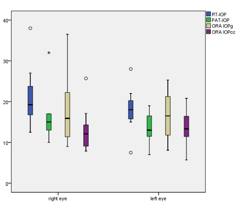
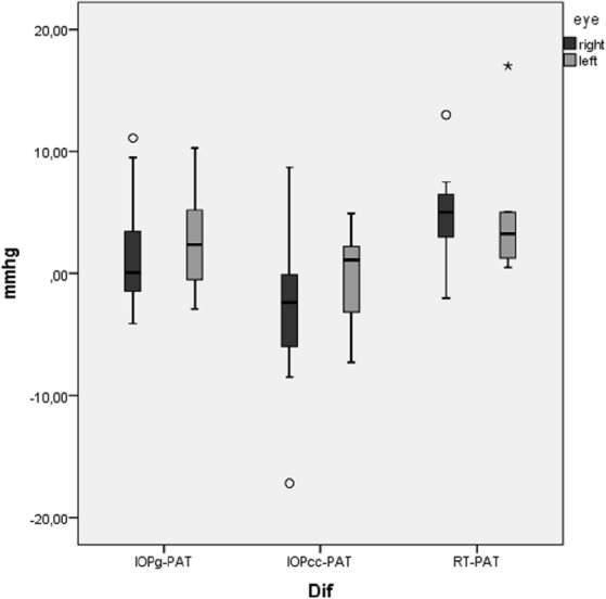
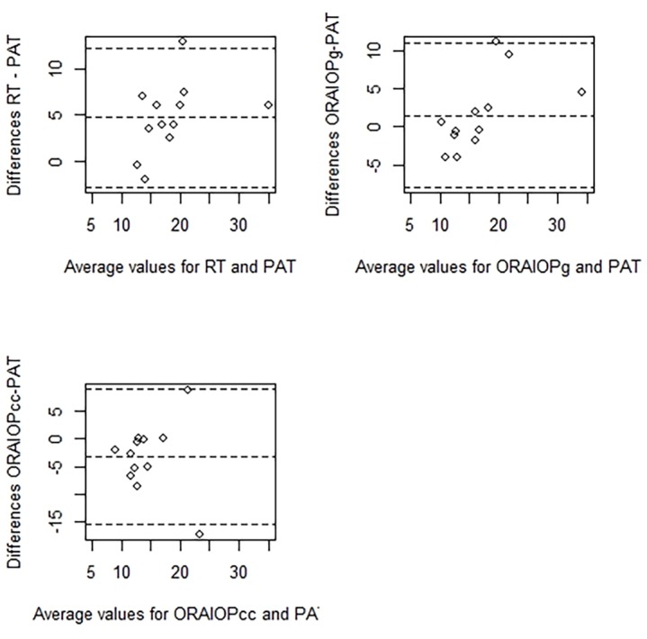
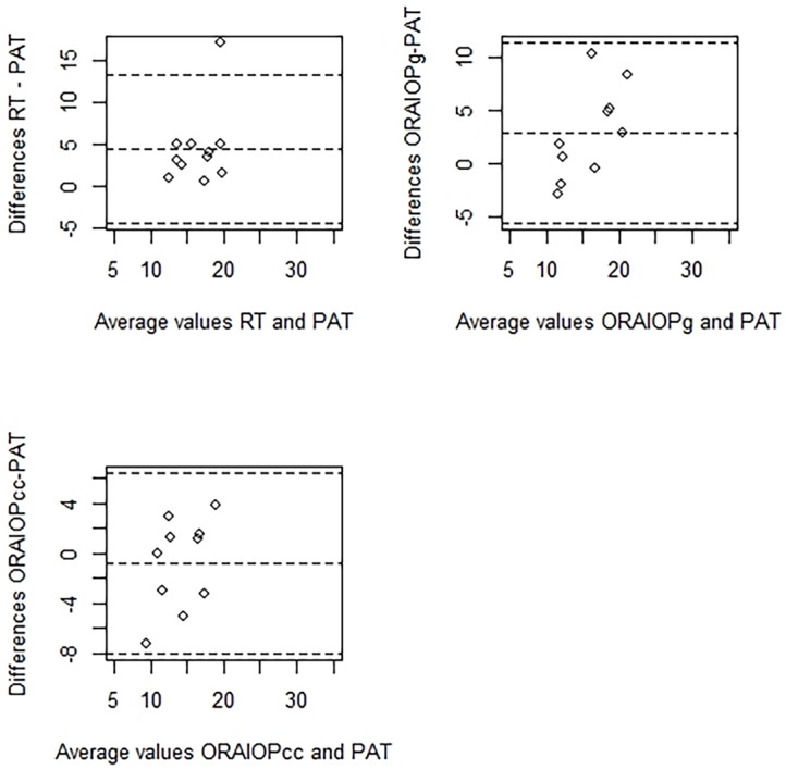
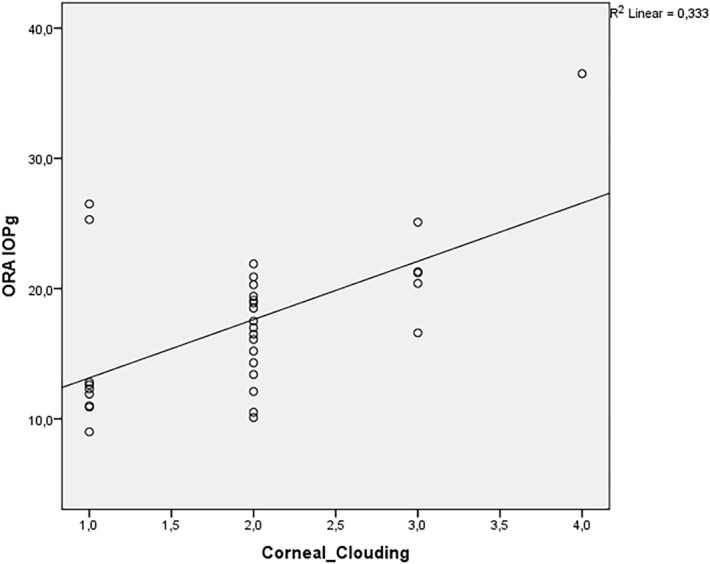
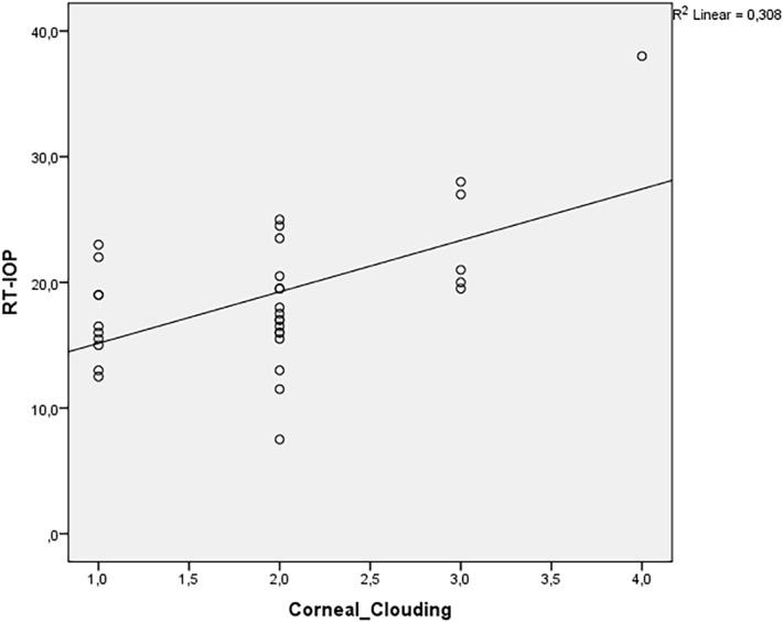
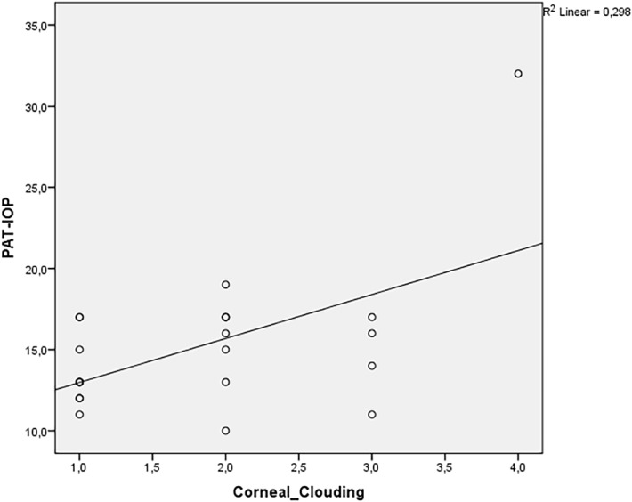
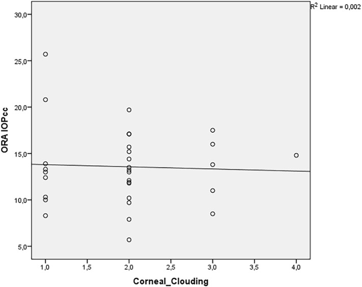
Results: 17 patients fulfilled the inclusion criterion. In all 17 patients IOP measurements were performed with RT (34 eyes) and ORA (33 eyes), while PAT measurement was possible in only 12 (24 eyes) patients. The RT, corneal-compensated intraocular pressure (IOPcc) and Goldmann-correlated intraocular pressure (IOPg) differed relevantly from IOP assessed with PAT. Corneal clouding in MPS patients correlated positively with PAT, RT and IOPg (r = 0.3, 0.5, and 0.5 respectively), but not with IOPcc (r = 0.07). The MPS-related corneal clouding correlated positively with biomechanical corneal parameters assessed with ORA: corneal hysteresis (r = 0.77) and corneal resistance factor (r = 0.77) either.
Conclusions: RT and ORA measurements were tolerated better than applanation tonometry in MPS patients. IOP measurements assessed with RT and ORA differed relevantly from PAT. Corneal-compensated IOP assessed with ORA seems to be less affected by the MPS-related corneal clouding than applanation or rebound tonometry. RT and ORA measurements should be preferred for IOP assessment in patients with MPS.
Conflict of interest statement
Figures
Similar articles
-
Influence of Corneal Opacity on Intraocular Pressure Assessment in Patients with Lysosomal Storage Diseases.PLoS One. 2017 Jan 12;12(1):e0168698. doi: 10.1371/journal.pone.0168698. eCollection 2017. PLoS One. 2017. PMID: 28081172 Free PMC article. Clinical Trial.
-
Intraocular pressure and biomechanical corneal properties measure by ocular response analyser in patients with primary congenital glaucoma.Acta Ophthalmol. 2016 Aug;94(5):e293-7. doi: 10.1111/aos.12912. Epub 2015 Dec 9. Acta Ophthalmol. 2016. PMID: 26647905
-
Influence of cornea on intraocular pressure measurement by ICARE PRO and ORA.Cesk Slov Oftalmol. 2019 Summer;75(3):111-118. doi: 10.31348/2019/3/1. Cesk Slov Oftalmol. 2019. PMID: 31779459 English.
-
Comparison of intraocular pressure measured by ocular response analyzer and Goldmann applanation tonometer after corneal refractive surgery: a systematic review and meta-analysis.BMC Ophthalmol. 2020 Jan 10;20(1):23. doi: 10.1186/s12886-019-1288-6. BMC Ophthalmol. 2020. PMID: 31924174 Free PMC article.
-
[Contemporary possibilities of intraocular pressure measurement].Cesk Slov Oftalmol. 2013 Oct;69(4):175-80. Cesk Slov Oftalmol. 2013. PMID: 24437996 Review. Czech.
Cited by
-
Influence of Corneal Opacity on Intraocular Pressure Assessment in Patients with Lysosomal Storage Diseases.PLoS One. 2017 Jan 12;12(1):e0168698. doi: 10.1371/journal.pone.0168698. eCollection 2017. PLoS One. 2017. PMID: 28081172 Free PMC article. Clinical Trial.
-
Mucopolysaccharidosis Type I: A Review of the Natural History and Molecular Pathology.Cells. 2020 Aug 5;9(8):1838. doi: 10.3390/cells9081838. Cells. 2020. PMID: 32764324 Free PMC article. Review.
-
Ophthalmological Findings in Mucopolysaccharidoses.J Clin Med. 2019 Sep 14;8(9):1467. doi: 10.3390/jcm8091467. J Clin Med. 2019. PMID: 31540112 Free PMC article. Review.
-
Mucopolysaccharidosis.Taiwan J Ophthalmol. 2023 Nov 28;13(4):443-450. doi: 10.4103/tjo.TJO-D-23-00137. eCollection 2023 Oct-Dec. Taiwan J Ophthalmol. 2023. PMID: 38249505 Free PMC article. Review.
-
Glaucoma in mucopolysaccharidoses.Orphanet J Rare Dis. 2021 Jul 15;16(1):312. doi: 10.1186/s13023-021-01935-w. Orphanet J Rare Dis. 2021. PMID: 34266471 Free PMC article. Review.
References
-
- Baehner F, Schmiedeskamp C, Krummenauer F, Miebach E, Bajbouj M et al. (2005) Cumulative incidence rates of the mucopolysaccharidoses in Germany. J Inherit Metab Dis 28(6):1011–7. - PubMed
-
- Summers CG, Ashworth JL (2011) Ocular manifestations as key features for diagnosing mucopolysaccharidoses. Rheumatology 50:30–40. - PubMed
-
- Ashworth JL, Biswas S, Wraith E, Lloyd IC (2006) The ocular features of the mucopolysaccharidoses. Eye 20(5):553–63. - PubMed
Publication types
MeSH terms
LinkOut - more resources
Full Text Sources
Other Literature Sources
Medical
