Comparative Study
. 2015 Aug 28;10(8):e0137133.
doi: 10.1371/journal.pone.0137133. eCollection 2015.

Comparative Genomic, MicroRNA, and Tissue Analyses Reveal Subtle Differences between Non-Diabetic and Diabetic Foot Skin

Affiliations
Comparative Study

Comparative Genomic, MicroRNA, and Tissue Analyses Reveal Subtle Differences between Non-Diabetic and Diabetic Foot Skin

Horacio A Ramirez et al. PLoS One. .

Abstract

Diabetes Mellitus (DM) is a chronic, severe disease rapidly increasing in incidence and prevalence and is associated with numerous complications. Patients with DM are at high risk of developing diabetic foot ulcers (DFU) that often lead to lower limb amputations, long term disability, and a shortened lifespan. Despite this, the effects of DM on human foot skin biology are largely unknown. Thus, the focus of this study was to determine whether DM changes foot skin biology predisposing it for healing impairment and development of DFU. Foot skin samples were collected from 20 patients receiving corrective foot surgery and, using a combination of multiple molecular and cellular approaches, we performed comparative analyses of non-ulcerated non-neuropathic diabetic foot skin (DFS) and healthy non-diabetic foot skin (NFS). MicroRNA (miR) profiling of laser captured epidermis and primary dermal fibroblasts from both DFS and NFS samples identified 5 miRs de-regulated in the epidermis of DFS though none reached statistical significance. MiR-31-5p and miR-31-3p were most profoundly induced. Although none were significantly regulated in diabetic fibroblasts, miR-29c-3p showed a trend of up-regulation, which was confirmed by qPCR in a prospective set of 20 skin samples. Gene expression profiling of full thickness biopsies identified 36 de-regulated genes in DFS (>2 fold-change, unadjusted p-value ≤ 0.05). Of this group, three out of seven tested genes were confirmed by qPCR: SERPINB3 was up-regulated whereas OR2A4 and LGR5 were down-regulated in DFS. However no morphological differences in histology, collagen deposition, and number of blood vessels or lymphocytes were found. No difference in proliferative capacity was observed by quantification of Ki67 positive cells in epidermis. These findings suggest DM causes only subtle changes to foot skin. Since morphology, mRNA and miR levels were not affected in a major way, additional factors, such as neuropathy, vascular complications, or duration of DM, may further compromise tissue's healing ability leading to development of DFUs.


Conflict of interest statement

Competing Interests: The authors have declared that no competing interests exist.

Figures

Fig 1
Fig 1. Differential expression of miRs reveals subtle changes between diabetic and non-diabetic foot epidermis.
A. Clustering analysis of NFS and DFS, which groups samples based on how similar is their miR expression, revealed no specific segregation of NFS or DFS and consistenly the heatmap did not show any distinguishable pattern in miR expression between the groups. B. Five microRNAs were found significantly de-regulated in laser captured epidermis from DFS as compared to NFS (t-test, unadjusted p<0.05). However, none were found statistically significant after multiple testing correction (FDR<0.05).
Fig 2
Fig 2. microRNA profiling of diabetic (DFF) and non-diabetic (NFF) foot fibroblasts shows induction of miR-29c.
A. Primary cells were generated from samples of diabetic foot (n = 4) and non-diabetic foot skin (n = 4) and miR profiling clustering analysis of DFF and NFF (miRs de-regulated > 2 fold) shows no specific segregation of DFF and NFF, similar to the pattern seen in the epidermis. B. Relative miR-29c expression in a prospective set of samples shows a trend of induction in DFF compared to NFF (Median, n = 8 per group, t-test, p = 0.09).
Fig 3
Fig 3. miR-31-3p, miR-31-5, and miR-29c-3p expression shows up-regulation in full thickness skin samples.
A. Both miR-31-3p and miR-31-5p show up-regulation in DFS compared to NFS, similar to what was observed in the PCR arrays generated from epidermis. However, they did not reach statistical significance when tested in a larger set of full thickness biopsies (miR-31-3p p = 0.07, miR-31-5p p = 0.31). B. Relative miR-29c-3p expression in full thickness skin biopsies confirms statistically significant up-regulation in prospectively collected samples (p = 0.043). Graphs show median and sample distribution. N = 10 samples per group. Mann Whitney U test was performed to determine whether differences between groups were statistically significant. * p<0.05.
Fig 4
Fig 4. Differential gene expression between diabetic and non-diabetic foot skin reveals specific small sets of genes regulated in DFS.
A. Heatmap showing the distinct clustering of NFS and DFS of differentially expressed genes. B. PCR validation of differentially regulated genes in full thickness biopsies. All the genes tested followed the same trend of up- or down-regulation observed from the microarray data, SERPINB3, LGR5, and OR2A4 reached statistical significance. Plots indicate sample distribution and median. (*p<0.05, N = 10 samples per group).
Fig 5
Fig 5. Differentially expressed genes between non-diabetic and diabetic full thickness skin biopsies.
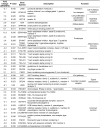
A list of genes that are regulated >2-fold in diabetic foot skin when compared to non-diabetic foot skin is presented along with their roles in different cellular processes.
Fig 6
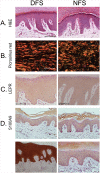
Fig 6. Morphological and immunohistochemical evaluation shows no substantial differences between DFS and NFS.
A. Histological assessment by H&E staining shows similar skin morphology between DFS and NFS. B. Collagen fiber alignment and thickness assessed by picrosirius red staining under polarized light shows no difference between DFS and NFS. C. LEPR immuno-peroxidase staining shows signal throughout the epidermis of both DFS and NFS with no difference in localization or staining intensity. D. S100A9 immuno-peroxidase staining of DFS shows variable expression whereby 67% of DFS showed similar expression to NFS (upper panel), while the other 33% showed much greater expression compared to NFS (D, lower panel).
Fig 7
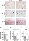
Fig 7. Similar numbers of lymphocytes, blood vessels and proliferating keratinocytes are found in DFS and NFS.
Immunostaining and quantification for CD45, a lymphocyte marker, (A, D), CD31, an endothelial cell marker, (B, E) and Ki67, a proliferation marker (C,F), reveal no differences in the number of lymphocytes, blood vessels and proliferating keratinocytes found in DFS and NFS. Ki67 immuno-peroxidase staining for proliferating cells (C) shows that proliferative keratinocytes were located in the basal layer of the skin, as expected. No significant differences were found between DFS and NFS in numbers of CD45+ cells (D), number of CD31+ (E) or Ki67 positive keratinocytes (F). Bar graphs indicate mean and SD.

References

    1. Shaw JE, Sicree RA, Zimmet PZ. Global estimates of the prevalence of diabetes for 2010 and 2030. Diabetes research and clinical practice. 2010;87(1):4–14. 10.1016/j.diabres.2009.10.007 . - DOI - PubMed
    1. Perez MI, Kohn SR. Cutaneous manifestations of diabetes mellitus. Journal of the American Academy of Dermatology. 1994;30(4):519–31; quiz 32–4. . - PubMed
    1. Van Hattem S, Bootsma AH, Thio HB. Skin manifestations of diabetes. Cleveland Clinic journal of medicine. 2008;75(11):772, 4,, 6–7 passim. . - PubMed
    1. Brem H, Tomic-Canic M. Cellular and molecular basis of wound healing in diabetes. The Journal of clinical investigation. 2007;117(5):1219–22. Epub 2007/05/04. 10.1172/JCI32169 - DOI - PMC - PubMed
    1. Reiber GE, Boyko E.J., and Smith D.G. Lower extremity foot ulcers and amputations in diabetes In: Stern MIHaMP, editor. Diabetes in America. Bethesda, Maryland, USA.: U. S. Government Printing Office; 1995. p. 409–28.

Publication types