

Pushing, pulling, and squeezing our way to understanding mechanotransduction
- PMID: 26318086
- PMCID: PMC4761538
- DOI: 10.1016/j.ymeth.2015.08.019
Pushing, pulling, and squeezing our way to understanding mechanotransduction
Abstract
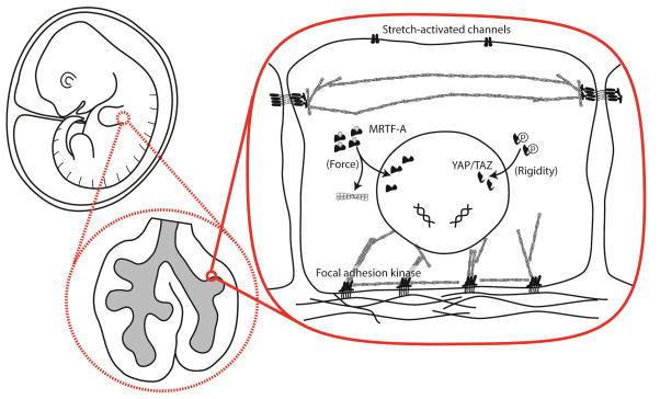
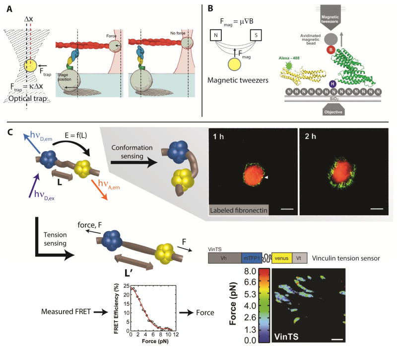
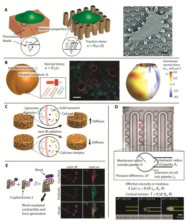
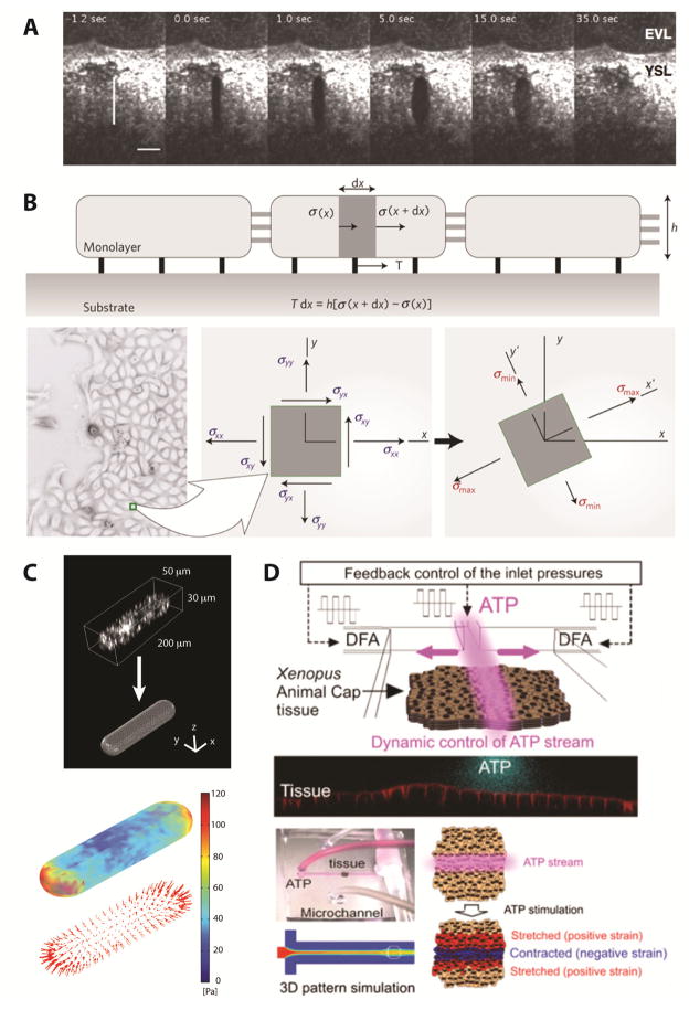
Mechanotransduction is often described in the context of force-induced changes in molecular conformation, but molecular-scale mechanical stimuli arise in vivo in the context of complex, multicellular tissue structures. For this reason, we highlight and review experimental methods for investigating mechanotransduction across multiple length scales. We begin by discussing techniques that probe the response of individual molecules to applied force. We then move up in length scale to highlight techniques aimed at uncovering how cells transduce mechanical stimuli into biochemical activity. Finally, we discuss approaches for determining how these stimuli arise in multicellular structures. We expect that future work will combine techniques across these length scales to provide a more comprehensive understanding of mechanotransduction.
Keywords: Mechanical stress; Morphogenesis.
Copyright © 2015 Elsevier Inc. All rights reserved.
Figures
Similar articles
-
Combining mechanical and optical approaches to dissect cellular mechanobiology.J Biomech. 2010 Jan 5;43(1):45-54. doi: 10.1016/j.jbiomech.2009.09.008. Epub 2009 Oct 12. J Biomech. 2010. PMID: 19819457 Free PMC article. Review.
-
Investigating piconewton forces in cells by FRET-based molecular force microscopy.J Struct Biol. 2017 Jan;197(1):37-42. doi: 10.1016/j.jsb.2016.03.011. Epub 2016 Mar 12. J Struct Biol. 2017. PMID: 26980477 Review.
-
Extending the Capabilities of Molecular Force Sensors via DNA Nanotechnology.Crit Rev Biomed Eng. 2020;48(1):1-16. doi: 10.1615/CritRevBiomedEng.2020033450. Crit Rev Biomed Eng. 2020. PMID: 32749116 Free PMC article. Review.
-
FRET-based Molecular Tension Microscopy.Methods. 2016 Feb 1;94:33-42. doi: 10.1016/j.ymeth.2015.07.010. Epub 2015 Jul 22. Methods. 2016. PMID: 26210398 Review.
-
[Applications of FRET technology in the study of mechanotransduction].Sheng Wu Yi Xue Gong Cheng Xue Za Zhi. 2013 Dec;30(6):1362-7. Sheng Wu Yi Xue Gong Cheng Xue Za Zhi. 2013. PMID: 24645627 Review. Chinese.
Cited by
-
E-cadherin and LGN align epithelial cell divisions with tissue tension independently of cell shape.Proc Natl Acad Sci U S A. 2017 Jul 18;114(29):E5845-E5853. doi: 10.1073/pnas.1701703114. Epub 2017 Jul 3. Proc Natl Acad Sci U S A. 2017. PMID: 28674014 Free PMC article.
-
Active biomaterials for mechanobiology.Biomaterials. 2021 Jan;267:120497. doi: 10.1016/j.biomaterials.2020.120497. Epub 2020 Oct 26. Biomaterials. 2021. PMID: 33129187 Free PMC article. Review.
-
Actuated 3D microgels for single cell mechanobiology.Lab Chip. 2022 May 17;22(10):1962-1970. doi: 10.1039/d2lc00203e. Lab Chip. 2022. PMID: 35437554 Free PMC article.
-
Taking the strain: quantifying the contributions of all cell behaviours to changes in epithelial shape.Philos Trans R Soc Lond B Biol Sci. 2017 May 19;372(1720):20150513. doi: 10.1098/rstb.2015.0513. Philos Trans R Soc Lond B Biol Sci. 2017. PMID: 28348250 Free PMC article. Review.
-
Quantitative reconstruction of time-varying 3D cell forces with traction force optical coherence microscopy.Sci Rep. 2019 Mar 11;9(1):4086. doi: 10.1038/s41598-019-40608-4. Sci Rep. 2019. PMID: 30858424 Free PMC article.
References
-
- Chen CS, Tan J, Tien J. Annu Rev Biomed Eng. 2004;6:275–302. - PubMed
Publication types
MeSH terms
Grants and funding
LinkOut - more resources
Full Text Sources
Other Literature Sources
