. 2015 Nov 6;290(45):26882-26898.
doi: 10.1074/jbc.M115.676890. Epub 2015 Aug 28.

Structural Basis for Substrate Specificity in Adenosylcobalamin-dependent Isobutyryl-CoA Mutase and Related Acyl-CoA Mutases

Affiliations

Structural Basis for Substrate Specificity in Adenosylcobalamin-dependent Isobutyryl-CoA Mutase and Related Acyl-CoA Mutases

Marco Jost et al. J Biol Chem. .

Abstract

Acyl-CoA mutases are a growing class of adenosylcobalamin-dependent radical enzymes that perform challenging carbon skeleton rearrangements in primary and secondary metabolism. Members of this class of enzymes must precisely control substrate positioning to prevent oxidative interception of radical intermediates during catalysis. Our understanding of substrate specificity and catalysis in acyl-CoA mutases, however, is incomplete. Here, we present crystal structures of IcmF, a natural fusion protein variant of isobutyryl-CoA mutase, in complex with the adenosylcobalamin cofactor and four different acyl-CoA substrates. These structures demonstrate how the active site is designed to accommodate the aliphatic acyl chains of each substrate. The structures suggest that a conformational change of the 5'-deoxyadenosyl group from C2'-endo to C3'-endo could contribute to initiation of catalysis. Furthermore, detailed bioinformatic analyses guided by our structural findings identify critical determinants of acyl-CoA mutase substrate specificity and predict new acyl-CoA mutase-catalyzed reactions. These results expand our understanding of the substrate specificity and the catalytic scope of acyl-CoA mutases and could benefit engineering efforts for biotechnological applications ranging from production of biofuels and commercial products to hydrocarbon remediation.

Keywords: acyl-CoA mutase; adenosylcobalamin (AdoCbl); enzyme catalysis; metalloenzyme; substrate specificity; x-ray crystallography.


Figures

FIGURE 1.
FIGURE 1.
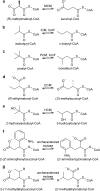
Reversible interconversions catalyzed by characterized (a–e) and proposed (f and g) acyl-CoA mutases. See main text for details. Two variants of HCM, HCM1 and HCM2, use (S)-3-hydroxybutyryl-CoA and (R)-3-hydroxybutyryl-CoA, respectively. R group in g denotes alkyl groups. Stereochemistry of compounds in f and g is not unambiguously established (with the exception of 2-(1′-methylpentyl)succinyl-CoA and (2′-methylhexyl)malonyl-CoA described previously (20)).
FIGURE 2.
FIGURE 2.
Synthetic scheme for pivalyl-CoA synthesis. DMAP is dimethylaminopyridine.
FIGURE 3.
FIGURE 3.
Overall structure of IcmF protomer and comparison of substrate-bound and substrate-free structures. a, overall structure of chain A of an IcmF dimer, shown in ribbon representation colored by domain. Cbl-binding domain is in yellow; TIM barrel substrate-binding domain is in green, and G-protein domain is in cyan. The linker region between the G-protein and substrate-binding domains is hidden for clarity. TIM barrel and Cbl-binding domain β-sheets are shown in darker colors for emphasis. Isobutyryl-CoA is threaded through the TIM barrel. Bound AdoCbl, GDP·Mg2+, and isobutyryl-CoA are shown in ball-and-stick representation with Cbl carbons in pink, 5′-dAdo carbons in cyan, cobalt in purple, GDP carbons in brown, Mg2+ in orange, and isobutyryl-CoA carbons in yellow. b, comparison of isobutyryl-CoA-bound (green) and substrate-free IcmF (lilac, PDB code 4XC6), revealing nearly identical structures. Selected residues contributing to substrate binding are shown in stick representation. AdoCbl and isobutyryl-CoA of the substrate-bound structure are shown as in a. AdoCbl of the substrate-free structure is shown in lilac.
FIGURE 4.
FIGURE 4.
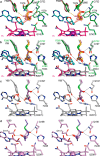
Overall mode of substrate binding in IcmF and comparison with MCM and HCM1. a, IcmF bound to Cbl (pink carbons; cobalt, purple), 5′-dAdo (cyan carbons), and isobutyryl-CoA (yellow carbons). 2FoFc omit electron density (orange mesh) contoured at 1.0 σ is shown around the substrates and 5′-dAdo. IcmF is shown with the chain A TIM barrel substrate-binding domain in green and the Cbl-binding domain in yellow. The Cbl-coordinating His-39 as well as selected residues that bind substrate are shown as sticks, with hydrogen bonds, ionic interactions, and π-π interactions shown as black dashed lines. The 5′-dAdo C5′ and the locations of hydrogen abstraction on the substrates are shown as spheres, and red dashed lines connect the C5′ to the Cbl cobalt and the substrate carbons. b, IcmF bound to n-butyryl-CoA (orange carbons) displayed as in a. c, IcmF bound to pivalyl-CoA (maroon carbons) displayed as in a. d, IcmF bound to isovaleryl-CoA (light pink carbons) displayed as in a. e, comparison of isobutyryl-CoA-bound IcmF (green) and methylmalonyl-CoA-bound MCM (gray, PDB code 4REQ), revealing nearly identical structures and substrate binding modes. IcmF shown as in a. MCM-bound methylmalonyl-CoA, Cbl, and 5′-dAdo are shown with gray carbons. f, comparison of isobutyryl-CoA-bound IcmF (green) and hydroxyisobutyryl-CoA-bound HCM1 (lilac, PDB code 4R3U), revealing similar structures and substrate binding modes. IcmF shown as in a. HCM1-bound hydroxyisobutyryl-CoA, Cbl, and 5′-dAdo are shown with lilac carbons.
FIGURE 5.
FIGURE 5.
Differences of substrate-binding domains of IcmF dimer. a, isobutyryl-CoA binding to IcmF chain A of the IcmF dimer, which is in a catalytically active conformation. Coloring as in Fig. 4. b, isobutyryl-CoA binding to IcmF chain B, which is in a catalytically inactive conformation. IcmF (green carbons), isobutyryl-CoA (yellow carbons), and 2FoFc electron density (orange mesh), contoured at 1.0 σ, are shown as in Fig. 4a. Note that there is no electron density past the 5′-phosphate of the isobutyryl-CoA nucleotide moiety; therefore, additional atoms were not modeled. The nucleotide portion is bound by few specific interactions, as indicated by black dashed lines. Other interactions between IcmF and isobutyryl-CoA are disrupted because of the conformational change in IcmF chain B compared with IcmF chain A. c, different conformations of substrate-binding domains of IcmF chains A (dark green) and B (gray) isobutyryl-CoA-bound IcmF. Chains are superposed by TIM barrel β-strands. Cbl of chain A is shown as in Fig. 4a, Cbl of chain B is shown with carbons in black. Distances between corresponding Cα atoms are indicated in Å.
FIGURE 6.
FIGURE 6.
5′-dAdo conformational changes in IcmF and glutamate mutase. a, 2FoFc omit electron density (orange mesh) contoured at 1.0 σ around Cbl and 5′-dAdo of n-butyryl-CoA bound IcmF. 5′-dAdo can be modeled in the C3′-endo conformation (cyan carbons) and in the C2′-endo conformation (light blue carbons). In the C2′-endo conformation, the C5′ is close to the Cbl cobalt, whereas in the C3′-endo conformation, the C5′ is pointed toward the substrate (orange carbons, dashed red line). Cbl is shown with carbons in pink and cobalt in purple. b, glutamate mutase active site (PDB code 1I9C) (33), revealing the presence of two 5′-dAdo conformers, C2′-endo (pink carbons) and C3′-endo (purple carbons), in the presence of glutamate (gray carbons). As in IcmF, the 5′-dAdo C5′ is close to the Cbl cobalt in the C2′-endo conformation (dashed red line) and pointed toward the location of hydrogen abstraction on the substrate in the C3′-endo conformation (dashed red line). Cbl is shown with carbons in light pink and Co in purple. c, comparison of the 5′-dAdo C3′-endo conformations in IcmF (cyan carbons) and glutamate mutase (purple carbons). Dashed red lines connect the 5′-dAdo C5′ and the corresponding substrate. Cbl is shown as in b. In both proteins, 5′-dAdo is stabilized by interactions (dashed black lines) to amino acid side chains (IcmF in green and glutamate mutase in pink). IcmF Gln-865 contributes to 5′-dAdo binding, but the corresponding Arg-66 in glutamate mutase does not. d, comparison of the 5′-dAdo C2′-endo conformations in IcmF (light blue carbons) and glutamate mutase (pink carbons). Protein side chains and Cbl colored as in c. Again, 5′-dAdo is stabilized by specific interactions (dashed black lines) to amino acid side chains. IcmF Tyr-779 contributes to 5′-dAdo binding, but the corresponding Pro-218 in glutamate mutase (hidden for clarity) does not. IcmF Asn-901 corresponds to glutamate mutase Lys-326 but does not contribute to 5′-dAdo binding.
FIGURE 7.
FIGURE 7.
IcmF active site and comparison with MCM and HCM1 in wall-eyed stereo view. a, overlay of four substrate-bound IcmF structures, revealing similar substrate orientation. Shown are isobutyryl-CoA (yellow carbons), n-butyryl-CoA (orange carbons), pivalyl-CoA (maroon carbons), and isovaleryl-CoA (light pink carbons). The locations of hydrogen abstraction are shown as spheres, located within 3.6 Å of the 5′-dAdo (C3′-endo conformation, cyan carbons) C5′ atom (cyan sphere), as indicated by the dashed red lines. Residues in the substrate-binding site are shown with dark green carbons. The third methyl group of pivalyl-CoA clashes with Phe-598 (yellow dashed line), leading to a small rotation of the side chain (maroon carbons) in this structure. Hydrogen bonds from Gln-732 and His-780 to the thioester carbonyl are indicated as dashed black lines. Cbl is shown with carbons in pink and cobalt in purple. b, overlay of substrate-bound IcmF and MCM (26) (PDB code 4REQ). Isobutyryl-CoA, n-butyryl-CoA, IcmF-bound 5′-dAdo, IcmF-bound Cbl, and IcmF residues are shown as in a. The locations of hydrogen atom abstraction in both methylmalonyl-CoA (gray carbons) and succinyl-CoA (purple carbons) overlay with those of IcmF substrates (spheres). MCM-bound 5′-dAdo, MCM-bound Cbl, and MCM substrate-binding residues are shown with gray carbons. Gln-197 and His-244 are conserved in MCM and IcmF, whereas IcmF Phe-598 is replaced by MCM Tyr-89 and IcmF Gln-742 is replaced by Arg-209, putatively accounting for the switch in substrate binding specificity. Hydrogen bonds and ionic interactions are shown as dashed black lines. c, active site of substrate-bound MCM, shown as in b, highlighting interactions to substrate carboxylate groups. Only interactions to methylmalonyl-CoA are shown for clarity. d, active site of substrate-bound HCM1 (PDB code 4R3U) (29), highlighting interactions to substrate hydroxyl groups. Hydroxyisobutyryl-CoA is shown in lilac and (S)-3-hydroxybutyryl-CoA in blue. Only interactions to hydroxyisobutyryl-CoA are shown for clarity. Locations of hydrogen atom abstraction are shown as spheres.
FIGURE 8.
FIGURE 8.
Stereochemical course of isobutyryl-CoA mutase reaction. The chemical mechanism shown at the bottom was established based on stereochemical studies (57). Following Co–C bond homolysis (step not shown), the 5′-dAdo radical abstracts a hydrogen atom (red) from the pro-S methyl group of isobutyryl-CoA (blue). The isobutyryl-CoA radical rearranges to the n-butyryl-CoA radical, which then re-abstracts the hydrogen atom from 5′-deoxyadenosine. The hydrogen atom ends up in the pro-S position. In the reverse reaction, the 5′-dAdo radical abstracts the pro-S hydrogen from n-butyryl-CoA. The structures of IcmF bound to isobutyryl-CoA (left) and n-butyryl-CoA (right) support the proposed stereochemistry. Isobutyryl-CoA (yellow carbons) positions its pro-S methyl group next to the 5′-dAdo group (cyan carbons), whereas n-butyryl-CoA positions its pro-S hydrogen (white sticks) toward the 5′-dAdo group. The red dashed line connects the 5′-dAdo C5′ to the closest hydrogen atom. Hydrogens are modeled based on ideal geometry. Cobalamin is shown with pink carbons and cobalt as a purple sphere.
FIGURE 9.
FIGURE 9.
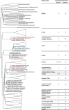
Simplified phylogenetic tree of acyl-CoA mutase substrate-binding domains. The sequences of substrate-binding domains cluster into distinct groups according to their substrate specificity. Larger clusters of characterized mutases are simplified as triangles. Clusters of thus far uncharacterized mutases are shown with organisms as indicated. The amino acid identities in the specificity determinant positions are indicated on the right (see main text and Fig. 11 for explanation of specificity determinant positions). IcmF from C. metallidurans is highlighted in blue, and two uncharacterized mutases and the most recently characterized PCM from X. autotrophicus are highlighted in red and discussed in the main text. The tree was rooted by midpoint rooting. Full tree is shown in Fig. 10 and in the supplemental material.
FIGURE 10.
FIGURE 10.
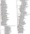
Full phylogenetic tree of acyl-CoA mutase substrate-binding domains (left side, top half of tree; right side, bottom half of tree). Red numbers represent certainty of branch assignment. A larger version of this figure is provided as supplemental material.
FIGURE 11.
FIGURE 11.
Sequence alignment of important regions of acyl-CoA mutases. Sequences were selected to represent most characterized acyl-CoA mutase classes, using sequences of structurally characterized (P. freudenreichii (24, 25), Homo sapiens MCM (27), and C. metallidurans IcmF (28)) or biochemically characterized mutases (P. freudenreichii MCM and H. sapiens MCM, C. metallidurans IcmF (15, 16), S. cinnamonensis ICM (57, 71), Aquincola tertiaricarbonis HCM1 (13), Rhodobacter sphaeroides ECM (11), and X. autotrophicus PCM (17)) when available. Two sequences from each of the two clusters of uncharacterized mutases were included, as well as two additional mutases encoded in the genome of A. aromaticum. Sequence determinant positions are highlighted by red boxes, positions proposed to distinguish MCMs and ECMs are highlighted by green boxes, and the unique AGGGGG stretch of uncharacterized mutases from A. aromaticum and Azoarcus toluclasticus is highlighted by a blue box. Other conserved catalytically important residues are labeled. Strict residue conservation is indicated by a yellow highlight, sequence similarity is indicated in red. Beginning of each sequence stretch is numbered on the left.
FIGURE 12.
FIGURE 12.
Homology model of uncharacterized A. aromaticum mutase and comparison with MCM. a, structure of succinyl-CoA-bound MCM (PDB code 4REQ) (26), shown with protein, 5′-dAdo, and Cbl carbons in gray and succinyl-CoA carbons in violet. Cobalt is shown in purple. Succinyl-CoA is bound by specific hydrogen bonds and electrostatic interactions (black dashed lines). b, homology model of A. aromaticum mutase in the same orientation as MCM and with 5′-dAdo and Cbl shown as in a. Protein residues are shown with carbons in yellow. Substrate (pink carbons) is modeled into the active site by adding the aminophenyl group to MCM-bound succinyl-CoA without altering the positioning of succinyl-CoA. Interactions to the thioester carbonyl and the carboxylate are conserved (black dashed lines). Replacement of MCM Gln-330 by Ala-307 creates a cavity in the active site that could accommodate the aminophenyl group. Ala-307 is relatively close to the phenyl group (2.2 Å), but this region likely has significant flexibility because of the following stretch of Gly residues. In addition, replacement of MCM Phe-287 by Asn-263 in the uncharacterized mutase positions a potential hydrogen bonding partner for the amino group in the active site (dashed red line).

Similar articles

Cited by

References

    1. Halpern J. (1985) Mechanisms of coenzyme B12-dependent rearrangements. Science 227, 869–875 - PubMed
    1. Frey P. A. (2010) in Comprehensive Natural Products II Chemistry and Biology (Mander L., and Liu H. W., eds) pp. 501–546, Elsevier, Oxford, UK
    1. Banerjee R. (2003) Radical carbon skeleton rearrangements: catalysis by coenzyme B12-dependent mutases. Chem. Rev. 103, 2083–2094 - PubMed
    1. Cracan V., and Banerjee R. (2012) Novel B12-dependent Acyl-CoA mutases and their biotechnological potential. Biochemistry 51, 6039–6046 - PMC - PubMed
    1. Cracan V., and Banerjee R. (2013) Cobalt and corrinoid transport and biochemistry. Met. Ions Life Sci. 12, 333–374 - PubMed

Publication types

MeSH terms

LinkOut - more resources