Review
. 2015 Dec;23(12):2059-2085.
doi: 10.1016/j.joca.2015.08.008. Epub 2015 Aug 28.

Atlas of radiographic features of osteoarthritis of the ankle and hindfoot

Affiliations
Review

Atlas of radiographic features of osteoarthritis of the ankle and hindfoot

V B Kraus et al. Osteoarthritis Cartilage. 2015 Dec.

Abstract

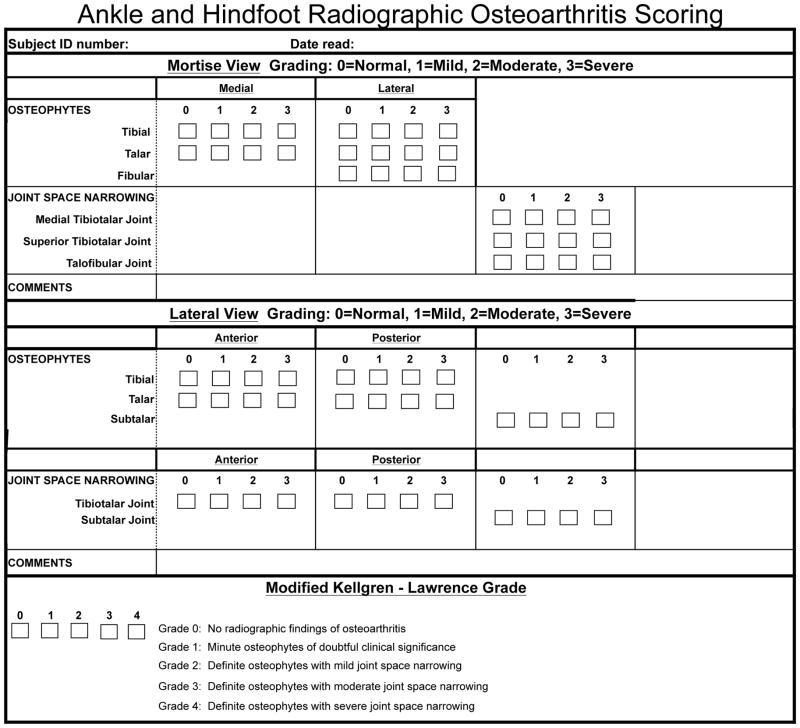
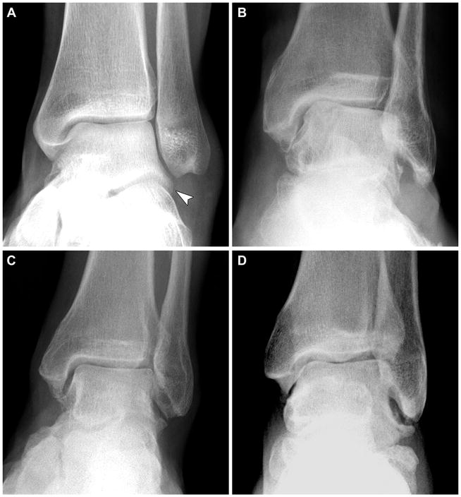
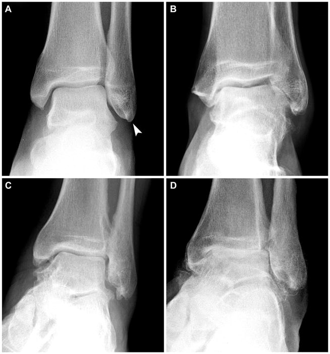
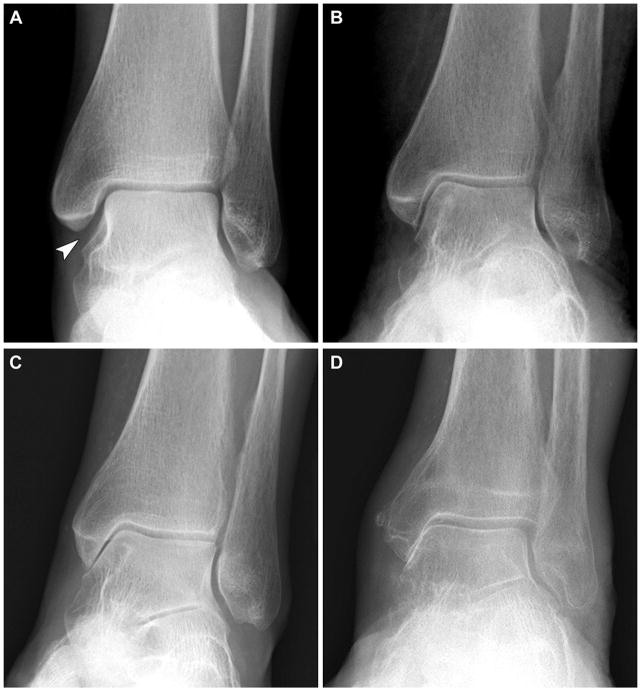
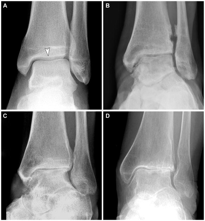
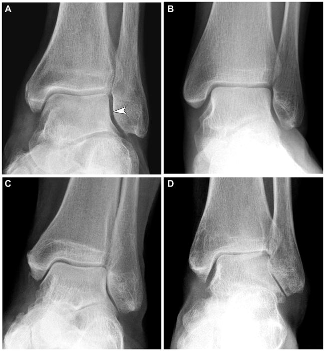
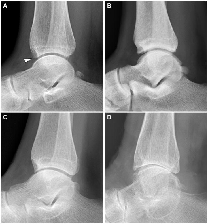
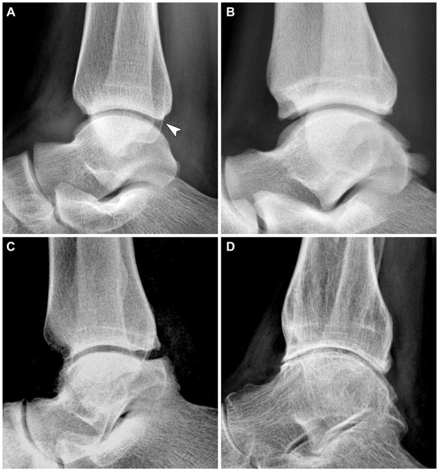
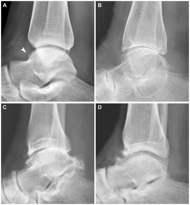
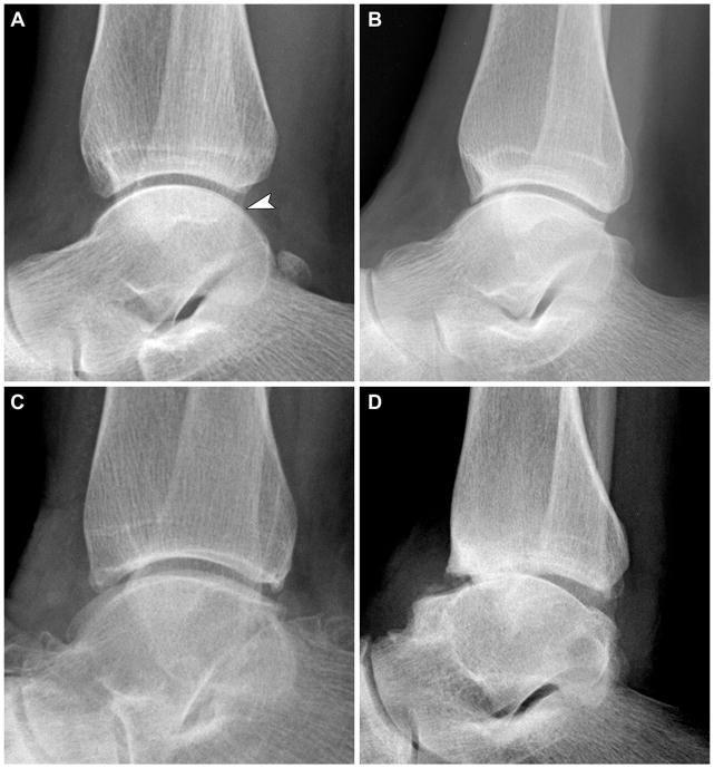
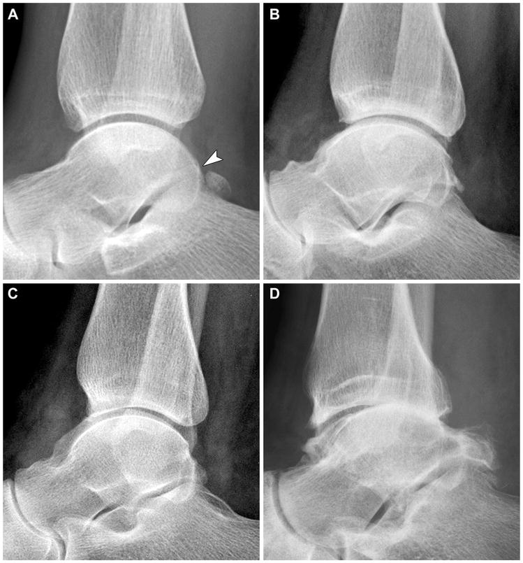
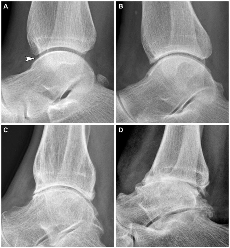
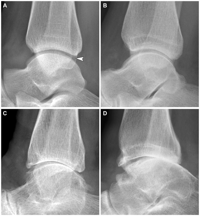
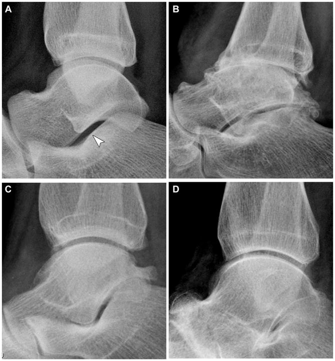
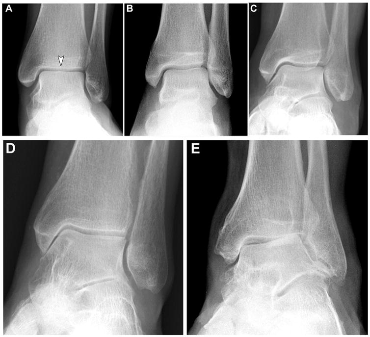
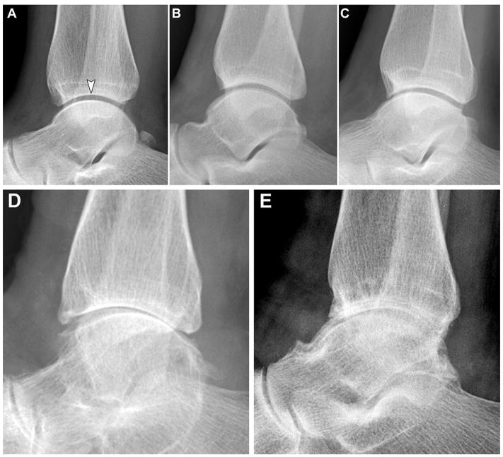
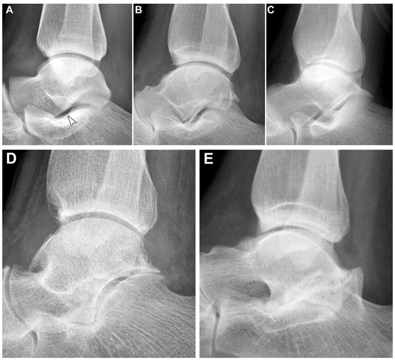
Objective: To develop a radiographic atlas of osteoarthritis (OA) for use as a template and guide for standardized scoring of radiographic features of OA of the ankle and hindfoot joints.

Method: Under Institutional Review Board approval, ankle and hindfoot images were selected from a cohort study and from among cases that underwent ankle radiography during a 6-month period at Duke University Medical Center. Missing OA pathology was obtained through supplementation of cases with the assistance of a foot and ankle specialist in Orthopaedic surgery and a musculoskeletal radiologist. Images were obtained and reviewed without patient identifying information. Images went through multiple rounds of review and final images were selected by consensus of the study team. For intra-rater and inter-rater reliability, the kappa statistic was calculated for two readings by three musculoskeletal radiologists, a minimum of two weeks apart, of ankle and hindfoot radiographs from 30 anonymized subjects.

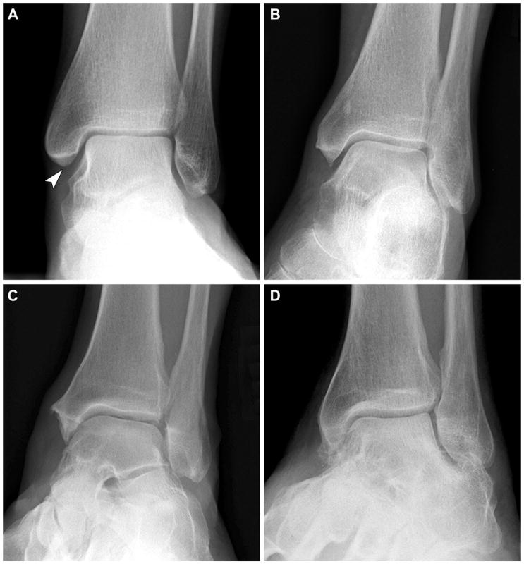
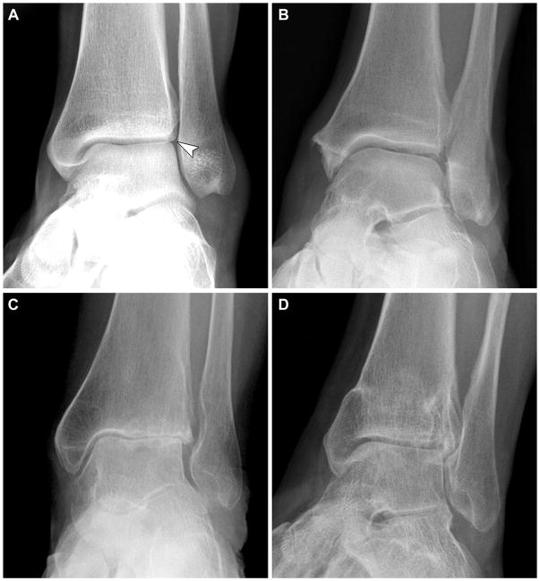
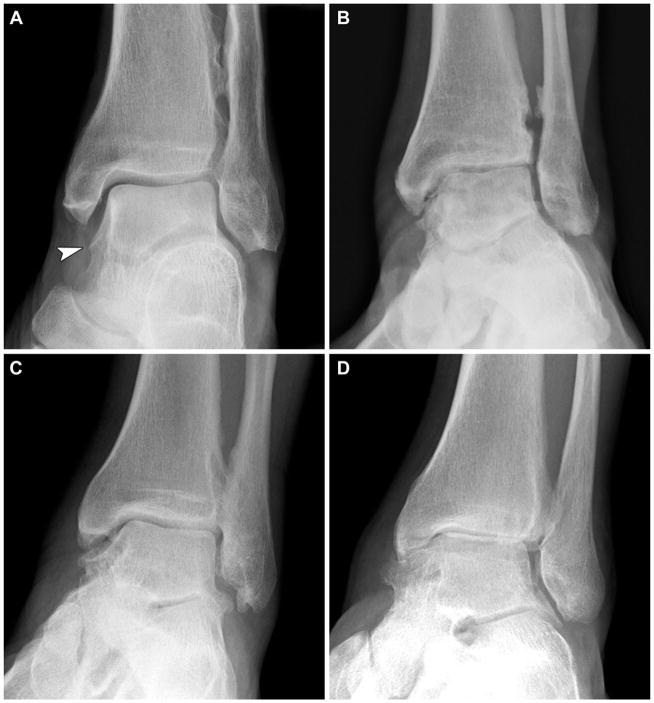
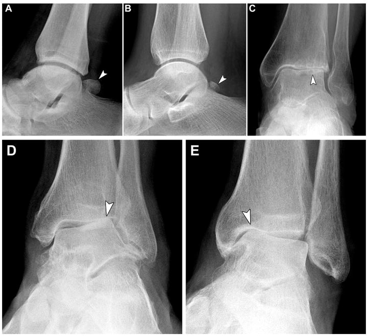
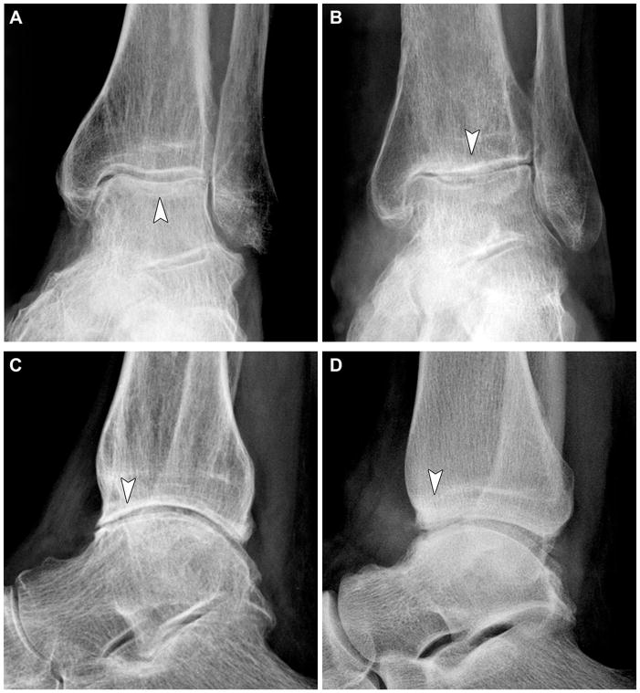
Results: The atlas demonstrates individual radiographic features (osteophyte and joint space narrowing (JSN)) and Kellgren-Lawrence grade for all aspects of the talocrural (ankle joint proper) and talocalcaneal (subtalar) joints. Reliability of scoring based on the atlas was quite good to excellent for most features indicated. Additional examples of ankle joint findings are illustrated including sclerosis, os trigonum, subchondral cysts and talar tilt.

Conclusions: It is anticipated that this atlas will assist with standardization of scoring of ankle and hindfoot OA by basic and clinical OA researchers.

Keywords: Ankle; Atlas; Hindfoot; Osteoarthritis; Radiograph; Subtalar.


Conflict of interest statement

Conflicts of Interest:

No authors have any conflict of interest or competing interests with respect to this work.

Figures

Figure 1
Figure 1
Sample scoring form to facilitate grading of ankle and hindfoot radiographs for features demonstrated in atlas.
Figure 2
Figure 2
Ankle Joint - Mortise view: (A) grade 0 normal, (B) grade 1 medial tibial osteophyte, (C) grade 2 medial tibial osteophyte, and (D) grade 3 medial tibial osteophyte.
Figure 3
Figure 3
Ankle Joint - Mortise view: (A) grade 0 normal, (B) grade 1 lateral tibial osteophyte, (C) grade 2 lateral tibial osteophyte, and (D) grade 3 lateral tibial osteophyte.
Figure 4
Figure 4
Ankle Joint - Mortise view: (A) grade 0 normal, (B) grade 1 medial talar osteophyte, (C) grade 2 medial talar osteophyte, and (D) grade 3 medial talar osteophyte.
Figure 5
Figure 5
Ankle Joint - Mortise view: (A) grade 0 normal, (B) grade 1 lateral talar osteophyte, (C) grade 2 lateral talar osteophyte, and (D) grade 3 lateral talar osteophyte.
Figure 6
Figure 6
Ankle Joint - Mortise view: (A) grade 0 normal, (B) grade 1 distal fibular osteophyte, (C) grade 2 distal fibular osteophyte, and (D) grade 3 distal fibular osteophyte.
Figure 7
Figure 7
Ankle Joint - Mortise view: (A) grade 0 normal, (B) grade 1 medial tibiotalar joint space narrowing, (C) grade 2 medial tibiotalar joint space narrowing, and (D) grade 3 medial tibiotalar joint space narrowing.
Figure 8
Figure 8
Ankle Joint - Mortise view: (A) grade 0 normal, (B) grade 1 superior tibiotalar joint space narrowing, (C) grade 2 superior tibiotalar joint space narrowing, and (D) grade 3 superior tibiotalar joint space narrowing.
Figure 9
Figure 9
Ankle Joint - Mortise view: (A) grade 0 normal, (B) grade 1 talofibular joint space narrowing, (C) grade 2 talofibular joint space narrowing, and (D) grade 3 talofibular joint space narrowing.
Figure 10
Figure 10
Ankle Joint - Lateral view: (A) grade 0 normal, (B) grade 1 anterior tibial osteophyte, (C) grade 2 anterior tibial osteophyte, and (D) grade 3 anterior tibial osteophyte.
Figure 11
Figure 11
Ankle Joint - Lateral view: (A) grade 0 normal, (B) grade 1 posterior tibial osteophyte, (C) grade 2 posterior tibial osteophyte, and (D) grade 3 posterior tibial osteophyte.
Figure 12
Figure 12
Ankle Joint - Lateral view: (A) grade 0 normal, (B) grade 1 anterior talar osteophyte, (C) grade 2 anterior talar osteophyte, and (D) grade 3 anterior talar osteophyte.
Figure 13
Figure 13
Ankle Joint - Lateral view: (A) grade 0 normal, (B) grade 1 posterior talar osteophyte, (C) grade 2 posterior talar osteophyte, and (D) grade 3 posterior talar osteophyte.
Figure 14
Figure 14
Subtalar Joint - Lateral view: (A) grade 0 normal, (B) grade 1 posterior talar osteophyte, (C) grade 2 posterior talar osteophyte, and (D) grade 3 posterior talar osteophyte.
Figure 15
Figure 15
Ankle Joint - Lateral view: (A) grade 0 normal, (B) grade 1 anterior tibiotalar joint space narrowing, (C) grade 2 anterior tibiotalar joint space narrowing, and (D) grade 3 anterior tibiotalar joint space narrowing.
Figure 16
Figure 16
Ankle Joint - Lateral view: (A) grade 0 normal, (B) grade 1 posterior tibiotalar joint space narrowing, (C) grade 2 posterior tibiotalar joint space narrowing, and (D) grade 3 posterior tibiotalar joint space narrowing.
Figure 17
Figure 17
Subtalar Joint - Lateral view: (A) grade 0 normal, (B) grade 1 subtalar joint space narrowing, (C) grade 2 subtalar joint space narrowing, and (D) grade 3 subtalar joint space narrowing.
Figure 18
Figure 18
Ankle Joint - Mortise view: (A) grade 0 normal, (B) Kellgren-Lawrence grade 1, (C) Kellgren-Lawrence grade 2, (D) Kellgren-Lawrence grade 3, and (E) Kellgren-Lawrence grade 4.
Figure 19
Figure 19
Ankle Joint - Lateral view: (A) grade 0 normal, (B) Kellgren-Lawrence grade 1, (C) Kellgren-Lawrence grade 2, (D) Kellgren-Lawrence grade 3, and (E) Kellgren-Lawrence grade 4.
Figure 20
Figure 20
Subtalar Joint - Lateral view: (A) grade 0 normal, (B) Kellgren-Lawrence grade 1, (C) Kellgren-Lawrence grade 2, (D) Kellgren-Lawrence grade 3, and (E) Kellgren-Lawrence grade 4.
Figure 21
Figure 21
Ankle: (A) os trigonum, (B) os trigonum, (C) subchondral cysts, (D) lateral talar tilt, (E) medial talar tilt.
Figure 22
Figure 22
Ankle: (A–D) examples of subchondral sclerosis.

Similar articles

Cited by

References

    1. McDaniel G, Renner JB, Sloane R, Kraus VB. Association of knee and ankle osteoarthritis with physical performance. Osteoarthritis Cartilage. 2011;19:634–638. - PMC - PubMed
    1. Kraus VB, Worrell TW, Renner JB, Coleman RE, Pieper CF. High prevalence of contralateral ankle abnormalities in association with knee osteoarthritis and malalignment. Osteoarthritis Cartilage. 2013;21:1693–1699. - PMC - PubMed
    1. Kuijt MT, Inklaar H, Gouttebarge V, Frings-Dresen MH. Knee and ankle osteoarthritis in former elite soccer players: a systematic review of the recent literature. J Sci Med Sport. 2012;15:480–487. - PubMed
    1. Rietveld AB. Dancers' and musicians' injuries. Clin Rheumatol. 2013;32:425–434. - PubMed
    1. Muehleman C, Margulis A, Bae WC, Masuda K. Relationship between knee and ankle degeneration in a population of organ donors. BMC Med. 2010;8:48. - PMC - PubMed

Publication types