. 2015 Nov:27:116-130.
doi: 10.1016/j.actbio.2015.08.040. Epub 2015 Aug 28.

Concentration-dependent rheological properties of ECM hydrogel for intracerebral delivery to a stroke cavity

Affiliations

Concentration-dependent rheological properties of ECM hydrogel for intracerebral delivery to a stroke cavity

Andre R Massensini et al. Acta Biomater. 2015 Nov.

Abstract

Biomaterials composed of mammalian extracellular matrix (ECM) promote constructive tissue remodeling with minimal scar tissue formation in many anatomical sites. However, the optimal shape and form of ECM scaffold for each clinical application can vary markedly. ECM hydrogels have been shown to promote chemotaxis and differentiation of neuronal stem cells, but minimally invasive delivery of such scaffold materials to the central nervous system (CNS) would require an injectable form. These ECM materials can be manufactured to exist in fluid phase at room temperature, while forming hydrogels at body temperature in a concentration-dependent fashion. Implantation into the lesion cavity after a stroke could hence provide a means to support endogenous repair mechanisms. Herein, we characterize the rheological properties of an ECM hydrogel composed of urinary bladder matrix (UBM) that influence its delivery and in vivo interaction with host tissue. There was a notable concentration-dependence in viscosity, stiffness, and elasticity; all characteristics important for minimally invasive intracerebral delivery. An efficient MRI-guided injection with drainage of fluid from the cavity is described to assess in situ hydrogel formation and ECM retention at different concentrations (0, 1, 2, 3, 4, and 8mg/mL). Only ECM concentrations >3mg/mL gelled within the stroke cavity. Lower concentrations were not retained within the cavity, but extensive permeation of the liquid phase ECM into the peri-infarct area was evident. The concentration of ECM hydrogel is hence an important factor affecting gelation, host-biomaterial interface, as well intra-lesion distribution.

Statement of significance: Extracellular matrix (ECM) hydrogel promotes constructive tissue remodeling in many tissues. Minimally invasive delivery of such scaffold materials to the central nervous system (CNS) would require an injectable form that exists in fluid phase at room temperature, while forming hydrogels at body temperature in a concentration-dependent fashion. We here report the rheological characterization of an injectable ECM hydrogel and its concentration-dependent delivery into a lesion cavity formed after a stroke based on MRI-guidance. The concentration of ECM determined its retention within the cavity or permeation into tissue and hence influenced its interaction with the host brain. This study demonstrates the importance of understanding the structure-function relationship of biomaterials to guide particular clinical applications.

Keywords: Biomaterial; Brain; Delivery; Extracellular matrix; Injection; Magnetic resonance imaging; Stereotactic; Stroke.


Figures

Fig. 1
Fig. 1
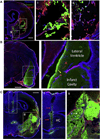
Considerations for the injection of biomaterials into a stroke cavity. A. Injection of an insufficient quantity or local concentration of an 2 mg/mL ECM preparation leads to a poor gelation within the cavity and hence does not afford a complete and homogenous coverage (2 weeks post-MCAo, 24 h post-injection). Although particles of ECM that formed accumulate at the border of damaged tissue (i), vast areas of host tissue and the cavity do not show any accumulation of ECM material (ii) indicating that concentration and volume of material is important to ensure proper coverage of the cavity. B. Trajectory for delivery through a needle requires careful planning based on in vivo non-invasive imaging. A trajectory for biomaterial delivery needs to avoid ventricular space, as it can lead to a puncture of the ventricular wall (yellow arrow) and the subsequent leakage of material into the ventricle. Such intraventricular leakage can lead to a decrease in biomaterial concentration in the cavity and an obstruction of cerebrospinal fluid (CSF) movement through the ventricle with potential damage to the choroid plexus. C. However, positioning of the injection tract to avoid the ventricle can damage critical neuroanatomical structures, such as the hippocampus, and lead to significant tissue tearing and backflow of biomaterial (i, red arrows). Placement of the cannula at the edge of the cavity can further damage already compromised tissue (white *), although it can deliver ECM material to the cavity. Nevertheless, an uneven distribution and heterogeneous concentration within the cavity (black *) can ensue with areas void of ECM containing extracellular fluid that has not been displaced (ii, white arrow). These examples indicate the need for appropriate neurosurgical planning of biomaterial delivery to ensure a homogenous and complete distribution of ECM throughout the stroke cavity (scale bars = 1 mm). (For interpretation of the references to colour in this figure legend, the reader is referred to the web version of this article.)
Fig. 2
Fig. 2
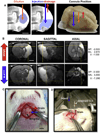
Injection–drainage of biomaterials and extracellular fluid. A. Delivery of material to the lesion cavity can be achieved by injection of a concentrate to be diluted in the extracellular fluid (ECF). For this, typically an injection site at the center of mass of the cavity is targeted (dotted green line = lesion cavity) [7,27]. However, this delivery method can lead to variations in material concentration and especially in case of in situ gelation can produce areas void of biomaterial [11]. In contrast, the creation of a second burr hole allows displaced ECF to be drained while biomaterial is delivered. Placement of these ideally target the lower parts of the cavity for injection (to facilitate displacement) and crucially the drainage cannula needs to be positioned at the most dorsal part of the cavity to fully exploit the Archimedes principle of fluid displacement. B. For injection–drainage, T2-weighted MRI scans were used to calculate the volume of the lesion cavity (hyperintense area), as well as to define coordinates for injection and drainage (AP = Anterior–Posterior; ML = Medio-Lateral; DV = Dorso-Ventral). C. Based on these coordinates, Burr holes were drilled into the skull at the appropriate location in relation to Bregma. D. Injection of a liquid hydrogel composed of extracellular matrix (ECM) through a needle/syringe fixed to the stereotactic device allowed injection of a volume equal to the lesion volume. As the biomaterial was denser than extracellular fluid (ECF), its injection led to the displacement of ECF from the cavity through the drainage cannula. Gelation of the ECM occurred inside the cavity, allowing adaptation to the topology of the lesion (scale bars = 2.5 mm). (For interpretation of the references to colour in this figure legend, the reader is referred to the web version of this article.)
Fig. 3
Fig. 3
Rheological characterization of ECM hydrogels. A. Viscosity of the ECM pre-gel at 10 °C was measured by applying a constant shear stress of 1 Pa. B. Representative curves of the ECM hydrogel gelation kinetics show the storage and loss modulus increase sigmoidally over time. Temperature is rapidly raised from 10 °C to 37 °C, and a small 0.5% oscillatory strain at a frequency of 1 rad/s was applied. C. Maximum storage modulus and loss modulus of the ECM hydrogel after gelation was complete at 37 °C. D. Gelation time of the ECM hydrogels to “50% gelation”, or time to 50% the maximum storage modulus. E–F. Representative graphs of the storage modulus, loss modulus, and complex viscosity of the ECM hydrogels at 4 mg/mL (E) and 8 mg/mL (F) plotted over angular frequencies on a log–log scale, measured at 37 °C by applying a small 0.5% oscillatory strain. *p ≤ 0.01 **p ≤ 0.001.
Fig. 4
Fig. 4
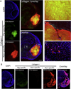
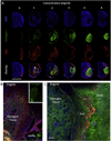
Detection of 8 mg/mL ECM hydrogel in the stroke cavity. A. The pig-specific collagen I antibody detects only the ECM hydrogel implanted into the stroke cavity, although there is some evidence of background autofluorescence and staining in damaged tissue. The non-specific collagen I antibody detects the implanted hydrogel with a very intense staining, but also stains host brain blood vessels as well as damaged tissue. The relatively high concentration of collagen I in the ECM hydrogel compared to the host brain is evidenced by the short exposure times required to acquire images. The core of the implant is detected equally by the pig-specific and the non-specific antibody, but there are differences in their staining pattern (i). Collagen I staining clearly allows a demarcation of the implant-host interface (ii). Only the non-specific collagen I antibody detects rat collagen present in the basement membrane of host blood vessels in intact tissue (iii). B. In a stroke brain injected with vehicle, there was no detection of pig-specific collagen I (only background fluorescence in the peri-infarct area). At the exposure time used for the ECM hydrogel (17 ms) there was no detection of any fluorescence of the non-specific antibody, but at a longer exposure time (200 ms), collagen I staining around the infarct cavity was evident. The pig-specific antibody can therefore be used to distinguish porcine-derived ECM implants versus rat host tissue with specific staining, but the ECM hydrogel can also be visualized macroscopically using the non-specific antibody by adjusting the exposure time for image acquisition (scale bars = 1 mm).
Fig. 5
Fig. 5
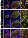
Detection of 8 mg/mL ECM hydrogel using ECM markers. ECM contains a variety of molecules that can be detected using immunohistochemistry. The higher concentrations of molecules compared to brain tissue afford its detection using different antibodies against collagen IV, hyaluronic acid, laminin and chondroitin sulfate. It is evident here that ECM molecules associated with glial scarring (collagen IV, chondroitin sulfate) and angiogenesis (laminin) are also highly expressed in the peri-infarct area and hence not ideal for a selective detection of ECM hydrogel. However, hyaluronic acid also emerged as a potential alternative marker to collagen I, both of which are abundantly present with the (UBM)-ECM preparations compared to brain tissue (scale bars = 500 µm).
Fig. 6
Fig. 6
Correspondence between pre-implant MRI and post-mortem distribution of ECM hydrogel in the stroke cavity. A. Coverage of the lesion using this approach is demonstrated by immunohistochemistry 24 h after injection. The lesion cavity is defined by glial scarring (GFAP), whereas the ECM hydrogel which contains high levels of collagen I can be detected using a collagen I antibody. It is remarkable that even within 24 h there is cell invasion into the material from the host (DAPI). B. An overlay of the fluorescent histology images with the pre-implantation MRI indicates that indeed a good coverage of the cavity has been achieved. Nevertheless, it is noteworthy that the material did not completely cover the hyperintense area on the MRI, as tissue remnants were present in this region (blue arrows). This subtle difference is not evident based on the histological assessment alone and indicates that further improvements in non-invasive imaging are required to better define microenvironments present within the infarct territory. However, if there is an overestimation of injection volume, the drainage of superfluous material will prevent a buildup in the cavity (scale bar = 2.5 mm). (For interpretation of the references to colour in this figure legend, the reader is referred to the web version of this article.)
Fig. 7
Fig. 7
Anterior–posterior images covering the tissue cavity caused by middle cerebral artery occlusion. A. The ECM hydrogel (8 mg/mL), as detected by collagen I staining (green), is fairly equally distributed throughout the cavity, defined by the lack of cells (DAPI staining in blue). Glial scarring (glial fibrillary acid protein in red) around the lesion cavity is also evident. The yellow dashed line indicates the point of injection, whereas the red dashed line depicts the position of the drainage cannula. In some areas, especially anteriorly (i), some permeation of ECM hydrogel into the host brain was evident (yellow *), whereas in other areas (red *), small “particulates” of ECM were present within non- gelled areas of the cavity. A lack of hydrogel in some edge regions (white *) of the host-biomaterial interface was also evident indicating that some further optimization (e.g. speed of injection) of biomaterial distribution within the lesion can further improve coverage (ii). B. The described approach produces fairly consistent coverage of the cavity, as can be seen in a further 4 examples. C. Still, further challenges for intracerebral delivery are apparent. Notably, the fragile peri-infarct tissue can be impacted by large volume injections of a high concentration of material (white arrows), whereas a denser material could also create a “clump” of material, leaving voids between host and material (red arrows), while a glial scar is forming (yellow arrows). By focusing on the tissue cavity, the peri-infarct tissue that is severely damaged (white *), but not lost, is not receiving biomaterial (red *). These aspects further highlight the importance of determining appropriate concentrations and speed of delivery to potentially further improve the delivery of biomaterials to the damaged brain (scale bars = 2.5 mm). (For interpretation of the references to colour in this figure legend, the reader is referred to the web version of this article.)
Fig. 8
Fig. 8
Concentration-dependent retention of ECM hydrogel in the lesion cavity. A. ECM hydrogel injection will only be retained within the lesion cavity if sufficient collagen I is present to afford gelation. Using the injection–drainage approach, it is possible to inject accurate concentrations of ECM into the cavity and determine retention of the ECM hydrogel. A vehicle injection (0 mg/mL) of PBS indicated no collagen I-dependent detection (33 ms) of material inside the lesion cavity (as delineated by Iba1 staining for microglia) or the host brain. At 1 and 2 mg/mL ECM material mostly dissipated into the host brain, whereas at 3 mg/mL, ECM hydrogel was formed and retained within the cavity with some material gelling in the peri-infarct tissue. Higher than 3 mg/mL concentrations resulted in a gelation within the lesion cavity with little to no ECM hydrogel permeation into adjacent host tissue (scale bar = 5 mm). B. At 1 mg/mL ECM injection, the injected material was only visible in the peri-infarct area. The pattern of distribution suggests permeation from the cavity. However, it is likely that some material was still present within the cavity and was lost upon sectioning due to a lack of structure (i.e. no gelation) (scale bar = 1 mm). C. At 3 mg/mL, gelation within the cavity occurred and this material was retained during sectioning, but it is also evident that some injected ECM material diffused into the damaged peri-infarct tissue potentially through the glial scar. Based on signal intensity, it is also possible to see a clear difference in collagen I content between 1 and 3 mg/mL. It is therefore important to note that not only is there a difference in “inductive” material being delivered, but the structural (i.e. gelation) properties of the ECM are also concentration dependent (scale bar = 500 µm).

References

    1. Modo M, Ambrosio F, Friedlander RM, Badylak SF, Wechsler LR. Bioengineering solutions for neural repair and recovery in stroke. Curr. Opin. Neurol. 2013;26:626–631. - PubMed
    1. Ashioti M, Beech JS, Lowe AS, Hesselink MB, Modo M, Williams SC. Multimodal characterisation of the neocortical clip model of focal cerebral ischaemia by MRI, behaviour and immunohistochemistry. Brain Res. 2007;1145:177–189. - PubMed
    1. Kondziolka D, Steinberg GK, Cullen SB, McGrogan M. Evaluation of surgical techniques for neuronal cell transplantation used in patients with stroke. Cell Transplant. 2004;13:749–754. - PubMed
    1. Meng F, Modo M, Badylak SF. Biologic scaffold for CNS repair. Regen. Med. 2014;9:367–383. - PubMed
    1. Cheng TY, Chen MH, Chang WH, Huang MY, Wang TW. Neural stem cells encapsulated in a functionalized self-assembling peptide hydrogel for brain tissue engineering. Biomaterials. 2013;34:2005–2016. - PubMed

Publication types