Review
. 2016 Jan 15:75:254-72.
doi: 10.1016/j.bios.2015.08.040. Epub 2015 Aug 20.

Towards detection and diagnosis of Ebola virus disease at point-of-care

Affiliations
Review

Towards detection and diagnosis of Ebola virus disease at point-of-care

Ajeet Kaushik et al. Biosens Bioelectron. .

Abstract

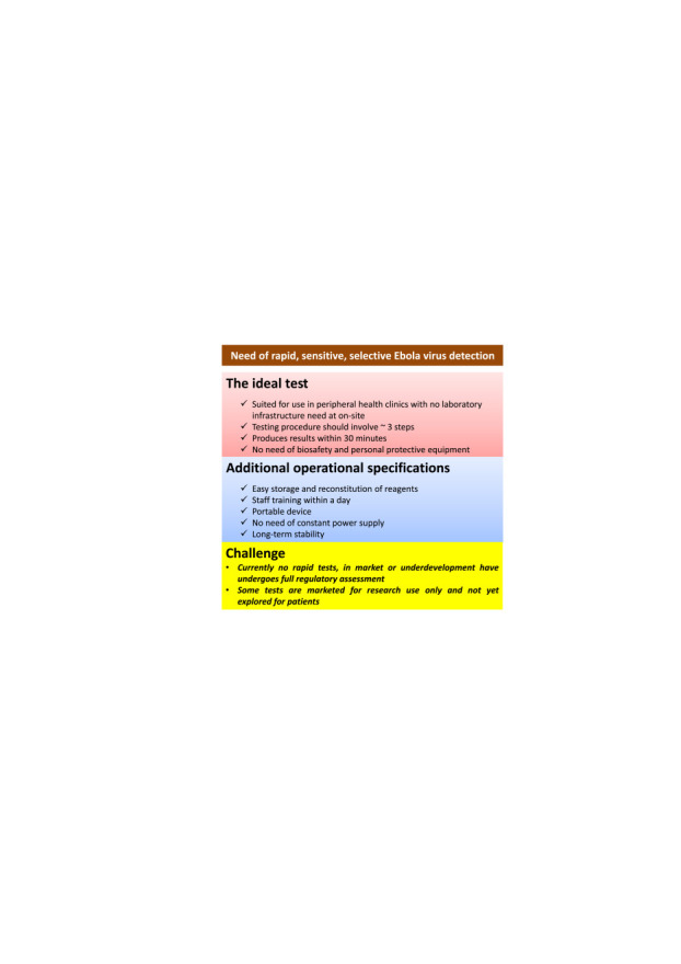
Ebola outbreak-2014 (mainly Zaire strain related Ebola virus) has been declared most widely spread deadly persistent epidemic due to unavailability of rapid diagnostic, detection, and therapeutics. Ebola virus disease (EVD), a severe viral hemorrhagic fever syndrome caused by Ebola virus (EBOV) is transmitted by direct contact with the body fluids of infected person and objects contaminated with virus or infected animals. World Health Organization (WHO) has declared EVD epidemic as public health emergency of international concern with severe global economic burden. At fatal EBOV infection stage, patients usually die before the antibody response. Currently, rapid blood tests to diagnose EBOV infection include the antigen or antibodies capture using ELISA and RNA detection using RT/Q-PCR within 3-10 days after the onset of symptoms. Moreover, few nanotechnology-based colorimetric and paper-based immunoassay methods have been recently reported to detect Ebola virus. Unfortunately, these methods are limited to laboratory only. As state-of-the art (SoA) diagnostics time to confirm Ebola infection, varies from 6h to about 3 days, it causes delay in therapeutic approaches. Thus developing a cost-effective, rapid, sensitive, and selective sensor to detect EVD at point-of-care (POC) is certainly worth exploring to establish rapid diagnostics to decide therapeutics. This review highlights SoA of Ebola diagnostics and also a call to develop rapid, selective and sensitive POC detection of EBOV for global health care. We propose that adopting miniaturized electrochemical EBOV immunosensing can detect virus level at pM concentration within ∼40min compared to 3 days of ELISA test at nM levels.

Keywords: Ebola Virus diseases; Ebola diagnostics; Ebola sensor; Ebola therapeutics; Point-of-care sensing.


Figures

Fig. 1
Fig. 1
Ebola virus taxonomy and classification.
Fig. 2
Fig. 2
Filamentous structure Ebola virus and Zaire Ebola strain Ebola virus with genomic presentation (Source: www.rcsb.org, www.emdatabank.org). Inset: genomic sequence of Ebola virus.(Choi and Croyle, 2013; Feldmann, 2014a; Nyakatura et al., 2015; Rivera and Messaoudi, 2015; Rougeron et al., 2015; Takada, 2012).
Fig. 3
Fig. 3
Microscopic view of EBOV Particle: (A) SEM of a single filamentous Ebola virus particle. (B) String-like EBOV particles are shedding from an infected cell in this electron micrograph. (C) SEM of filamentous Ebola virus particles attached to and budding from a chronically infected VERO E6 cell (blue) (25,000x magnification). (D) Ebola virus particles found both as extracellular particles and budding particles from chronically infected African green monkey kidney cells. (E) SEM of Ebola virus budding from the surface of a Vero cell (African green monkey kidney epithelial cell line). (F) EBOV Nucleocapsids and Virus Particles: TEM of EBOV nucleocapsids (small orange circles) and virus particles (larger orange filamentous forms) within infected African green monkey kidney cells. (G) Ebola-infected VERO E6 Cell: SEM of filamentous EBOV particles budding from an infected VERO E6 cell. (H) SEM of Ebola virions on the surface of a tetherin-expressing cell. http://www.sciencedaily.com/releases/2009/01/090127152838.htm. (Source: A-H NIAID of NIH). (I) Can robots help stop the Ebola outbreak? Germ-zapping robot could support war against Ebola (Sagripanti and Lytle, 2011). (For interpretation of the references to color in this figure legend, the reader is referred to the web version of this article.)
Fig. 4
Fig. 4
Entry pathway of Ebola Virus into host cell (Kawaoka, 2005). Upon binding to cell-surface receptors, Ebola gets internalized in endosome. Within endosome, endosomal proteases: cathepsin B and cathepsin L, slash the viral GP1 protein into N-terminal fragment and then cathepsin B digests it further into only GP2. GP2 aids in the fusion of viral envelope and endosomal membrane, releasing viral genome into the cytoplasm. Upon release the proteolysis of GP1 is prevented by CA074 (inhibitor) and therefore infection advances.
Fig. 5
Fig. 5
Illustration of Ebola Pathogenesis (Choi and Croyle, 2013), (Feldmann and Geisbert, 2011).
Fig. 6
Fig. 6
Illustration of clinical phase of EVD.
Fig. 7
Fig. 7
Salient features of Ebola outbreak-2014 (Frieden et al., 2014).
Fig. 8
Fig. 8
Exploring the need to develop rapid and sensitive Ebola detection methods.
Fig. 9
Fig. 9
Methodologies used to detect Ebola virus concentration during Ebola outbreak-2014.
Fig. 10
Fig. 10
(A) Comparison of the results obtained in serum and urine samples infected by Zaire Ebola virus Mayinga (TCID50 1105 /mL), 1105/mL), and results were compared with those for corresponding unspiked samples. (B) Cross-reactivity with antigens for other Ebola virus species and Lake Victoria marburg virus Musoke. (C) Demonstration of Immuno-filtration assay based on photometric integrated with immune-filtration column at diseases location (Lucht et al., 2007).
Fig. 11
Fig. 11
IF staining patterns of Ebola virus NP-expressing HeLa cells. (a) anti-Ebola virus NP rabbit serum. (b) Positive staining with serum collected from a patient with Ebola virus infected convalescent phase, and (c) Positive IF staining with serum collected from an Ebola infected monkey.(Saijo et al., 2001).
Fig. 12
Fig. 12
(A) IF staining of HeLa cells expressed by Ebola virus NP with anti-Ebola virus-NP rabbit serum case No. 1, (B) EBO-R infected monkey serum, (C) normal rabbit serum, (D) Ebola virus uninfected monkey serum case 2, (E), normal HeLa cells treated anti-Ebola virus-NP rabbit serum case 1, and (F) Ebola virus infected monkey serum. (Ikegami et al. 2002).
Fig. 13
Fig. 13
(A) Immunohistopathological analysis of in-situ hybridization and ultrastructural characteristics of fatal Ebola hemorrhagic fever. (B) Hepatic histopathological features and viral immunostaining in a fatal case of Ebola virus infection, (C) Splenic histopathological features and immunohistochemical characteristics in fatal cases of filovirus infection, (D) Cutaneous immunohistochemical and ultrastructural features in fatal cases of Ebola virus, (E) Gastrointestinal tract histopathological features and immunohistochemistry in a fatal case of Ebola hemorrhagic fever, (F) Renal histopathological features and immunohistochemistry in Ebola and Marburg hemorrhagic fevers, (G) Histopathological and immunohistochemical features in the testis of a fatal case of Ebola virus infection, (H) Bone marrow in a fatal case of Ebola virus infection (Martin et al. 2015).
Fig. 14
Fig. 14
(A) Schematic illustration of nanobarcodes 4G1R, 2G1R, 1G1R, 1G2R and 1G4R decoded based on fluorescence intensity ratio. (B) Nanobarcode colors (2-6 bar codes are 4G1R, 2G1R, 1G1R, 1G2R and 1G4R, respectively while 1 & 7 are 488–labeled starting oligonucleotide component and Bodipy 630/650–labeled starting oligonucleotide component) in an agarose gel on illumination using strong UV light. (C) Multiple target detection, (D) Dot-blotting detection of multiple DNA targets with nanobarcodes. (Li et al., 2005).
Fig. 15
Fig. 15
Immunofluorescence image of staining of co-cultured HeLa-CD4-LTRlacZ target cells with HeLa cells transfected with respect to HIV-1 Tat (A), Tat and HIV-1 Env (B), Tat and Ebola virus-GP (C). Confocal microscopy fields of co-cultures of CMTMR (red) labeled HeLa-CD4-LTRlacZ cells and CTG labeled (green) HeLa cells (D), HIV-1 Env (E), Ebola virus- GP (F) expression vectors. (Bar et al. 2006) (For interpretation of the references to color in this figure legend, the reader is referred to the web version of this article.).
Fig. 16
Fig. 16
(A) Schematic illustration of paper based genosensor consists of enzyme transcription and translation combined with engineered gene circuits to detect Ebola virus. (B) Prototype of Ebola RNA Sensors based Sudan and Zaire Strains sequencing of the Ebola Virus. 24 toehold switch-based RNA sensors were fabricated and tested-based on RNA segment windows (A–L) to detect Sudan and Zaire strains of Ebola virus at 570 nm absorption within 90 min at 30 °C (C) 240 paper-based reactions used to test the 24 sensors, yellow(control and un-triggered) and purple (activated toehold sensors). (D) Sequence specificity tested for 4 Sudan and 4 Zaire sensors.(E) Fold change of the color output rate of sensors SD and ZH over a titration of RNA concentrations.(Pardee et al. 2014).
Fig. 19
Fig. 19
Illustration of utilized specific Rx for Ebola
Fig. 17
Fig. 17
3D schematic illustration of resonance transmissions based opto-fluidic nanoplasmonic biosensors. (A) Immobilization and capturing of antibody to target virus, (B) VSV attaches only to the antibody immobilized sensor. (C) SEM of patterned SiNx membrane after Au deposition. (D) Au deposition result in suspended plasmonic nano-hole sensors without any lift-off process (inset: nanohole openings without clogging). (E) Immunosensing surface functionalization.(Yanik et al., 2010).
Fig. 18
Fig. 18
Illustration of microarray configuration and detection mechanism to detect Ebola virus using single particle interferometric reflectance imaging sensor (SP-IRIS). Specific antibodies immobilized onto microarray to detect virus via colorimetric based imaging as illustrated green, red, and blue spots represent anti-VSV, anti-EBOV, and anti-MARV probes, respectively (For interpretation of the references to color in this figure legend, the reader is referred to the web version of this article.).
Fig. 20
Fig. 20
Genomic structures of Ebola vaccine candidates-based on rVSV (Panel A) and cAd3 (Panel B) (Kanapathipillai, 2014, Kanapathipillai et al., 2014).
Fig. 21
Fig. 21
(A) Illustration of gene-trap insertions in the rVSV-GP-Ebola virus selected cell population. (B) Presentation of hypothetical model to understand the roles of CTSB, the HOPS complex and NPC1 during Ebola virus entry.(Carette et al. 2011).
Fig. 22
Fig. 22
(A) The Dimeric Structure of VP40 and related changed after Ebola virus infection. Dimeric crystal structure of VP40 EDN displayed with NTDs. This dimeric interface displayed A55, H61, F108, A113, and M116 (green) and L117 (yellow). (B) A cross-section of an Ebola virus virion is modeled by assembly of VP40 (NTD is in blue), And (B)2D modeling of 4 individual VP40 filaments derived from the assembly of VP40 hexamers.(Bornholdt et al., 2013) (For interpretation of the references to color in this figure legend, the reader is referred to the web version of this article.).
Fig. 23
Fig. 23
The top row shows the type of mutation of patient/raw identified with the Kissidougou Guinean sequence, with genomic locations indicated above and Cluster assignments are shown at the left. This informatics confirmed that Sierra Leone outbreak stemmed by the introduction of two genetically distinct viruses from Guinea around the same time. (Gire et al., 2014).
Fig. 24
Fig. 24
Illustration of filovirus genome in anti-genome (viral complementary) sense. 7 viral genes separated by intergenic regions and flanked by 50 and 30 UTRs (A), mini-genome and viral support protein plasmids used to drive the gLuc minigenome systems (B), and gLuc-expressing filovirus genomes (C)(Uebelhoer et al., 2014).
Fig. 25
Fig. 25
Illustration of GP monomer and MFL fragments.(Wang et al., 2014).
Fig. 26
Fig. 26
Schematic illustration of possible future prospects of miniaturized nano-enabling electrochemical Ebola sensor for POC application.

Similar articles

  • Analytical Validation of the ReEBOV Antigen Rapid Test for Point-of-Care Diagnosis of Ebola Virus Infection.
    Cross RW, Boisen ML, Millett MM, Nelson DS, Oottamasathien D, Hartnett JN, Jones AB, Goba A, Momoh M, Fullah M, Bornholdt ZA, Fusco ML, Abelson DM, Oda S, Brown BL, Pham H, Rowland MM, Agans KN, Geisbert JB, Heinrich ML, Kulakosky PC, Shaffer JG, Schieffelin JS, Kargbo B, Gbetuwa M, Gevao SM, Wilson RB, Saphire EO, Pitts KR, Khan SH, Grant DS, Geisbert TW, Branco LM, Garry RF. Cross RW, et al. J Infect Dis. 2016 Oct 15;214(suppl 3):S210-S217. doi: 10.1093/infdis/jiw293. Epub 2016 Aug 31. J Infect Dis. 2016. PMID: 27587634 Free PMC article.
  • Development of an antigen detection assay for early point-of-care diagnosis of Zaire ebolavirus.
    DeMers HL, He S, Pandit SG, Hannah EE, Zhang Z, Yan F, Green HR, Reyes DF, Hau D, McLarty ME, Altamura L, Taylor-Howell C, Gates-Hollingsworth MA, Qiu X, AuCoin DP. DeMers HL, et al. PLoS Negl Trop Dis. 2020 Nov 3;14(11):e0008817. doi: 10.1371/journal.pntd.0008817. eCollection 2020 Nov. PLoS Negl Trop Dis. 2020. PMID: 33141837 Free PMC article.
  • Lateral Flow Immunoassays for Ebola Virus Disease Detection in Liberia.
    Phan JC, Pettitt J, George JS, Fakoli LS 3rd, Taweh FM, Bateman SL, Bennett RS, Norris SL, Spinnler DA, Pimentel G, Sahr PK, Bolay FK, Schoepp RJ. Phan JC, et al. J Infect Dis. 2016 Oct 15;214(suppl 3):S222-S228. doi: 10.1093/infdis/jiw251. Epub 2016 Jul 20. J Infect Dis. 2016. PMID: 27443616 Free PMC article.
  • Strategies in Ebola virus disease (EVD) diagnostics at the point of care.
    Coarsey CT, Esiobu N, Narayanan R, Pavlovic M, Shafiee H, Asghar W. Coarsey CT, et al. Crit Rev Microbiol. 2017 Nov;43(6):779-798. doi: 10.1080/1040841X.2017.1313814. Epub 2017 Apr 25. Crit Rev Microbiol. 2017. PMID: 28440096 Free PMC article. Review.
  • Ebola virus disease.
    Jacob ST, Crozier I, Fischer WA 2nd, Hewlett A, Kraft CS, Vega MA, Soka MJ, Wahl V, Griffiths A, Bollinger L, Kuhn JH. Jacob ST, et al. Nat Rev Dis Primers. 2020 Feb 20;6(1):13. doi: 10.1038/s41572-020-0147-3. Nat Rev Dis Primers. 2020. PMID: 32080199 Free PMC article. Review.

Cited by

References

    1. Baden L.R., Kanapathipillai R., Campion E.W., Morrissey S., Rubin E.J., Drazen J.M. Ebola - an ongoing crisis. N. Engl. J. Med. 2014;371(15):1458–1459. - PubMed
    1. Bah E.I., Lamah M.-C., Fletcher T., Jacob S.T., Brett-Major D.M., Sall A.A., Shindo N., Fischer W.A., Lamontagne F., Saliou S.M., Bausch D.G., Moumié B., Jagatic T., Sprecher A., Lawler J.V., Mayet T., Jacquerioz F.A., Mendez Baggi M.F., Vallenas C., Clement C., Mardel S., Faye O., Faye O., Soropogui B., Magassouba N., Koivogui L., Pinto R., Fowler R.A. Vol. 372. 2014. Clinical Presentation of Patients with Ebola Virus Disease in Conakry, Guinea; pp. 40–47. (N. Engl. J. Med.). - PubMed
    1. Baize S., Leroy E., Georges A., GEORGES†COURBOT M.C., Capron M., Bedjabaga I., LANSOUD†SOUKATE J.,, Mavoungou E. Inflammatory responses in Ebola virus†infected patients. Clin. Exp. Immunol. 2002;128(1):163–168. - PMC - PubMed
    1. Baize S., Pannetier D., Oestereich L., Rieger T., Koivogui L., Magassouba N.F., Soropogui B., Sow M.S., Keïta S., De Clerck H., Tiffany A., Dominguez G., Loua M., Traoré A., Kolié M., Malano E.R., Heleze E., Bocquin A., Mély S., Raoul H., Caro Vr, Cadar Dn, Gabriel M., Pahlmann M., Tappe D., Schmidt-Chanasit J., Impouma B., Diallo A.K., Formenty P., Van Herp M., Günther S. Emergence of Zaire Ebola Virus Disease in Guinea. N. Engl. J. Med. 2014;371(15):1418–1425. - PubMed
    1. Bar S., Takada A., Kawaoka Y., Alizon M. Detection of cell-cell fusion mediated by Ebola virus glycoproteins. Journal of virology. 2006;80(6):2815–2822. - PMC - PubMed

Publication types

MeSH terms