. 2015 Dec;161(2):260-9.
doi: 10.1016/j.clim.2015.08.007. Epub 2015 Aug 25.

Interleukin-7 is required for CD4(+) T cell activation and autoimmune neuroinflammation

Affiliations

Interleukin-7 is required for CD4(+) T cell activation and autoimmune neuroinflammation

Brian R Lawson et al. Clin Immunol. 2015 Dec.

Abstract

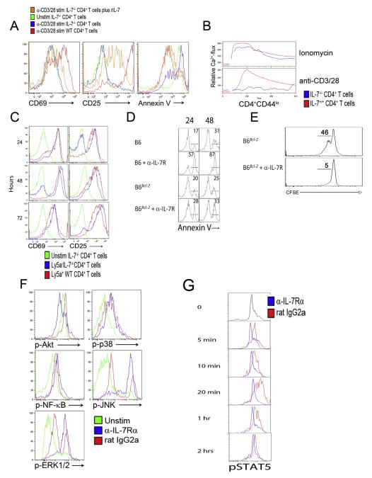
IL-7 is known to be vital for T cell homeostasis but has previously been presumed to be dispensable for TCR-induced activation. Here, we show that IL-7 is critical for the initial activation of CD4(+) T cells in that it provides some of the necessary early signaling components, such as activated STAT5 and Akt. Accordingly, short-term in vivo IL-7Rα blockade inhibited the activation and expansion of autoantigen-specific CD4(+) T cells and, when used to treat experimental autoimmune encephalomyelitis (EAE), prevented and ameliorated disease. Our studies demonstrate that IL-7 signaling is a prerequisite for optimal CD4(+) T cell activation and that IL-7R antagonism may be effective in treating CD4(+) T cell-mediated neuroinflammation and other autoimmune inflammatory conditions.

Keywords: EAE; IL-7; Signaling pathways; T cells.


Figures

Fig. 1
Fig. 1
IL-7 is required for CD4+ T cell activation. (A) Receptor-blocking anti-IL-7Rα antibodies inhibit antigen-induced upregulation of activation markers on naïve PLP-specific TCR transgenic CD4+ T cells. Shown are histograms of CD69 or CD25 cell-surface levels at 1, 2 and 7 days post-treatment with the indicated stimuli and antibodies. (B) Reduced PLP-specific CD4+ TCR transgenic T cell proliferation in the presence of anti-IL-7Rα antibodies and APCs. Proliferating cells were identified by FACS via CFSE-dilution. (C) Treatment with anti-IL-7Rα (blue) but not control (red) antibodies causes increased apoptosis of PLP-TCR transgenic CD4+ T cells 48 h following PLP stimulation. (D) The presence of anti-IL-7Rα (blue), but not control (red), antibodies inhibits upregulation of Bcl-2 in PLP-TCR transgenic CD4+ T cells stimulated with PLP up to 4 days. Day 4 is depicted. For panels A-D, splenocytes from PLP-TCR transgenic mice were pretreated with anti-IL-7Rα or isotype control antibodies (25 μg/ml each), cultured with or without IL-7 (10 ng/ml) as indicated, and stimulated with PLP (10 μg/ml). All FACS plots are gated on CD4+ T cells. Horizontal bars in A and B indicate activated or proliferating cells, respectively. The data are representative of 3–6 independent experiments.
Fig. 2
Fig. 2
STAT5 and Akt phosphorylation in CD4+ T cell activation requires IL-7. (A) Inefficient activation and increased apoptosis of TCR-stimulated CD4+ T cells from IL-7−/− mice are corrected by rIL-7 treatment. Splenocytes from WT (red) or IL-7−/− (blue) mice were activated with anti-CD3/CD28 antibodies for 48 h. CD69 and CD25 cell-surface levels and Annexin V positivity were determined on CD4+ T cells by FACS. Addition of rIL-7 (10 ng/ml, orange) prevented apoptosis (Annexin V upregulation) of IL-7−/− CD4+ T cells and restored activation. Green lines, unstimulated IL-7−/− CD4+ T cells. (B) CD4+ T cells from IL-7−/− mice (blue) mobilize Ca2+ after ionomycin but not anti-CD3/CD28 stimulation. CD4+ T cells from WT mice (red) were used as positive control. (C) Wild-type splenocytes restore TCR-activation of CD4+ T cells from IL-7−/− mice in mixed cultures. Splenocytes from Ly5aIL-7−/− and Ly5a+ WT mice were mixed 1:1 and stimulated with anti-CD3/CD28 antibodies. CD69 and CD25 expression on gated Ly5aIL-7−/− (blue) and Ly5a+ WT (red) CD4+ T cells was determined by FACS. Green lines, unstimulated IL-7−/− CD4+ T cells. (D) Transgenic Bcl-2-expression rescues CD4+ T cells from IL-7R blockade-dependent activation-induced cell death (AICD). WT or Bcl-2 transgenic C57BL/6 splenocytes were pretreated with anti-IL-7Rα or isotype control antibodies and stimulated with anti-CD3/CD28 antibodies. Annexin V expression was analyzed on CD4+ T cells every 24 h for 96 h (24 and 48 hour time points are depicted). Numbers indicate % cells in the respective gate. (E) The Bcl-2 transgene fails to restore TCR-induced CD4+ T cell proliferation in the presence of anti-IL-7Rα antibodies. (F) PLP-specific TCR transgenic CD4+ T cells show a stress phenotype in the presence of anti-IL-7Rα antibodies. Splenocytes from PLP-TCR transgenic mice were treated with anti-IL-7Rα or isotype antibodies and stimulated with PLP. Time points up to 2 h were analyzed by FACS for phosphorylation (activation) of Akt, p38, NF-κB p65, JNK, and Erk1/2. Histograms depict the 20 min time point, when isotype-treated control cells reached maximal activation. Blue, IL-7Rα antibody-treated CD4+ T cells; red, isotype-treated CD4+ T cells; green, unstimulated CD4+ T cells. (G) TCR stimulation induces STAT5 phosphorylation in an IL-7-dependent manner. Splenocytes from PLP-TCR transgenic mice were treated with anti-IL-7Rα or isotype antibodies (25 μg/ml each) and stimulated with PLP (10 μg/ml). CD4+ T cells were analyzed at the indicated time points by FACS for STAT5 phosphorylation (activation). Blue, IL-7Rα antibody-treated cells; red, isotype-treated cells. All data are representative of 3–5 independent experiments.
Fig. 3
Fig. 3
Constitutive STAT5 phosphorylation compensates for the lack of IL-7 signaling during CD4+ T cell activation. Purified CD4+CD25 T cells from C57BL/6 and STAT5-CA transgenic mice were stimulated with anti-CD3/CD28 for 72 h in the presence or absence of anti-IL-7Rα antibody (25 μg/ml). CD4+ T cells were analyzed by FACS for acquisition of the activation markers CD25 and CD69. Data are representative of 2 independent experiments.
Fig. 4
Fig. 4
IL-7Rα blockade in vivo induces preferential elimination of antigen-stimulated autoreactive CD4+ T cells. (A) Antigen-engaged PLP-specific CD4+ TCR transgenic T cells are mostly eliminated from LN and spleen in SJL mice undergoing EAE and treated for a short time with anti-IL-7Rα (blue line) compared to isotype control antibody (rat IgG2a, red line). Naive (CD44low CD62Lhi) Thy1.1 PLP-specific TCR transgenic CD4+ T cells (Thy1.1, CD90.1) were CFSE labeled and transferred into SJL recipients (Thy1.2) at day 10 post-EAE induction. These mice were then treated or not with anti-IL-7Rα antibody (200 μg, 2 injections/3 days, starting at the time of cell transfer), and LN and spleen cells were harvested and analyzed by FACS. n = 4–5 mice/group; p < 0.001. Insets depict plots at smaller Y-axis scale to better visualize CFSE dilution. (B) Anti-IL-7Rα antibody treatment of EAE mice induces apoptosis of transferred PLP-specific TCR transgenic CD4+ T cells. Undivided (CFSEhi) TCR transgenic CD4+ T cells from panel A were analyzed by FACS for Annexin V positivity. n = 4–5 mice/group; p < 0.001. (C) Anti-IL-7Rα antibody treatment does not induce loss of PLP-specific TCR transgenic CD4+ T cells in unimmunized (non-EAE) SJL recipients. Cell transfer and treatments were as detailed in panel A. (D) Effector PLP-specific CD4+ TCR transgenic T cells also require IL-7 signaling. Naive PLP-specific CD4+ TCR transgenic T cells (Thy1.1, CD90.1) were transferred into SJL recipients (Thy1.2) simultaneously with EAE induction (day 0). After disease appearance (day 10; EAE severity score ≥ 1), these mice were treated with anti-IL-7Rα or control antibodies (200 μg, 3 injections/5 days), and LN and spleen cells were harvested and analyzed by FACS using antibodies to CD4 and CD90.1 to identify donor cells. n = 3–4 mice/group; p < 0.01. Data are representative of 2–3 independent experiments.
Fig. 5
Fig. 5
Relapsing/remitting EAE is attenuated by IL-7Rα blockade. (A) Incidence and (B) severity of PLP-induced R-EAE in SJL mice is significantly reduced by administration of anti-IL-7Rα (open circles) compared to isotype control antibody (closed circles). n = 8–11 mice/group; p < 0.001. (C) Increased conversion of procaspases 3 and 8 to active caspases in CD4+ T cells obtained two weeks after R-EAE induction from anti-IL-7Rα antibody-treated mice (blue line) compared to controls (red line). n = 8–11 mice/group; p < 0.01. (D) Incidence and (E) severity of R-EAE induced in SJL mice by transfer of polarized TH1 cells were reduced by treatment with anti-IL-7Rα antibody (open circles) compared to isotype antibody (closed circles). n = 5–8 mice/group; p < 0.001. (F) Incidence and (G) severity of PLP-induced R-EAE is reduced if anti-IL-7Rα antibodies are administered at the start of acute disease (day 8). n = 8–11 mice/group; p < 0.001 from days 8–35. (H) Incidence and (I) severity of relapses in PLP-induced R-EAE mice are reduced when anti-IL-7Rα antibody treatment is initiated prior to the first relapse (day 20). n = 8–11 mice/group; p < 0.001 from days 20–35. R-EAE was induced in young female SJL mice either by injection of PLP peptide in CFA plus PTX (A, B, F, G, H, and I) or by adoptive transfer of CD4+ T cells obtained from PLP-immunized SJL donors and polarized in vitro with PLP and IL-12 to TH1 cell type (D and E). Arrows indicate initiation of anti-IL-7Rα or isotype control antibody treatment (200 μg, 3 times/week). Data are expressed as either daily group means (incidence) or mean ± SEM (EAE Severity Score), and are representative of 3–5 independent experiments.

Similar articles

Cited by

References

    1. Surh CD, Sprent J. Homeostasis of naive and memory T cells. Immunity. 2008;29:848–862. - PubMed
    1. Kittipatarin C, Khaled AR. Interlinking interleukin-7. Cytokine. 2007;39:75–83. - PMC - PubMed
    1. Palmer MJ, Mahajan VS, Trajman LC, Irvine DJ, Lauffenburger DA, Chen J. Interleukin-7 receptor signaling network: an integrated systems perspective. Cell. Mol. Immunol. 2008;5:79–89. - PMC - PubMed
    1. Jiang Q, Li WQ, Aiello FB, Mazzucchelli R, Asefa B, Khaled AR, Durum SK. Cell biology of IL-7, a key lymphotrophin. Cytokine Growth Factor Rev. 2005;16:513–533. - PubMed
    1. Wofford JA, Wieman HL, Jacobs SR, Zhao Y, Rathmell JC. IL-7 promotes Glut1 trafficking and glucose uptake via STAT5-mediated activation of Akt to support T-cell survival. Blood. 2008;111:2101–2111. - PMC - PubMed

Publication types

MeSH terms