. 2015 Aug 28;4(9):1163-79.
doi: 10.1242/bio.012468.

The evolution of the dystroglycan complex, a major mediator of muscle integrity

Affiliations

The evolution of the dystroglycan complex, a major mediator of muscle integrity

Josephine C Adams et al. Biol Open. .

Abstract

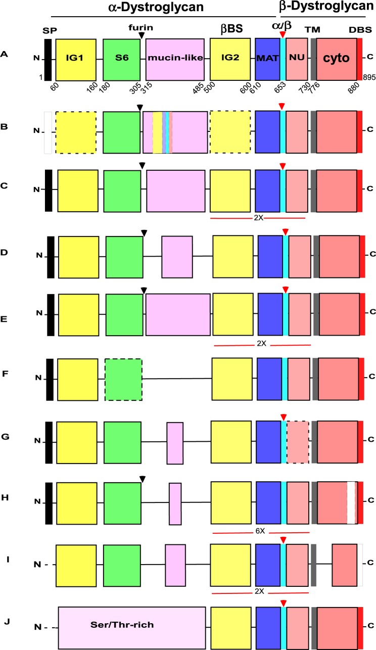
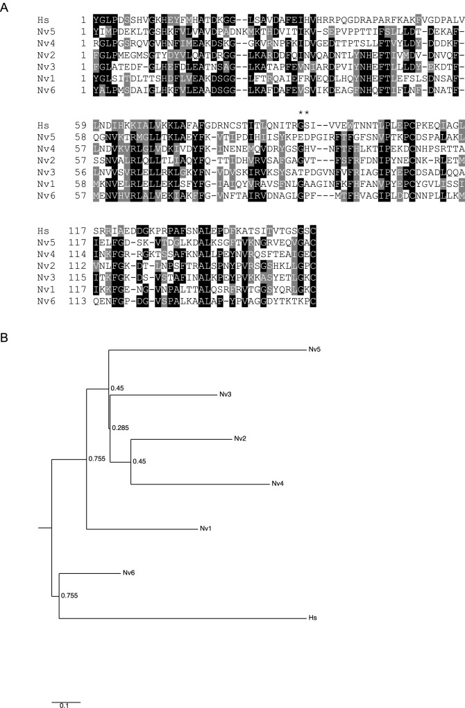
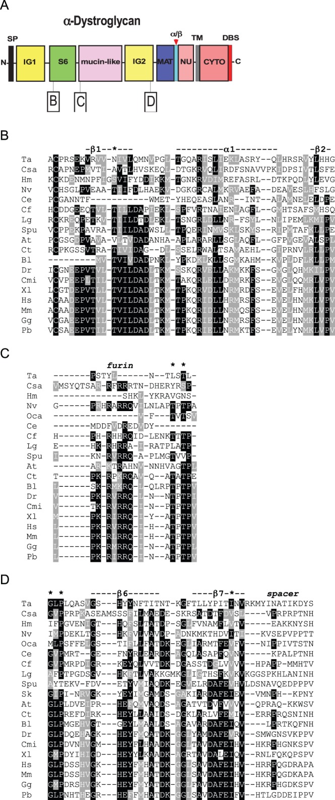
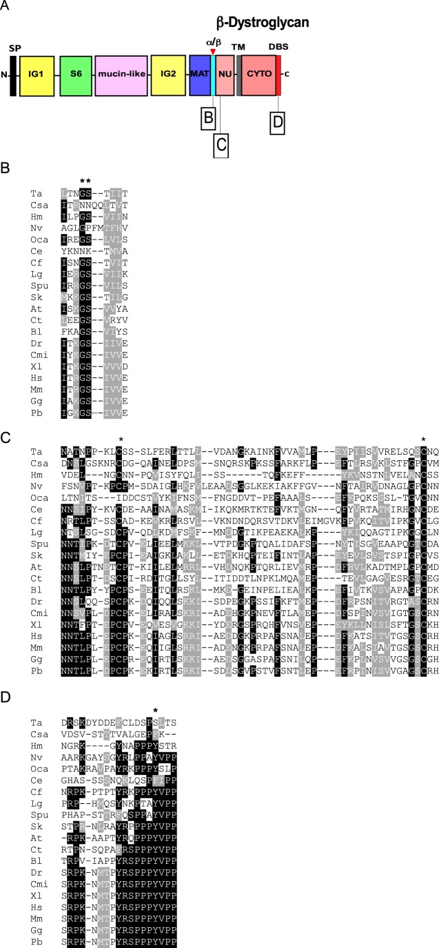
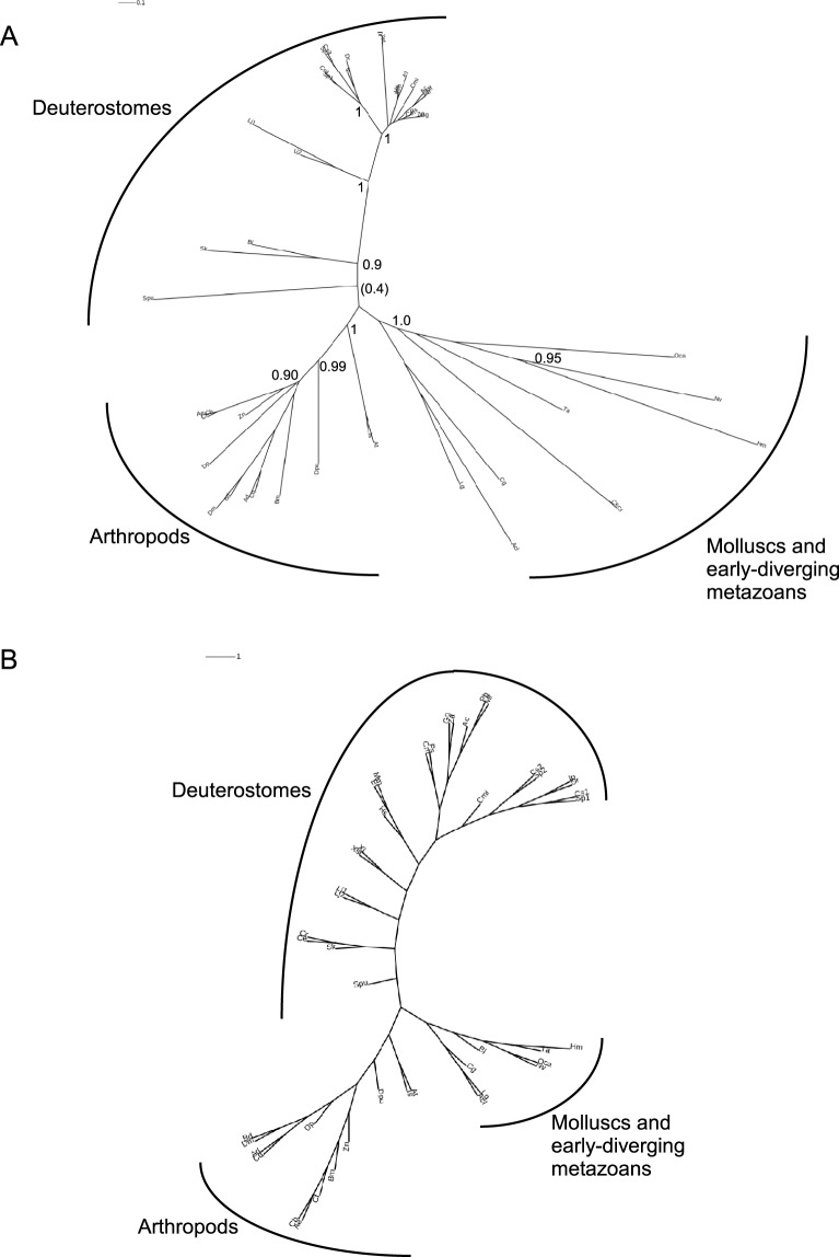
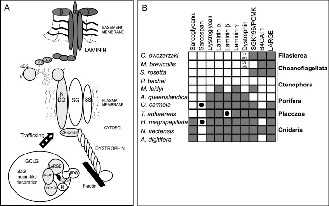
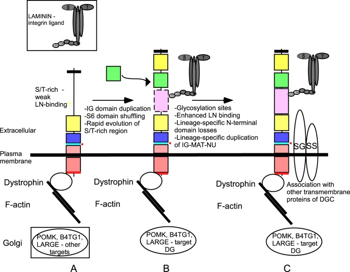
Basement membrane (BM) extracellular matrices are crucial for the coordination of different tissue layers. A matrix adhesion receptor that is important for BM function and stability in many mammalian tissues is the dystroglycan (DG) complex. This comprises the non-covalently-associated extracellular α-DG, that interacts with laminin in the BM, and the transmembrane β-DG, that interacts principally with dystrophin to connect to the actin cytoskeleton. Mutations in dystrophin, DG, or several enzymes that glycosylate α-DG underlie severe forms of human muscular dystrophy. Nonwithstanding the pathophysiological importance of the DG complex and its fundamental interest as a non-integrin system of cell-ECM adhesion, the evolution of DG and its interacting proteins is not understood. We analysed the phylogenetic distribution of DG, its proximal binding partners and key processing enzymes in extant metazoan and relevant outgroups. We identify that DG originated after the divergence of ctenophores from porifera and eumetazoa. The C-terminal half of the DG core protein is highly-conserved, yet the N-terminal region, that includes the laminin-binding region, has undergone major lineage-specific divergences. Phylogenetic analysis based on the C-terminal IG2_MAT_NU region identified three distinct clades corresponding to deuterostomes, arthropods, and mollusks/early-diverging metazoans. Whereas the glycosyltransferases that modify α-DG are also present in choanoflagellates, the DG-binding proteins dystrophin and laminin originated at the base of the metazoa, and DG-associated sarcoglycan is restricted to cnidarians and bilaterians. These findings implicate extensive functional diversification of DG within invertebrate lineages and identify the laminin-DG-dystrophin axis as a conserved adhesion system that evolved subsequent to integrin-ECM adhesion, likely to enhance the functional complexity of cell-BM interactions in early metazoans.

Keywords: Basement membrane; Dystroglycan; Dystroglycanopathy; Metazoa; Multicellularity; Protein domain analysis.


Conflict of interest statement

Competing interests

The authors declare no competing or financial interests.

Figures

Fig. 1.
Fig. 1.
Domain architectures of dystroglycans from different animal phyla. (A) The DG typical of vertebrates Callorhincus milii (elephant shark), Lethenteron japonicum (Cyclostomata), (Strongylocentrotus purpuratus (Echinoderma) and an annelid, Capitella teleta. (B-J) domain architectures of DG identified in (B) Urochordata (Ciona intestinalis) and Cephalochordata (Branchiostoma lanceolatum), (C) Hemichordata (Saccoglossus kowalevskii), (D) Mollusca, (Gastropods Lottia gigantea and Aplysia californica, Bivalve, Crassostrea gigas and cephalopod, Octopus vulgaris), (E) arthropod classes (Insecta and Hymenoptera, Camponotus floridanus and others) (see also supplementary material Fig. S1), (F) Nematoda (Caenorhabditis elegans and Caenorhabditis remanei), (G) Cnidaria, Hydra magnipapillata, (H) Cnidaria, Nematostella vectensis (see also supplementary material Fig. S2), (I) Placozoa (Trichoplax adhaerens), (J) Porifera (homoscleromorph Oscarella carmela) (see also supplementary material Fig. S3). Expansions of the IG2_MAT_NU module are indicated in C, E and I (2×) and in H (6×). Black arrowheads indicate the furin cleavage site. Red arrowheads indicate the Gly-Ser α/β maturation site. SP, signal peptide; IG1 and IG2, immunoglobulin-like domains; S6, S6-like domain; βBS, β-subunit binding site on the IG2 domain; MAT, C-terminal region of α-dystroglycan upstream of the Gly-Ser maturation site; NU, natively unfolded region that forms the N-terminal region of the ectodomain of β-dystroglycan; TM, transmembrane; cyto, cytoplasmic domain; DBS, dystrophin-binding site. The SP is reported as a black box if complete, or a white box if partial. Dotted lines indicate protein sequences that are incomplete at the N-terminal end. Dotted boxes around the IG domains of Urochordata (B) or the S6 domain of nematodes (F) indicate the divergence of these domains (less than 20% sequence identity). The dotted box for NU in H. magnipapillata DG (G) indicates the presence of two deletions in this region. The white box within the cytoplasmic domain of N. vectensis DG (H) indicates the presence of an insertion. In T. adhaerens DG (I), no DBS was detected (white box). Diagrams are not to scale. Accession codes and other details are in Table 2.
Fig. 2
Fig. 2
. Analysis of the multiple repetitions of the IG2_MAT_NU region in N. vectensis DG. (A) Multiple sequence alignment of the six IG2_MAT_NU modules of N. vectensis DG compared with this region of human DG. Alignments were prepared in MUSCLE 3.8 and are presented in Boxshade. Asterisks indicate the GS proteolysis site in human DG. At each position, black background includes identical residues; grey background indicates conservative substitutions, and white background indicates non-conserved residues. (B) Phylogenetic analysis of the six IG2_MAT_NU modules of N. vectensis DG (alignment of 153 positions) compared with the same region of human DG. The tree was prepared in PhyML with 200 cycles of boot-strapping. Numbers indicate bootstrap support values, with 1 as maximum. Scale=substitutions/site.
Fig. 3.
Fig. 3.
Multiple sequence alignments of functionally important regions from α-dystroglycan. The dataset includes representative species from the phyla in which DG was identified. (A) Schematic of α-DG and the regions presented in the alignments. Key as in Fig. 1. (B) Thr192 (*) and the surrounding secondary structure at the beginning of the S6 domain. (C) The furin cleavage site and the Thr-Pro-Thr motif (* *) at the beginning of the mucin-like region. (D) The last two β-strands (dashed line) of the IG2 domain, followed by a spacer region that precedes MAT. The conserved Gly563, Pro565 and Ile593 are also pinpointed by asterisks. Alignments were prepared as in Fig. 2. Codes for species names are as in Table 2.
Fig. 4
Fig. 4
. Multiple sequence alignments of functionally important regions from β-dystroglycan. Key as in Fig. 1. (A) Schematic of β-DG and the regions presented in the alignments. (B) The Gly-Ser α/β maturation site (**). (C) Region of the NU domain encompassing the two conserved Cys residues (*). (D) The dystrophin-binding site. The shade coding is as in Fig. 2. Codes for species names are as in Table 2. Tyr892 (*) is a phosphorylation site.
Fig. 5.
Fig. 5.
Phylogenetic analysis based on the IG2_MAT-NU region of dystroglycans. The IG2_MAT_NU regions from DGs from 46 species (245 positions) were aligned in PRANK and phylogenetic trees constructed (A) in PhyML with 200 cycles of boot-strapping, or (B) as a consensus tree in PROTPARS. Unrooted trees are presented with proportionate branch lengths. Scale bars=substitutions/site. In A, only bootstrap branch support values >0.95 are shown. Codes for species names are as in Table 2.
Fig. 6.
Fig. 6.
Evolution of dystroglycan-binding proteins. (A) Schematic of the interactions of dystroglycan with other members of the DGC and the modifying enzymes that act on DG. In the Golgi complex, α-dystroglycan is post-translationally modified at multiple Thr/Ser residues in its mucin-like region during its trafficking to the cell surface. CR, cysteine-rich domain; SG: sarcoglycan; SS: sarcospan. (B) The phylogenetic distributions of DG, dystroglycan-binding proteins of the DGC and DG-modifying enzymes in early-diverging metazoans and their closest unicellular relatives. Species are representative of the indicated phyla. Key: Grey squares, predicted protein identified, BLASTP e-value<1e−10; black circles, BLASTP e-value>1e−10 and <0.05; white squares, no homologue identified. See supplementary material Table S1 for accession numbers.
Fig. 7.
Fig. 7.
A model for the evolution of dystroglycan and the DGC. See text for details.

References

    1. Abedin M. and King N. (2008). The premetazoan ancestry of cadherins. Science 319, 946-948. 10.1126/science.1151084 - DOI - PubMed
    1. Adams J. C. (2013). The evolution of extracellular matrix: an over-view. In Evolution of Extracellular Matrix (ed. Keeley F. W. and Mecham R. P.), pp. 1-25. Berlin: Springer; 10.1007/978-3-642-36002-2 - DOI
    1. Barresi R. and Campbell K. P. (2006). Dystroglycan: from biosynthesis to pathogenesis of human disease. J. Cell Sci. 119, 199-207. 10.1242/jcs.02814 - DOI - PubMed
    1. Boffi A., Bozzi M., Sciandra F., Woellner C., Bigotti M. G., Ilari A. and Brancaccio, (2001). Plasticity of secondary structure in the N-terminal region of β-dystroglycan. Biochim. Biophys. Acta 1546, 114-121. 10.1016/S0167-4838(01)00131-5 - DOI - PubMed
    1. Bogdanik L., Framery B., Frölich A., Franco B., Mornet D., Bockaert J., Sigrist S. J., Grau Y. and Parmentier M.-L. (2008). Muscle dystroglycan organizes the postsynapse and regulates presynaptic neurotransmitter release at the Drosophila neuromuscular junction. PLoS ONE 3, e2084 10.1371/journal.pone.0002084 - DOI - PMC - PubMed