Review
. 2015 Dec;355(3):410-28.
doi: 10.1124/jpet.115.227330. Epub 2015 Aug 28.

Matrix Metalloproteinases as Regulators of Vein Structure and Function: Implications in Chronic Venous Disease

Affiliations
Review

Matrix Metalloproteinases as Regulators of Vein Structure and Function: Implications in Chronic Venous Disease

Elisabeth MacColl et al. J Pharmacol Exp Ther. 2015 Dec.

Abstract

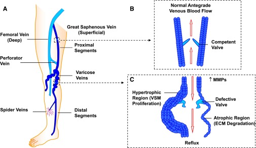
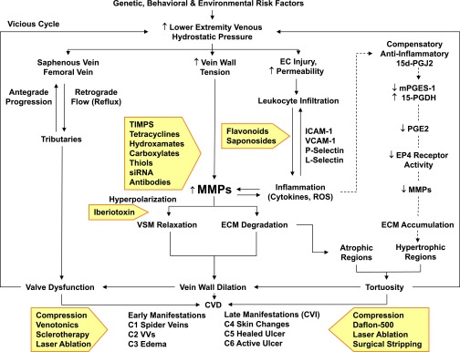
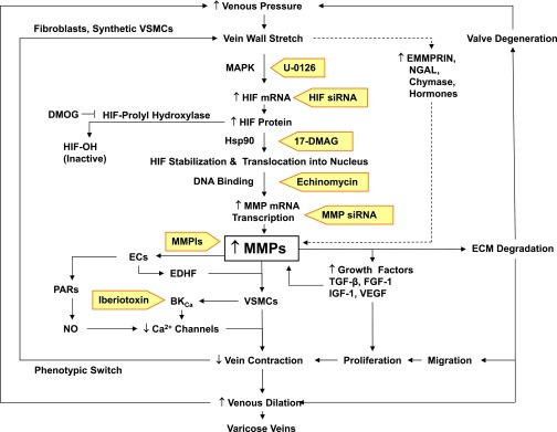
Lower-extremity veins have efficient wall structure and function and competent valves that permit upward movement of deoxygenated blood toward the heart against hydrostatic venous pressure. Matrix metalloproteinases (MMPs) play an important role in maintaining vein wall structure and function. MMPs are zinc-binding endopeptidases secreted as inactive pro-MMPs by fibroblasts, vascular smooth muscle (VSM), and leukocytes. Pro-MMPs are activated by various activators including other MMPs and proteinases. MMPs cause degradation of extracellular matrix (ECM) proteins such as collagen and elastin, and could have additional effects on the endothelium, as well as VSM cell migration, proliferation, Ca(2+) signaling, and contraction. Increased lower-extremity hydrostatic venous pressure is thought to induce hypoxia-inducible factors and other MMP inducers/activators such as extracellular matrix metalloproteinase inducer, prostanoids, chymase, and hormones, leading to increased MMP expression/activity, ECM degradation, VSM relaxation, and venous dilation. Leukocyte infiltration and inflammation of the vein wall cause further increases in MMPs, vein wall dilation, valve degradation, and different clinical stages of chronic venous disease (CVD), including varicose veins (VVs). VVs are characterized by ECM imbalance, incompetent valves, venous reflux, wall dilation, and tortuosity. VVs often show increased MMP levels, but may show no change or decreased levels, depending on the VV region (atrophic regions with little ECM versus hypertrophic regions with abundant ECM) and MMP form (inactive pro-MMP versus active MMP). Management of VVs includes compression stockings, venotonics, and surgical obliteration or removal. Because these approaches do not treat the causes of VVs, alternative methods are being developed. In addition to endogenous tissue inhibitors of MMPs, synthetic MMP inhibitors have been developed, and their effects in the treatment of VVs need to be examined.


Figures

Fig. 1.
Fig. 1.
The lower-extremity venous system and changes in VVs. The lower extremity has an intricate system of superficial and deep veins connected by perforator veins (A), and venous valves that allow blood flow in the antegrade direction toward the heart (B). Vein dysfunction may manifest as small spider veins and could progress to large dilated VVs with incompetent valves (C). VVs mainly show atrophic regions where an increase in MMPs increases ECM degradation, but could also show hypertrophic regions in which increased MMPs and ECM degradation would promote VSMC proliferation, leading to tortuosity, dilation, defective valves, and venous reflux (C).
Fig. 2.
Fig. 2.
Major subtypes and structures of MMPs. A typical MMP consists of a propeptide, a catalytic metalloproteinase domain, a linker peptide (hinge region), and a hemopexin domain. The propeptide has a cysteine switch PRCGXPD whose cysteine sulfhydryl (–SH) group chelates the active site Zn2+, keeping the MMP in the latent pro-MMP zymogen form. The catalytic domain contains the Zn2+-binding motif HEXXHXXGXXH; two Zn2+ ions (one catalytic and one structural); specific S1, S2, …, Sn and S1′, S2′, …, Sn′pockets, which confer specificity; and two or three Ca2+ ions for stabilization. Some MMPs show exceptions in their structures. Gelatinases have three type II fibronectin repeats in the catalytic domain. Matrilysins have neither a hinge region nor a hemopexin domain. Furin-containing MMPs such as MMP-11, -21, and -28 have a furin-like proprotein convertase recognition sequence in the propeptide C terminus. MMP-28 has a slightly different cysteine switch motif PRCGVTD. MT-MMPs typically have a transmembrane domain and a cytosolic domain. MMP-17 and -25 have a glycosylphosphatidylinositol (GPI) anchor. MMP-23 lacks the consensus PRCGXPD motif, has a cysteine residue located in a different sequence ALCLLPA, may remain in the latent inactive proform through its type II signal anchor, and has a cysteine-rich region and an immunoglobulin-like proline-rich region. aa, amino acid.
Fig. 3.
Fig. 3.
MMP-substrate interaction. For simplicity, MMP-3 is used as a prototype, and only the catalytic domain is shown, with the rest of the MMP structure interrupted by squiggles. (A) In preparation for substrate binding, an incoming H2O molecule is polarized between the free MMP basic Glu202 and acidic Zn2+. Zn2+ is localized in the MMP HEXXHXXGXXH motif by binding imidazole rings from three histidines (His201, His205, His211), whereas Met219 in the conserved XBMX Met-turn serves as a hydrophobic base to support the structure surrounding the catalytic Zn2+. (B) Using H+ from free H2O, the substrate carbonyl binds to Zn2+, forming a Michaelis complex. MMP S1, S2, S3, ..., Sn pockets on the left side of Zn2+ and primed S1′, S2′, S3′, ..., Sn′ pockets on the right side of Zn2+ confer binding specificity to substrate P1, P2, P3, ..., Pn and P1′, P2′, P3′, ..., Pn′ substituents, respectively. S1 and S3 are located away from the catalytic center, whereas S2 is close to Zn2+. (C) Substrate-bound H2O is freed. Zn2+-bound O from Glu-bound H2O performs a nucleophilic attack on substrate carbon, and Glu202 abstracts a proton from Glu-bound H2O to form an N–H bond with substrate N, thus forming a tetrahedral intermediate. (D) Free H2O is taken up, and the second proton from Glu-bound H2O is transferred to the substrate, forming another N–H bond. This allows the substrate scissile C–N bond to break, releasing the N portion of the substrate and forming an MMP-carboxylate complex. Another free H2O is taken up, releasing the remaining carboxylate portion of the substrate and the free MMP (A). The positions of conserved His and Glu vary in different MMPs.
Fig. 4.
Fig. 4.
Pathophysiology and management of CVD. Certain genetic, behavioral, and environmental risk factors cause an increase in hydrostatic pressure in the lower-extremity saphenous and femoral veins, leading to venous reflux and valve dysfunction. Increased hydrostatic pressure also increases vein wall tension, leading to increases in MMPs, and could also cause EC injury, increased permeability, leukocyte infiltration, and increased levels of adhesion molecules, inflammatory cytokines, and ROS, leading to further increases in MMPs. Increased MMPs may cause VSM hyperpolarization and relaxation as well as ECM degradation, leading to vein wall dilation, valve dysfunction, and progressive increases in venous hydrostatic pressure (vicious cycle). Increased MMPs generally promote ECM degradation, particularly in atrophic regions. Other theories (indicated by dashed arrows) suggest a compensatory anti-inflammatory pathway involving prostaglandins and their receptors that leads to decreased MMPs and thereby ECM accumulation, particularly in hypertrophic regions of VVs. Persistent valve dysfunction and progressive vein wall dilation and tortuosity lead to different stages of CVD and CVI. Current therapies of CVD and CVI (presented in shaded arrows) include physical, pharmacological, and surgical approaches. Inhibitors of the activity or action of MMPs (also presented in shaded arrows) may provide potential tools for the management of CVD/CVI. 15d-PGJ2, 15-deoxy-delta-12,14-prostaglandin J2; mPGES-1, membrane-associated prostaglandin E synthase-1; 15-PGDH, 15-hydroxyprostaglandin dehydrogenase; VCAM-1, vascular cell adhesion molecule-1.
Fig. 5.
Fig. 5.
Mechanisms linking increased venous pressure to increased MMP expression and VVs. Increased venous pressure causes vein wall stretch, which increases HIF mRNA and protein levels and, in turn, MMP levels. Increased wall stretch may also increase other MMP inducers such as EMMPRIN, neutrophil gelatinase–associated lipocalin (NGAL), chymase, and hormones. Increased MMPs may activate PARs in ECs, leading to nitric oxide (NO) production and venous dilation. MMPs may also stimulate ECs to release EDHF, which in turn opens BKCa channels in VSM, leading to hyperpolarization, decreased Ca2+ influx, and decreased vein contraction. Loss of contractile function in VSM could cause a phenotypic switch to synthetic VSMCs. MMPs may also increase the release of growth factors, leading to VSMC proliferation. MMPs also cause ECM degradation, leading to VSMC migration, proliferation, further decreases in vein contraction and increases in venous dilation, and VVs. MMP-induced ECM degradation may also cause valve degeneration, leading to further increases in venous pressure. As indicated in shaded arrows, inhibitors of MMP synthesis [U0126, HIF small interfering RNA (siRNA), 17-dimethylaminoethylamino-17-demethoxygeldanamycin (17-DMAG), echinomycin, MMP small interfering RNA], activity (MMPIs), or actions (iberiotoxin) may provide new tools for management of VVs. DMOG, dimethyloxaloylglycine (an experimental inhibitor of HIF-prolyl hydroxylase); FGF, fibroblast growth factor; Hsp90, heat shock protein 90; IGF, insulin-like growth factor; NGAL, neutrophil gelatinase–associated lipocalin.
Fig. 6.
Fig. 6.
TIMP-MMP interaction. TIMP-1 and MMP-3 are used as prototypes. (A) TIMP is an ∼190 amino acid (aa) protein, with an N-terminal domain (loops L1, L2, and L3) and C-terminal domain (loops L4, L5, and L6), which fold independently as a result of six disulfide bonds between 12 specific Cys residues. The N-terminal Cys1-Thr-Cys-Val4 and Glu67-Ser-Val-Cys70 are connected via a disulfide bond between Cys1 and Cys70 and are essential for MMP inhibition, as they enter the MMP active site and bidentately chelate the MMP Zn2+. The carbonyl oxygen and α-amino nitrogen in the TIMP Cys1 coordinate with the MMP Zn2+, which is localized in the MMP molecule via the three histidines in the HEXXHXXGXXH motif. The TIMP α-amino group then expels Zn2+-bound H2O by binding the MMP H2O binding site and forming an H bond with carboxylate oxygen from conserved MMP Glu202 (E in the HEXXHXXGXXH sequence). (B) TIMP Thr2 side chains then enter the MMP S1′ pocket in a manner similar to that of a substrate P1′substituent, largely determining the affinity to MMP. The Thr2 –OH group could also interact with Glu202, further contributing to expelling Zn2+-bound H2O and preventing substrate degradation. Additionally, the TIMP Cys3, Val4, and Pro5 interact with MMP S2′, S3′, and S4′ pockets in a P2′-, P3′-, and P4′-like manner, further preventing substrate binding or degradation. The amino acids involved in Zn2+- and pocket-binding vary in different TIMPs.

Similar articles

Cited by

References

    1. Aimes RT, Quigley JP. (1995) Matrix metalloproteinase-2 is an interstitial collagenase. Inhibitor-free enzyme catalyzes the cleavage of collagen fibrils and soluble native type I collagen generating the specific 3/4- and 1/4-length fragments. J Biol Chem 270:5872–5876. - PubMed
    1. Aravind B, Saunders B, Navin T, Sandison A, Monaco C, Paleolog EM, Davies AH. (2010) Inhibitory effect of TIMP influences the morphology of varicose veins. Eur J Vasc Endovasc Surg 40:754–765. - PubMed
    1. Arbiser JL, Petros J, Klafter R, Govindajaran B, McLaughlin ER, Brown LF, Cohen C, Moses M, Kilroy S, Arnold RS, et al. (2002) Reactive oxygen generated by Nox1 triggers the angiogenic switch. Proc Natl Acad Sci USA 99:715–720. - PMC - PubMed
    1. Aremu MA, Mahendran B, Butcher W, Khan Z, Colgan MP, Moore DJ, Madhavan P, Shanik DG. (2004) Prospective randomized controlled trial: conventional versus powered phlebectomy. J Vasc Surg 39:88–94. - PubMed
    1. Asanuma K, Magid R, Johnson C, Nerem RM, Galis ZS. (2003) Uniaxial strain upregulates matrix-degrading enzymes produced by human vascular smooth muscle cells. Am J Physiol Heart Circ Physiol 284:H1778–H1784. - PubMed

Publication types

Substances