Review
. 2015 Nov-Dec;17(11-12):727-31.
doi: 10.1016/j.micinf.2015.08.004. Epub 2015 Aug 28.

Host-pathogen reorganisation during host cell entry by Chlamydia trachomatis

Affiliations
Review

Host-pathogen reorganisation during host cell entry by Chlamydia trachomatis

Andrea Nans et al. Microbes Infect. 2015 Nov-Dec.

Abstract

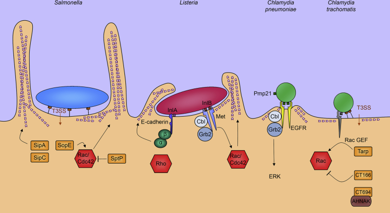
Chlamydia trachomatis is obligate intracellular bacterial pathogen that remains a significant public health burden worldwide. A critical early event during infection is chlamydial entry into non-phagocytic host epithelial cells. Like other Gram-negative bacteria, C. trachomatis uses a type III secretion system (T3SS) to deliver virulence effector proteins into host cells. These effectors trigger bacterial uptake and promote bacterial survival and replication within the host cell. In this review, we highlight recent cryo-electron tomography that has provided striking insights into the initial interactions between Chlamydia and its host. We describe the polarised structure of extracellular C. trachomatis elementary bodies (EBs), and the supramolecular organisation of T3SS complexes on the EB surface, in addition to the changes in host and pathogen architecture that accompany bacterial internalisation and EB encapsulation into early intracellular vacuoles. Finally, we consider the implications for further understanding the mechanism of C. trachomatis entry and how this might relate to those of other bacteria and viruses.

Keywords: Chlamydia; Cryo-electron tomography; Cytoskeleton; Entry; Type III secretion.


Figures

Fig. 1
Fig. 1
Polarised structure of the Chlamydia trachomatis elementary body in contact with the host cell. Three-dimensional surface representation of a Chlamydia trachomatis elementary body in contact with the host cell, generated from segmentation of a cryo-electron tomogram. Cellular plasma membrane (orange), bacterial outer membrane (green), inner membrane (cyan), inner membrane invagination (blue), T3SS (red), ribosomes (purple) and DNA nucleoid (yellow) are shown.
Fig. 2
Fig. 2
Diverse interactions between EBs and host cells. Confocal micrographs of cultured RPE1 cells expressing LifeAct to visualise actin filaments (red) 30 min after infection with C. trachomatis LGV2 labelled with AlexaFluor 488 (green). Left panel shows membrane ruffles, right panel shows filopodial capture. Scale bars, 1 μm.
Fig. 3
Fig. 3
Comparison of chlamydial entry pathways and the archetypal ‘trigger’ and ‘zipper’ mechanisms of bacterial entry. Schematic summarising the mechanisms of cell entry by Salmonella typhimurium, Listeria monocytogenes, Chlamydia pneumoniae and Chlamydia trachomatis. Salmonella (blue) is the archetypal example of the ‘trigger’ mechanism. Salmonellae deliver T3SS effectors, Salmonella invasion proteins (Sips) and Salmonella outer proteins (Sops) (orange), which cooperate to induce actin reorganisation by directly binding and manipulating actin or via reversible stimulation of Rho-family GTPases Cdc42 and Rac1 (red). Listeria monocytogenes (red) is the archetypal example of the ‘zipper’ mechanism. Listeria uses surface internalins (InlA, InlB; brown) to bind cognate receptors (E-cadherin, Met; dark blue) to stimulate actin reorganisation via signalling through adaptor proteins (catenins, green; Grb2, Cbl, violet) and Rho-family GTPases (red). Chlamydia pneumoniae elementary bodies (green) utilise the species-specific Pmp21 surface protein to engage and stimulate signalling via epidermal growth factor receptor (EGFR, yellow) to promote bacterial entry. Chlamydia trachomatis elementary bodies (green) engage multiple receptors and deliver T3SS effectors including Tarp and CT166 (orange) to reversibly stimulate the Rho-family GTPase Rac1 (red) and trigger bacterial internalisation. CT694 engages host AHNAK to promote cytoskeletal reorganisation.

References

    1. Latest World Health Organisation figures.
    1. Hu V.H., Holland M.J., Burton M.J. Trachoma: protective and pathogenic ocular immune responses to Chlamydia trachomatis. PLoS Negl Trop Dis. 2013;7:e2020. - PMC - PubMed
    1. Cocchiaro J.L., Valdivida R.H. New insights into Chlamydia intracellular survival mechanisms. Cell Microbiol. 2009;11:1571–1578. - PMC - PubMed
    1. Huang Z., Chen M., Li K., Dong X., Han J., Zhang Q. Cryo-electron tomography of Chlamydia trachomatis gives a clue to the mechanism of outer membrane changes. J Electron Microsc (Tokyo) 2010;59:237–241. - PubMed
    1. Hackstadt T., Todd W.J., Caldwell H.D. Disulphide-mediated interactions of the chlamydial outer membrane protein: role in the differentiation of Chlamydiae. J Bacteriol. 1985;161:25–31. - PMC - PubMed

MeSH terms

Substances