. 2015 Sep 22;6(28):25402-17.
doi: 10.18632/oncotarget.4517.

KLF8 promotes tumorigenesis, invasion and metastasis of colorectal cancer cells by transcriptional activation of FHL2

Affiliations

KLF8 promotes tumorigenesis, invasion and metastasis of colorectal cancer cells by transcriptional activation of FHL2

Qingqing Yan et al. Oncotarget. .

Abstract

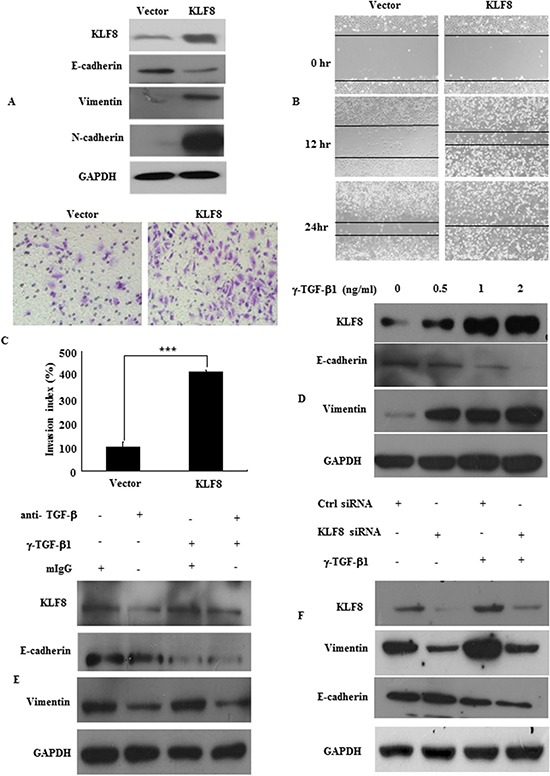
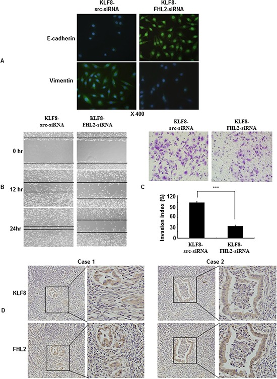
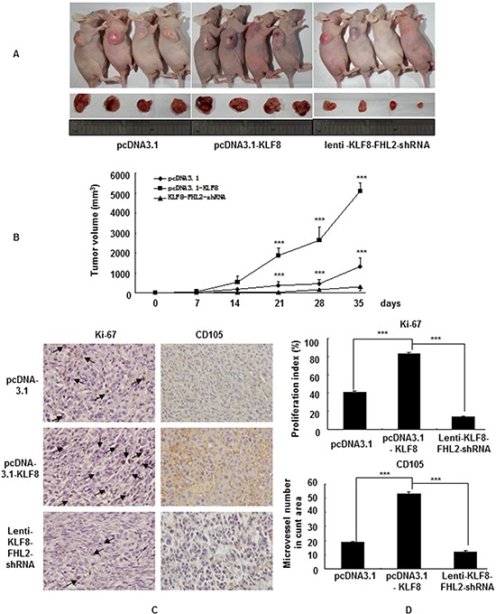
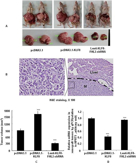
The transcription factor Krüppel-like factor (KLF)8 plays an important role in the formation of several human tumors, including colorectal cancer. We recently identified four-and-a-half LIM protein 2 (FHL2) as a critical inducer of the epithelial-to-mesenchymal transition (EMT) and invasion. However, the molecular mechanism by which KLF8 affects FHL2-mediated tumor proliferation, EMT and metastasis remains unknown. Here, we showed that KLF8 overexpression promoted EMT and metastatic phenotypes. KLF8 expression was stimulated by transforming growth factor (TGF)-β1. Moreover, KLF8 acted as a potential EMT inducer by stimulating vimentin expression and inducing a loss of E-cadherin in stable KLF8-transfected cells. KLF8 overexpression induced a strong increase in FHL2 expression, and a positive correlation between the expression patterns of KLF8 and FHL2 was observed in CRC cells. Promoter reporter and chromatin immunoprecipitation (ChIP) assays demonstrated that KLF8 directly bound to and activated the human FHL2 gene promoter. However, siRNA-mediated repression of FHL2 in KLF8-overexpressing cells reversed the EMT and the proliferative and metastatic phenotypes. In vivo, KLF8 promoted FHL2-mediated proliferation and metastasis via orthotopic implantation. Taken together, this work identified KLF8-induced FHL2 activation as a novel and critical signaling mechanism underlying human breast/colorectal cancer invasion and metastasis.

Keywords: EMT; FHL2; KLF8; colorectal cancer; metastasis.


Conflict of interest statement

CONFLICTS OF INTEREST

The authors declare no conflict of interest.

Figures

Figure 1
Figure 1. KLF8 upregulation promotes EMT
A. KLF8, E-cadherin, vimentin, N-cadherin expression in stable transfectants in LoVo cells, detected by western blot. B. Images of the wound closure of monolayer LoVo stable transfectants. Original magnification, 10×. C. Invasive potential of LoVo stable transfectants transfected with the vector, ***, p < 0.001. D. KLF8 expression in LoVo cells treated with the indicated concentrations of recombinant γ-TGF-β1 for 48 h. The protein levels of KLF8, E-cadherin and vimentin were detected by western blot. E. LoVo cells were treated with recombinant TGF-β1 (γ-TGF-β1; 2 ng/ml) in the presence of neutralizing anti-TGF-β1 antibody (α-TGF-β, 2 μg/ml) or mouse IgG (mIgG) for 48 h. The expression of KLF8, E-cadherin and vimentin was detected by western blot. F. Stable KLF8 transfectants were seeded in 6-well plates overnight and treated with γ-TGF-β1 (2 ng/ml) for an additional 48 h. The expression of KLF8, E-cadherin and vimentin was detected by western blot. All figures are representative of four independent experiments with similar findings.
Figure 2
Figure 2. Expression profiles of KLF8 and FHL2 in colon cancer
A. KLF8 and FHL2 expression was detected in colon cancer cell lines by western blot. GAPDH was used as the internal control. B. Double staining of KLF8 and FHL2 in LoVo cells by an indirect immunofluorescence, with the nuclei counterstained by Hoechst 33258 (original magnification, 400×). C. FHL2 (a, c) and KLF8 (b, d) expression in normal or cancerous colon tissue specimen was detected by immunohistochemistry. These figures are representative of the patients. D. Average scores of the two proteins in normal and cancerous colon tissues. ***, p < 0.001 between normal and cancer tissues. E. Positive staining for KLF8 and FHL2 was quantified, and their correlation was analyzed using Spearman's correlation method. Scale bars, 20 μm in B and 100 μm in C.
Figure 3
Figure 3. KLF8 directly binds to the promoter of FHL2
A. The luciferase (Luc) reporter constructs contained the FHL2 promoter with three potential KLF8 binding sites upstream of a luciferase gene. B. Chromatin immunoprecipitation (ChIP) was performed using an anti-KLF8 antibody or control IgG. The FHL2 promoter region to which KLF8 binds was significantly enriched after immunoprecipitation by an anti-KLF8 antibody. PCR products following ChIP were run on an ethidium-stained gel. C. KLF8 activates the FHL2 promoter (FHL2p) after co-transfection in LoVo cells. Luciferase activity was measured 48 h after transfection. Luciferase activity is expressed as the ratio of the promoter reporter activity to the control vector luciferase activity. RLU, relative luciferase units. ***, p < 0.001; ****, p > 0.05 D. Primer sequences used for site-directed mutations. A 136-bp (containing GT-box 1) FHL2 promoter fragment was cloned into pGL3 basic to generate pLuc55-wt. Site-directed mutagenesis of GT-box 1 in pLuc55 was performed to generate pLuc55-mt. The locations of GT-box 1 are labeled in bold, and the mutated nucleotides were underlined. wt, wildtype and mt, mutant. Diagram showing the GT-box 1 status in the two constructs. ■, wildtype GT-box 1 and □, mutated GT-box 1. E. Transcriptional activities of reporters in the transient transfections. Dual luciferase assay were performed, and the results are expressed as fold induction of RLU. ***, p < 0.001; ****, p > 0.05.
Figure 4
Figure 4. KLF8 promotes FHL2-mediated cell proliferation and morphology in CRC
A. Stable KLF8 transfectants were transfected with FHL2 siRNA, and FHL2 expression was detected by western blot 48 h later. B. LoVo/KLF8-src siRNA and LoVo/KLF8-FHL2 siRNA cells seeded in 96-well tissue culture plates in triplicate and cultured in complete medium for 24, 48 and 72 h were evaluated. Cell proliferation was assessed using the WST-1 assay. **, p < 0.01. C. Morphology of LoVo/vector, LoVo/KLF8, LoVo/KLF8-src siRNA and LoVo/KLF8-FHL2 siRNA cells, visualized by phase-contrast microscopy. D. LoVo cells stained with rhodamine-phallotoxin for 48 h to identify F-actin filaments were visualized under fluorescent microscopy. All experiments were repeated 2 to 3 times with similar findings. E. EMT biomarkers, including E-cadherin, N-cadherin, vimentin, and FHL2, were detected by western blot 48 h after transfection. Scale bars represent 20 μm in C and D.
Figure 5
Figure 5. KLF8 is required for FHL2-mediated EMT and metastatic phenotypes
A. Immunofluorescence and microscopic visualization of E-cadherin (blue) and vimentin (green) staining in KLF8 src siRNA and KLF8-FHL2-siRNA cells. B. For the wound-healing experiments, cells were analyzed with live-cell microscopy. Original magnification, 10×. C. LoVo stable KLF8 transfectants were transfected with FHL2 siRNA 48 h later, and the invasive ability of the cells decreased. ***, p < 0.001. The experiments were repeated at least three times. D. Representative IHC images are shown for FHL2 and KLF8 expression in lymph node metastatic cancer tissues. Scale bars, 20 μm in A and 100 μm in D.
Figure 6
Figure 6. KLF8 promotes FHL2-mediated cell proliferation in CRC in vivo
A. LoVo cells (5 × 10⁷) were injected subcutaneously in the right flanks of nude mice. One week after tumor cell injection, equal amounts of vector, KLF8 or KLF8-FHL2-shRNA cells were directly injected into the tumors at three different positions. Images shown were captured on day 35 after injection. B. Tumor size was measured weekly after tumor cell inoculation in each group. ***, p < 0.001, vector vs. KLF8 and KLF8 vs. KLF8-FHL2-shRNA, respectively. C. FHL2 knockdown significantly inhibited KLF8-induced proliferation (Ki-67, ***, p < 0.001, vector vs. KLF8 and KLF8 or KLF8-FHL2-shRNA, respectively), and a considerable decrease of tumor vessel density (CD105, ***, p < 0.001, vector vs. KLF8 and KLF8 or KLF8-FHL2-shRNA) was observed by immunohistochemistry. Scale bars represent 100 μm.
Figure 7
Figure 7. KLF8 is required for FHL2-mediated EMT and liver metastasis in orthotopic tumors after intrasplenic injection
A. Necropsy images of mice with orthotopically implanted CRC, with metastatic loci marked by arrows. B. The mice were sacrificed, and metastatic cancer tissues were stained with H&E. Scale bars, 100 μm. C. Tumor volumes (mean ± standard error; ***, p < 0.001, vector vs. KLF8; KLF8 vs. KLF8-FHL2-siRNA) in mice measured on the last day of the experiment, at autopsy (n = 2). D. The expression of E-cadherin in tumors derived from LoVo cells was determined by qPCR. ***, p < 0.001, vector vs. KLF8; KLF8 vs. KLF8-FHL2-siRNA.

Similar articles

Cited by

References

    1. Jackson SP, MacDonald JJ, Lees-Miller S, Tjian R. GC box binding induces phosphorylation of Sp1 by a DNA-dependent protein kinase. Cell. 1990;63:155–65. - PubMed
    1. Chen X, Johns DC, Geiman DE, Marban E, Dang DT, Hamlin G, Sun R, Yang VW. Krüppel-like factor 4 (gut-enriched Krüppel-like factor) inhibits cell proliferation by blocking G1/S progression of the cell cycle. J Biol Chem. 2001;276:30423–8. - PMC - PubMed
    1. Guo Z, Zhang W, Xia G, Niu L, Zhang Y, Wang X, Zhang Y, Jiang B, Wang J. Sp1 upregulates the four and half lim 2 (FHL2) expression in gastrointestinal cancers through transcription regulation. Mol Carcinog. 2010;49:826–36. - PubMed
    1. Hong JS, Kim SW, Koo JS. Sp1 up-regulates cAMP-response-element-binding protein expression during retinoic acid-induced mucous differentiation of normal human bronchial epithelial cells. Biochem J. 2008;410:49–61. - PMC - PubMed
    1. Abdelrahim M, Smith R, 3rd, Burghardt R, Safe S. Role of Sp proteins in regulation of vascular endothelial growth factor expression andproliferation of pancreatic cancer cells. Cancer Res. 2004;64:6740–9. - PubMed

Publication types

MeSH terms