

XRCC2 as a predictive biomarker for radioresistance in locally advanced rectal cancer patients undergoing preoperative radiotherapy
- PMID: 26320178
- PMCID: PMC4741669
- DOI: 10.18632/oncotarget.4975
XRCC2 as a predictive biomarker for radioresistance in locally advanced rectal cancer patients undergoing preoperative radiotherapy
Abstract
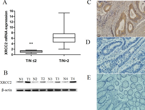
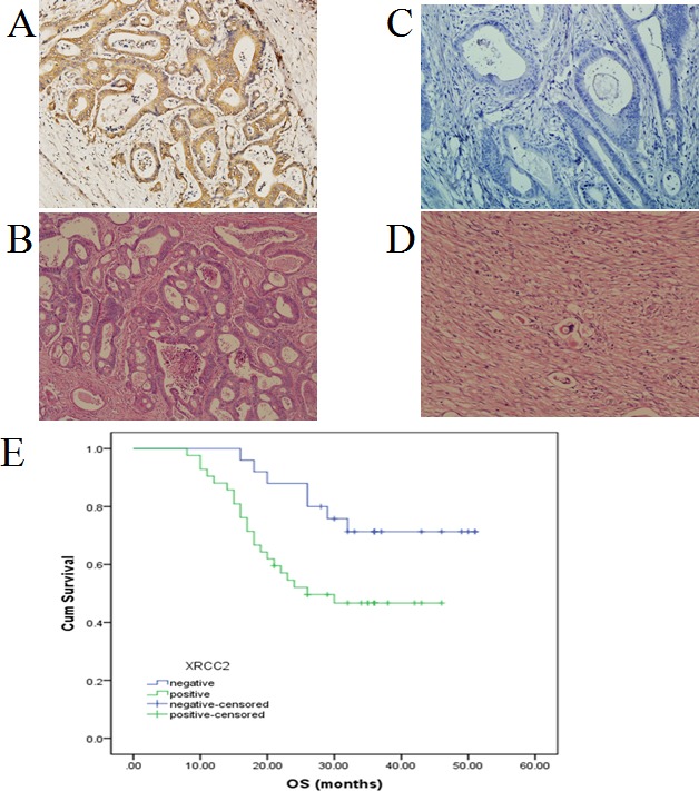
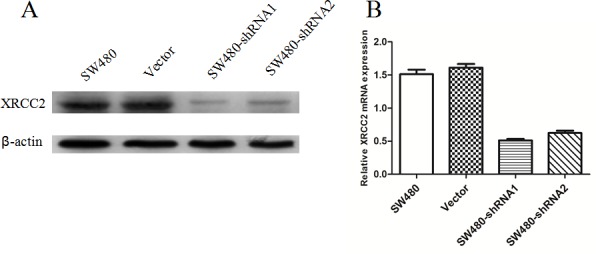
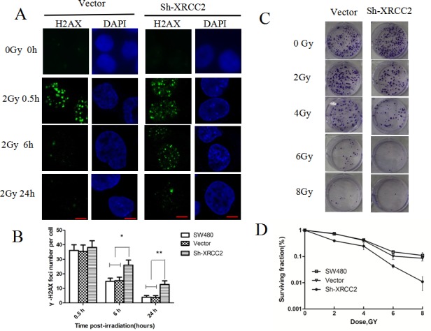
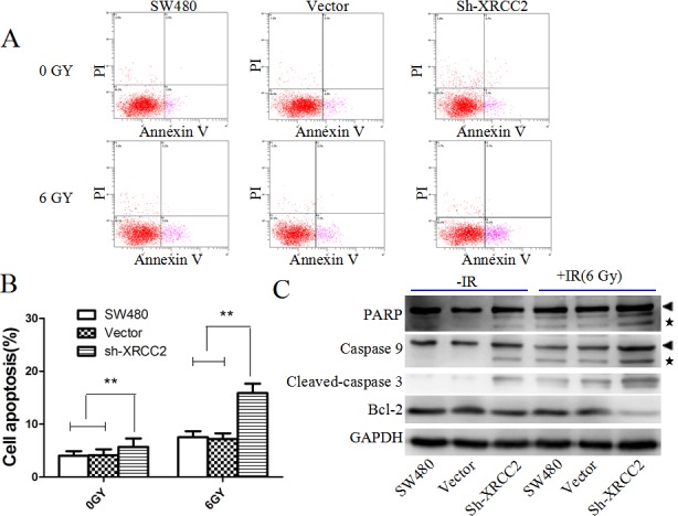
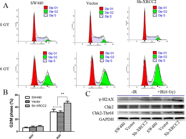
XRCC2 has been shown to increase the radioresistance of some cancers. Here, XRCC2 expression was investigated as a predictor of preoperative radiotherapy (PRT) treatment response in locally advanced rectal cancer (LARC). XRCC2 was found to be overexpressed in rectal cancer tissues resected from patients who underwent surgery without PRT. In addition, overall survival for LARC patients was improved in XRCC2-negative patients compared with XRCC2-positive patients after treatment with PRT (P < 0.001). XRCC2 expression was also associated with an increase in LARC radioresistance. Conversely, XRCC2-deficient cancer cells were more sensitive to irradiation in vitro, and a higher proportion of these cells underwent cell death induced by G2/M phase arrest and apoptosis. When XRCC2 was knocked down, the repair of DNA double-strand breaks caused by irradiation was impaired. Therefore, XRCC2 may increases LARC radioresistance by repairing DNA double-strand breaks and preventing cancer cell apoptosis. Moreover, the present data suggest that XRCC2 is a useful predictive biomarker of PRT treatment response in LARC patients. Thus, inhibition of XRCC2 expression or activity represents a potential therapeutic strategy for improving PRT response in LARC patients.
Keywords: Clinical Section; XRCC2; preoperative radiotherapy; radioresistance; rectal cancer.
Conflict of interest statement
There is no conflict of interest.
Figures
Similar articles
-
XRCC2-Deficient Cells are Highly Sensitive to 5-Fluorouracil in Colorectal Cancer.Cell Physiol Biochem. 2017;43(3):1207-1219. doi: 10.1159/000481762. Epub 2017 Oct 5. Cell Physiol Biochem. 2017. PMID: 28977800
-
Phosphatidylinositol 3-kinase CB association with preoperative radiotherapy response in rectal adenocarcinoma.World J Gastroenterol. 2014 Nov 21;20(43):16258-67. doi: 10.3748/wjg.v20.i43.16258. World J Gastroenterol. 2014. PMID: 25473181 Free PMC article.
-
PSMB8 as a Candidate Marker of Responsiveness to Preoperative Radiation Therapy in Rectal Cancer Patients.Int J Radiat Oncol Biol Phys. 2017 Aug 1;98(5):1164-1173. doi: 10.1016/j.ijrobp.2017.03.023. Epub 2017 Mar 22. Int J Radiat Oncol Biol Phys. 2017. PMID: 28721901
-
Neoadjuvant chemotherapy without radiotherapy for locally advanced rectal cancer.Future Oncol. 2014 Nov;10(14):2243-57. doi: 10.2217/fon.14.127. Future Oncol. 2014. PMID: 25471037 Review.
-
Mechanisms of microRNA action in rectal cancer radiotherapy.Chin Med J (Engl). 2022 Sep 5;135(17):2017-2025. doi: 10.1097/CM9.0000000000002139. Chin Med J (Engl). 2022. PMID: 35943251 Free PMC article. Review.
Cited by
-
Molecular differentiation between complete and incomplete responders to neoadjuvant therapy in rectal cancer.Res Sq [Preprint]. 2024 Jul 1:rs.3.rs-4456000. doi: 10.21203/rs.3.rs-4456000/v1. Res Sq. 2024. PMID: 39011117 Free PMC article. Preprint.
-
Tissue-Based Markers as a Tool to Assess Response to Neoadjuvant Radiotherapy in Rectal Cancer-Systematic Review.Int J Mol Sci. 2022 May 27;23(11):6040. doi: 10.3390/ijms23116040. Int J Mol Sci. 2022. PMID: 35682714 Free PMC article.
-
Radiotherapy resistance: identifying universal biomarkers for various human cancers.J Cancer Res Clin Oncol. 2022 May;148(5):1015-1031. doi: 10.1007/s00432-022-03923-4. Epub 2022 Feb 3. J Cancer Res Clin Oncol. 2022. PMID: 35113235 Free PMC article. Review.
-
Aberrant Expression of RAD52, Its Prognostic Impact in Rectal Cancer and Association with Poor Survival of Patients.Int J Mol Sci. 2020 Mar 4;21(5):1768. doi: 10.3390/ijms21051768. Int J Mol Sci. 2020. PMID: 32143539 Free PMC article.
-
Novel significant stage-specific differentially expressed genes in hepatocellular carcinoma.BMC Cancer. 2019 Jul 5;19(1):663. doi: 10.1186/s12885-019-5838-3. BMC Cancer. 2019. PMID: 31277598 Free PMC article.
References
-
- de Campos-Lobato LF, Stocchi L, da Luz Moreira A, Geisler D, Dietz DW, Lavery IC, Fazio VW, Kalady MF. Pathologic complete response after neoadjuvant treatment for rectal cancer decreases distant recurrence and could eradicate local recurrence. Ann Surg Oncol. 2011;18:1590–1598. - PubMed
-
- Huh JW, Kim HR, Kim YJ. Clinical prediction of pathological complete response after preoperative chemoradiotherapy for rectal cancer. Dis Colon Rectum. 2013;56:698–703. - PubMed
-
- Baker B, Salameh H, Al-Salman M, Daoud F. How does preoperative radiotherapy affect the rate of sphincter-sparing surgery in rectal cancer? Surg Oncol. 2012;21:e103–109. - PubMed
-
- Minsky BD. Progress in the treatment of locally advanced clinically resectable rectal cancer. Clin Colorectal Cancer. 2011;10:227–237. - PubMed
Publication types
MeSH terms
Substances
LinkOut - more resources
Full Text Sources
Other Literature Sources
