. 2015 Oct 6;6(30):29111-28.
doi: 10.18632/oncotarget.5059.

The importance of microenvironment: the role of CCL8 in metastasis formation of melanoma

Affiliations

The importance of microenvironment: the role of CCL8 in metastasis formation of melanoma

Tamás Barbai et al. Oncotarget. .

Abstract

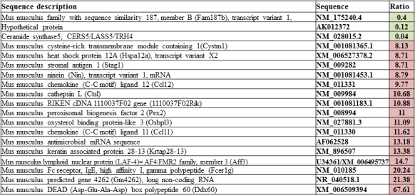
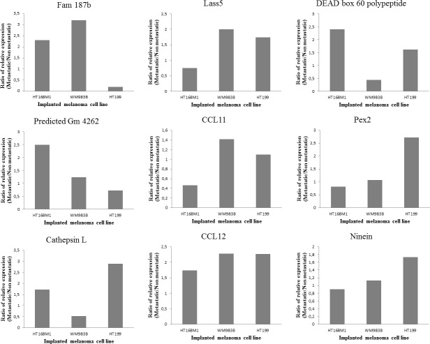
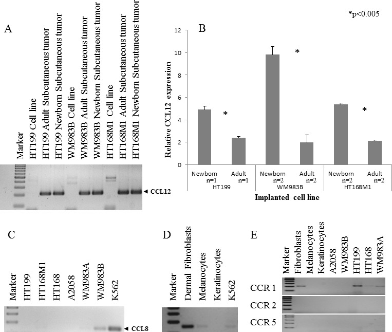
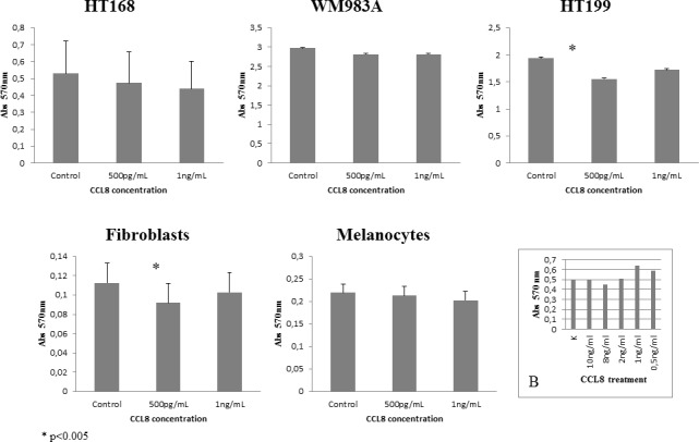
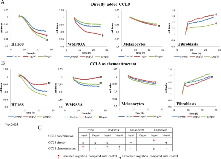
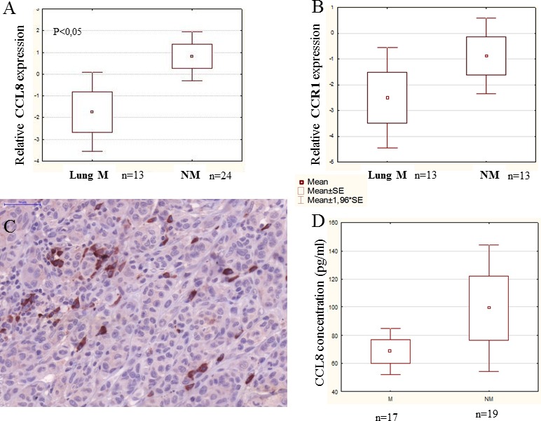
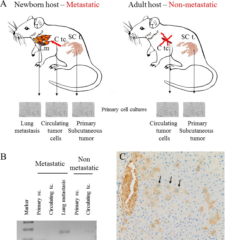
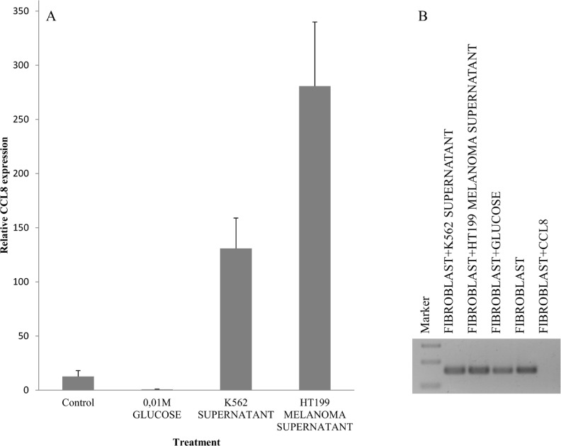
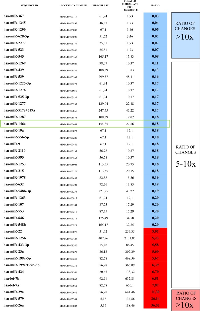
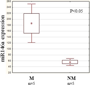
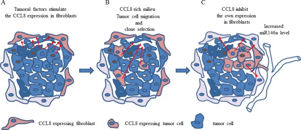
We have attempted to characterize the changes occurring on the host side during the progression of human melanoma. To investigate the role of tumor microenvironment, we set up such an animal model, which was able to isolate the host related factors playing central role in metastasis formation. One of these 'factors', CCL12, was consequently selected and its behavior was examined alongside its human homologue (CCL8). In our animal model, metastasis forming primary melanoma in the host exhibited increased level of CCL12 mRNA expression. In clinical samples, when examining the tumor and the host together, the cumulative (tumor and host) CCL8 expression was lower in the group in which human primary melanoma formed lung metastasis compared to non-metastatic primary tumors. We could not detect significant difference in CCL8 receptor (CCR1) expression between the two groups. Increased migration of the examined tumor cell lines was observed when CCL8 was applied as a chemoattractant. The tumor cells and their interactions can be influenced the expression of CCL8 by dermal fibroblasts, as a significant change in the metastatic microenvironment. Furthermore, we examined changes in miRNA profile resulted by CCL8 and miR146a appears to be a promising prognostic marker for following this process.

Keywords: CCL8; melanoma metastasis; miR146a; microenvironment.


Conflict of interest statement

COMPETING INTERESTS

There is no competing interest affecting the authors.

Figures

Figure 1
Figure 1. Gene sets from microarray experiment
Single cell suspension of HT168M1 human melanoma cell line was semiorthotopically implanted into newborn SCID mice. The RNA expression pattern of this, metastatic version of the primary tumor was investigated via Agilent Mouse Oligo Microarray Platform. The stromal expression changes were compared to healthy, age matched controls and the nineteen genes showing the largest expressional changes were selected for further validation.
Figure 2
Figure 2. Validation of microarray data
Three different human melanoma cell lines (HT199, HT168M1, WM983B) were semiorthotopically implanted at the same time into newborn (metastatic version) and adult (non-metastatic version) SCID mice. Lung metastases were detected only in the newborn host. Nine out of the previously selected 19 genes were suitable for host specific PCR. Their relative expression was measured in xenotransplanted metastatic and non-metastatic animal models. In every case, changes in expression of the target gene in the host of the metastatic and non-metastatic version of the primary tumor were compared with the expression level of the beta microglobulin housekeeping gene. The results were displayed as the ratio of expression levels in the two hosts, with the changes considered significant if the ratio was higher than 1.5-fold.
Figure 3
Figure 3. Qualitative and quantitative expression of CCL12 and CCL8
A. The qualitative CCL12 pattern showed that the host specific PCR was appropriate to discriminate between the implanted human melanoma cell line and the experimental tumor samples. B. Three human melanoma cell lines (HT199, WM983B, HT168M1) were implanted into newborn and adult SCID mice. A more than 1.5 fold change of CCL12 relative expression was detected between the primary subcutaneous tumor from the newborn (metastatic model) and adult (non-metastatic model) animals at RNA level. Data presented are mean values ± SD. C. CCL8 expression was investigated in six different human melanoma cell lines and was detected in only one of them (WM983B). D. The expression of CCL8 (human homologue of CCL12) was demonstrated in non-tumoral (dermal fibroblast, melanocyte) cells. The positive control was the ubiquitously CCL8 expressing K562 leukemia cell line. E. The expression of CCL8 receptors (CCR1, CCR2 and CCR5) was analyzed in normal cells (dermal fibroblasts, melanocytes, keratinocytes) and tumor cell lines (HT199, A2058, WM983A, WM983B, HT168). CCR1 expression was only detected in fibroblasts and three different melanoma cell lines (HT199, HT168, WM983A).
Figure 4
Figure 4. Effect of CCL8 on cell viability
The effect of CCL8 on cell viability was detected in the presence of two different concentrations (500 pg/ml and 1 ng/ml) of human recombinant CCL8 and was compared to untreated control, after 12 hours of treatment. The viability changes were measured by MTT test, where absorbance was detected on 570 nm proportionate to the number of living cells. Every tumor cell line and fibroblasts carried the CCL8 receptor, CCR1. Significant differences were observed in HT199 (tumor cell line) and dermal fibroblasts (host cell) at the concentration of 500 pg/ml, where viability was reduced in both cases. Data presented are mean values ± SD of absorbance obtained from 18 parallel samples in one representative experiment. *p < 0.005. B. Concentration range of CCL8 treatment on HT168 cell line.
Figure 5
Figure 5. Effect of CCL8 on migration
A. Cells were treated with recombinant human CCL8, added directly to the cells in two different concentrations. Graphs represent the migration activity of cells 5 hours after treatment related to18 hours of data collection. In case of melanocytes the control line and the line of 10 ng/ul runs together. B. CCL8 was applied as a chemoattractant in two different concentrations, where cells migrated towards the chemokine source through a transwell membrane. Graphs represent the migration activity of cells 5 hours after treatment related to 18 hours of data collection. Data presented are cell index. *p < 0.005. C. Summarized results represent that directly added CCL8 inhibited tumor cell migration while as a chemoattractant it increased cell motility. The migration of non-tumor cells was either not altered, or was inhibited by CCL8 as a chemoattractant (fibroblasts in low concentration).
Figure 6
Figure 6. CCL8 expression in human primary melanoma samples
Human melanoma samples were divided into two groups: non-metastatic primary tumor (NM - no metastases detected during the five year follow-up) and metastatic primary tumor (M - the patients developed metastases within five years). Patients of the metastatic group had distant organ metastases in the lung, liver, brain, bones, skin and lymph nodes. A. Relative expression of CCL8 in clinical samples was measured by real time PCR and significant differences were detected between non-metastatic and lung metastatic primary human melanomas, the latter expressing lower quantities of CCL8. B. There was no difference in CCR1 expression level between the two groups (lung metastatic vs. non-metastatic). C. Heterogeneous expression of CCL8 protein was detected in the human melanoma primary tumor samples by immunohistochemistry (magnification: 20x). D. Systemic appearance of CCL8 in serum of melanoma patients was analyzed by ELISA kit. Thirty-six samples were analyzed and CCL8 protein was detected. Differences found between metastatic and non-metastatic samples were not stasistically significant, because deviation of the datas.
Figure 7
Figure 7. CCL8 expression of tumor cells from different stages of the metastatic cascade
A. Animal model of human melanoma progression. Primary tumors from adult and newborn hosts (SC t.: subcutaneously implanted tumor) were removed along with lung metastases (Lm), which were formed only in newborn mice. Primary cell cultures were created from all of the above tumors and from the circulating tumor cells (C tc.) of newborn and adult mice. B. CCL8 expression was qualitatively detected after mRNA isolation from the metastases of the metastatic system (newborn) and from the circulating tumor cells of the non-metastatic system (adult). C. CCL8 expressing tumor cell clones were visualized at protein level in the metastatic (newborn) primary xenograft tumor by immunohistochemistry (magnification: 20x).
Figure 8
Figure 8. Simulation of host-tumor cooperation
Human dermal fibroblast cells were treated with HT199 melanoma and K562 leukemia cell-free supernatants (to simulate the effect of secreted tumoral factors) and pure, recombinant human CCL8. We used untreated cells and samples treated with 0.01M glucose solution as controls. CCL8 relative expression was measured by real time PCR. A. Increase in CCL8 expression was observed after melanoma and leukemia supernatant treatment. B. In contrast, pure CCL8 treatment terminated the continuous expression of CCL8.
Figure 9
Figure 9. CCL8 specific miRNA pattern
Human dermal fibroblasts were treated for 12 hours with human recombinant CCL8. MicroRNA quantitative profile was analyzed by nCounter miRNA Expression Assay Kit from NanoString. The changes were compared to miRNA levels measured in untreated control fibroblasts. MicroRNAs which showed fivefold or higher difference between the two groups were accepted, thus we created a CCL8 specific microRNA pattern. miR146a, which was localized in the middle range of our list, has already been described to participate in the regulation of CCL8, according to the literature [37].
Figure 10
Figure 10. miR146a expression in human primary melanoma
We investigated the quantitative expression of miR146a in randomly selected 5 metastatic and 5 non-metastatic primary melanomas. A significantly higher miR146a expression level was measured in the metastatic group (independent of the localization of the metastasis) than in the non-metastatic group. Data presented are mean values ± SE.
Figure 11
Figure 11. The hypothetical effect of CCL8 in host-tumor system
A. Soluble factors produced by tumor cells stimulate the CCL8 expression of dermal fibroblasts in the stroma. B. The increasing concentration of CCL8 and its chemoattractant ability results in an increased migration of tumor cells to the stroma. It also takes part in the selection of potentially CCL8 expressing tumor cells harboring metastatic potential. C. In this case the increasing CCL8 concentration can regulate its own expression in dermal fibroblasts. The change in miR146a expression (overexpressed in dermal fibroblasts in this case) serves as an appropriate marker for this process.

Similar articles

Cited by

References

    1. Slominski AT, Carlson JA. Melanoma resistance: a bright future for academicians and a challenge for patient advocates. Mayo Clin Proc. 2014;89:429–433. 2014. - PMC - PubMed
    1. Psaila B, Lyden D. The metastatic niche: adapting the foreign soil. Nat Rev Cancer. 2009;9:285–93. - PMC - PubMed
    1. Folkman J. Role of angiogenesis in tumor growth and metastasis. Semin Oncol. 2002;29:15–8. - PubMed
    1. Taipale J, Keski-Oja J. Growth factors in the extracellular matrix. FASEB J. 1997;11:51–59. - PubMed
    1. Weigelt B, Bissell MJ. Unraveling the microenvironmental influences on the normal mammary gland and breast cancer. Semin Cancer Biol. 2008;18:311–21. - PMC - PubMed

Publication types

MeSH terms