. 2015 Oct 1;195(7):3119-28.
doi: 10.4049/jimmunol.1402670. Epub 2015 Aug 28.

Negative Regulation of Memory Phenotype CD8 T Cell Conversion by Adhesion and Degranulation-Promoting Adapter Protein

Affiliations

Negative Regulation of Memory Phenotype CD8 T Cell Conversion by Adhesion and Degranulation-Promoting Adapter Protein

Jessica K Fiege et al. J Immunol. .

Abstract

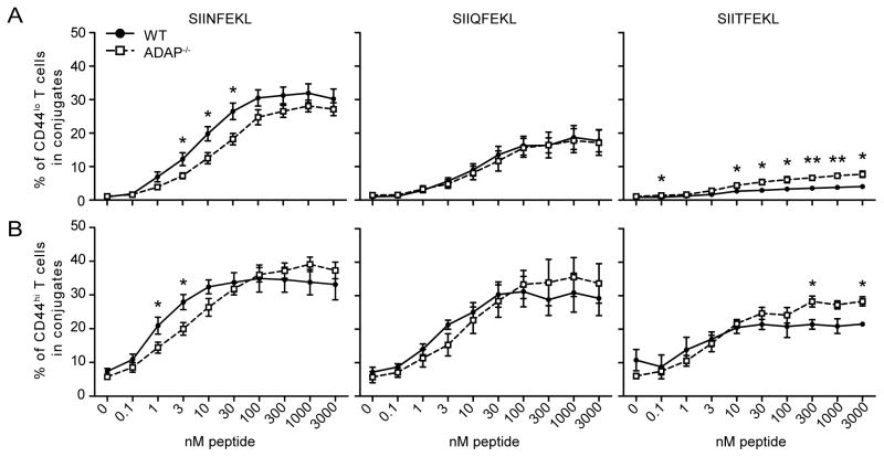
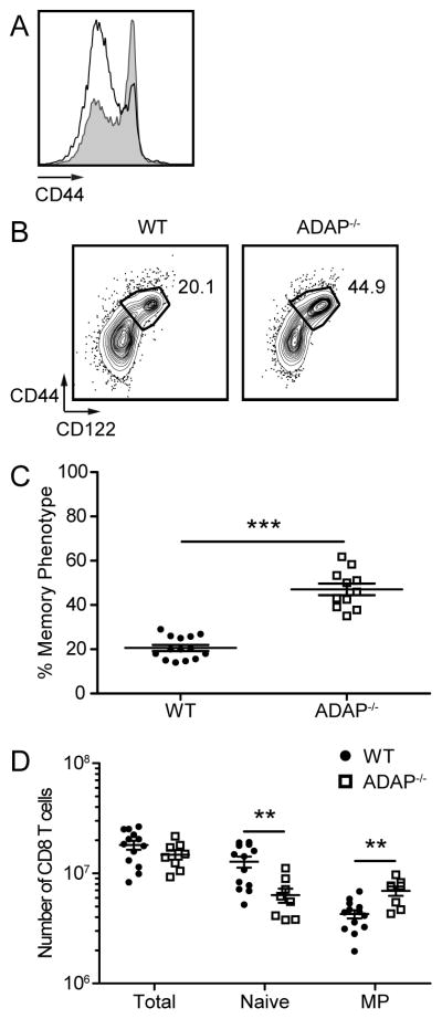
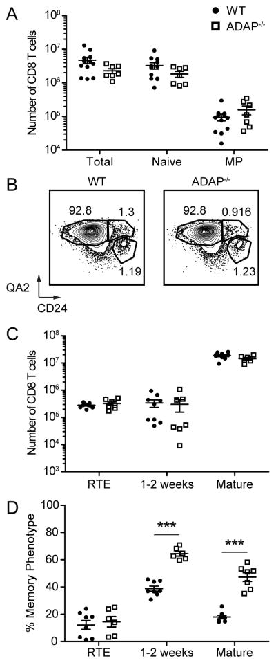
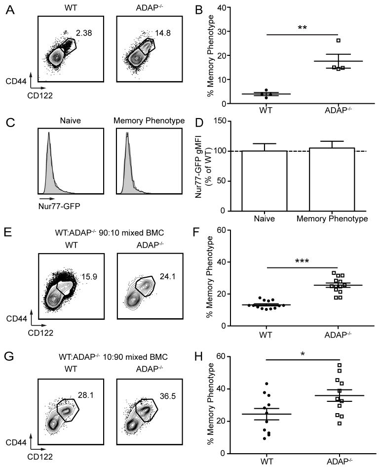
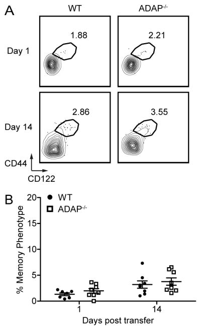
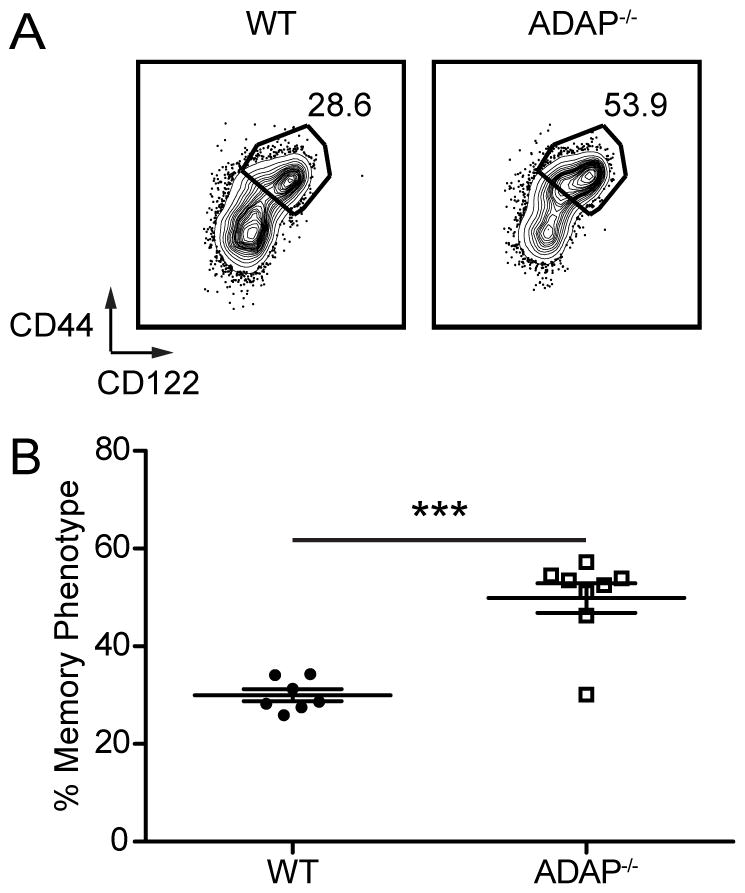
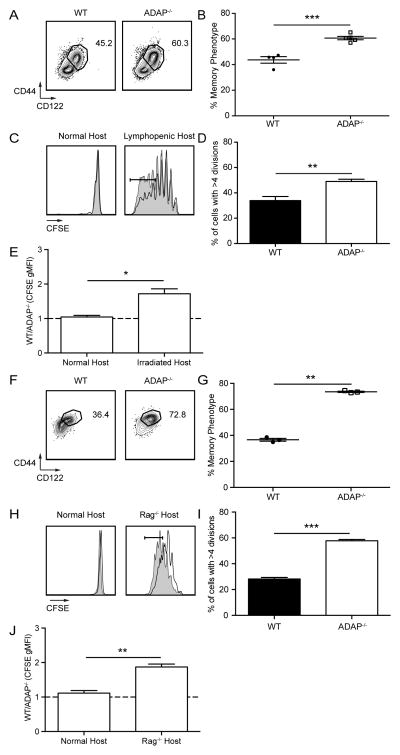
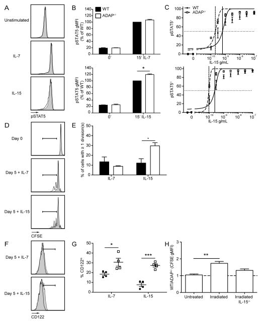
The maintenance of T cell repertoire diversity involves the entry of newly developed T cells, as well as the maintenance of memory T cells generated from previous infections. This balance depends on competition for a limited amount of homeostatic cytokines and interaction with self-peptide MHC class I. In the absence of prior infection, memory-like or memory phenotype (MP) CD8 T cells can arise from homeostatic cytokine exposure during neonatal lymphopenia. Aside from downstream cytokine signaling, little is known about the regulation of the conversion of naive CD8 T cells to MP CD8 T cells during acute lymphopenia. We have identified a novel negative regulatory role for adhesion and degranulation-promoting adapter protein (ADAP) in CD8 T cell function. We show that in the absence of ADAP, naive CD8 T cells exhibit a diminished response to stimulatory Ag, but an enhanced response to weak agonist-altered peptide ligands. ADAP-deficient mice exhibit more MP CD8 T cells that occur following thymic emigration and are largely T cell intrinsic. Naive ADAP-deficient CD8 T cells are hyperresponsive to lymphopenia in vivo and exhibit enhanced activation of STAT5 and homeostatic Ag-independent proliferation in response to IL-15. Our results indicate that ADAP dampens naive CD8 T cell responses to lymphopenia and IL-15, and they demonstrate a novel Ag-independent function for ADAP in the suppression of MP CD8 T cell generation.


Figures

Figure 1
Figure 1. ADAP modulates T-APC interactions across multiple TCR affinities
Conjugate assays between WT or ADAP−/− OT-I T cells and B6 splenocytes were performed as described in Materials and Methods. T:APC conjugate formation of naïve CD44lo OT-I T cells (A) and MP CD44hi OT-I T cells (B) with B6 splenocytes pulsed with the indicated concentrations of N4 (left), Q4 (center) or T4 (right) peptides. Results are averages from duplicate or quadruplicate wells from independent experiments (± SEM): seven experiments (N4), four experiments (Q4), and three experiments (T4).
Figure 2
Figure 2. Increased memory phenotype CD8 T cells in the absence of ADAP
Cells from spleens were obtained from WT and ADAP−/− adult mice (aged 8–12 weeks), and the expression of CD44 and CD122 was analyzed on CD8+ CD4 TCR-β + cells. (A) CD44 staining from WT (black) and ADAP−/− (grey shaded) cells. (B) Contour plots of bulk CD8 T cells. Numbers represent the percentage of cells with the phenotype of memory (CD44hi CD122hi). (C) The percentage of CD44hi CD122hi cells in the spleens of WT (black circles) and ADAP−/− (open squares) mice. (D) Absolute number of total CD8+ CD4 TCR-β+, CD44lo CD122lo, and CD44hi CD122hi cells from the spleen. The results (C and D) are compiled from at least five independent experiments (± SEM), with at least two mice per experiment.
Figure 3
Figure 3. Memory phenotype in the absence of ADAP occurs after CD8 T cells exit the thymus
Cells from thymus (A) or spleen (B–D) were obtained from WT and ADAP−/− unimmunized 8–12 week old adult mice and the expression of surface molecules was analyzed on CD8+ CD4 TCR-β+ cells. (A) Absolute number of total CD8SP TCR-βhi, CD44lo CD122lo, and CD44hi CD122hi cells from the thymus. (B) CD24 and QA2 staining from WT and ADAP−/− spleens. Numbers represent the percentage of RTE, 1–2 week post egress and mature CD8 T cells. Recent thymic emigrants (RTEs) are defined as CD24hi Qa2lo. Cells that have emigrated between 1–2 weeks prior are defined as CD24hi Qa2hi. Mature cells are defined as CD24lo Qa2hi. (C) Absolute number of RTE, 1–2 week post egress and mature CD8 T cells. (D) The percentage of MP from RTE, 1–2 week post egress and mature CD8 T cell populations. The results (A) are compiled from at least five independent experiments (± SEM) with at least two mice per experiment, and (C and D) are compiled from at least three independent experiments with at least three mice per experiment.
Figure 4
Figure 4. Memory phenotype in the absence of ADAP is not due to CD8 T cell self-reactivity, and is specific to ADAP-deficient CD8 T cells
(A) CD44 and CD122 staining of spleens from WT and ADAP−/− Rag2−/− P14-Tg mice. Numbers represent the percentage of MP cells. Analyzed cells are CD8+ CD4 Vα2+ cells. (B) Percentage of MP WT and ADAP−/− Rag2−/− P14-Tg T cells. (C) Spleens from littermate polyclonal WT and ADAP−/− mice expressing the Nur77-GFP reporter were harvested and the expression of Nur77-GFP was analyzed by flow cytometry in CD8+CD44lo/CD122lo (Naïve) and CD8+CD44hi/CD122hi (MP) cells. Representative histograms depict Nur77-GFP expression for WT (black line) overlaid with ADAP−/− (grey shaded). (D) Quantification of Nur77-GFP fluorescence of naïve and MP CD8+ T cells from 4 separate littermate pairs of WT and ADAP−/− mice analyzed in two independent experiments. Values are normalized to Nur77-GFP expression observed in WT mice. (E–H) WT (CD45.1/2) and ADAP−/− (CD45.2) bone marrow cells at the indicated ratios were transferred into lethally irradiated CD45.1 recipients. Mixed bone marrow chimeras were harvested 8–12 weeks after transfer of donor marrow. (E–F) WT: ADAP−/−, 90:10 ratio, (G–H) WT: ADAP−/−, 10:90 ratio. (E and G) CD44 and CD122 staining of CD8+ CD4 TCR-β + donor cells from recipient spleens. Numbers represent the percentage of MP cells. (F and H) Percentage of CD44hi CD122hi donor cells. The results (B) are compiled from three independent experiments (± SEM) with at least two mice per experiment, (D) is compiled from three independent experiments, each with at least one littermate pair, and (F and G) are compiled from three independent experiments, each with at least 4 mice.
Figure 5
Figure 5. ADAP-deficient mature naïve CD8 T cells do not more readily convert to memory phenotype in the steady state
Naïve CD8 T cells from pLNs were isolated from WT (CD45.2) and ADAP−/− (CD45.1/2) adult mice, mixed at a 1:1 ratio and transferred into CD45.1 recipient mice (1 × 10⁵ cells/mice). (A) CD44 and CD122 staining of transferred cells from recipient spleens. Numbers represent the percentage of MP CD8 T cells. (B) Percentage of CD44hi CD122hi donor cells. The results (B) are compiled from two independent experiments, each with four mice (± SEM).
Figure 6
Figure 6. Neonatal lymphopenic generation of memory phenotype CD8 T cells is greater in the absence of ADAP
Cells from the spleen of 3 week old WT or ADAP−/− mice were harvested and analyzed for MP. (A) CD44 and CD122 staining. Numbers represent the percentage of MP CD8 T cells. (B) Percentage of CD8+ TCR-beta+ QA2hi CD24lo MP cells. The results (B) are compiled from two independent experiments (± SEM), with at least three mice in each group.
Figure 7
Figure 7. Mature naïve CD8 T cells convert to memory phenotype in the absence of ADAP in response to lymphopenic signals
(A–E) Naïve CD8 T cells from pLNs were isolated from WT (CD45.2) and ADAP−/− (CD45.2) adult mice, labeled with CFSE, and separately transferred into normal or sub-lethally irradiated CD45.1 recipients. Recipient spleens were harvested 10 days after transfer. (A) CD44 and CD122 staining of transferred cells from lymphopenic spleens. Numbers represent the percentage of MP CD8 T cells. (B) Percentage of CD44hi CD122hi donor cells from lymphopenic recipient spleens. (C) CFSE staining from WT (black) or ADAP−/− (grey shaded) donor cells. (D) Percentage of donor cells that had undergone >4 cell divisions. (E) Ratio of WT:ADAP−/− CFSE gMFI in normal or lymphopenic recipient spleens. (F–J) Naïve OT-I CD8 T cells from pLNs were isolated from WT (CD45.1) and ADAP−/− (CD45.1/2) adult mice, labeled with CFSE, and co-transferred into normal or Rag1−/− CD45.2 recipients. Recipient spleens were harvested 7 days after transfer. (F) CD44 and CD122 staining of transferred cells from Rag1−/− spleens. Numbers represent the percentage of MP CD8 T cells. (G) Percentage of CD44hi CD122hi donor cells from Rag1−/− recipient spleens. (H) CFSE staining from WT (black) or ADAP−/− (grey shaded) donor cells. (I) Percentage of donor cells that had undergone >4 cell divisions. (J) Ratio of WT:ADAP−/− CFSE gMFI in normal or Rag1−/− recipient spleens. The results (B and D) are representative of three independent experiments (± SEM), with at least four mice of each condition. The results (G and I) are representative of three independent experiments (± SEM), with at least three mice of each condition. The results (E and J) are compiled from three independent experiments (± SEM).
Figure 8
Figure 8. Mature naïve CD8 T cells are more responsive to IL-15 in the absence of ADAP
(A–C) Bulk splenocytes were obtained from WT and ADAP−/− adult mice and stained for intracellular pSTAT5. (A) Histograms of WT (black) or ADAP−/− (grey shaded) pSTAT5 staining on CD8+ CD122lo cells after addition of 1 ng/mL of IL-7 or IL-15 for 15 minutes. (B) STAT5 phosphorylation was measured by flow cytometry and quantified as the population gMFI staining normalized to WT IL-7 or IL-15 stimulated samples from CD8+ CD122lo cells. (C) Dose response to IL-15 (0.1–300 ng/mL) for 15 minutes in CD8+ CD122lo cells (top) and CD8+ CD122hi cells (bottom) LogEC50 for CD8+ CD122lo cells was 6.3 × 10⁻¹⁰ (WT) and 2.0 × 10⁻¹⁰ (ADAP−/−), For CD8+ CD122hi cells, the LogEC50 was 2.4 × 10⁻¹⁰ (WT) and 9.4 × 10⁻¹¹ (ADAP−/−). (D–G) Naïve CD8 T cells were isolated from WT (CD45.1/2) or ADAP−/− (CD45.2) OT-I adult mice, mixed at a 1:1 ratio, and labeled with CFSE. WT and ADAP−/− cells were co-cultured in the presence of 10 ng/mL of IL-7 or IL-15 for 5 days. (D) CFSE staining of WT and ADAP−/− CD8 T cells at day 0 or 5 after addition of cytokine. Gate indicates cells that have undergone 1 or more divisions. (E) Percentage of donor cells that have undergone one or more divisions. (F) CD122 staining of WT and ADAP−/− CD8 T cells at day 5 after addition of cytokine. Gate indicates cells that are CD122hi. (G) Percentage of CD8 T cells with expression of CD122hi. (H) Naïve OT-I CD8 T cells from pLNs were isolated from WT (CD45.1) and ADAP−/− (CD45.1/2) adult mice, labeled with CFSE, and co-transferred into untreated or sub-lethally irradiated CD45.2 or IL-15−/− CD45.2 recipients. Recipient spleens were harvested 7 days after transfer. Ratio of WT:ADAP−/− CFSE gMFI in untreated or sub-lethally irradiated wild-type or IL-15−/− recipient spleens. The results (B, C, E, G and H) are compiled from three independent experiments (± SEM).

Similar articles

Cited by

References

    1. Takada K, Jameson SC. Naive T cell homeostasis: from awareness of space to a sense of place. Nat Rev Immunol. 2009;9:823–832. - PubMed
    1. Min B, McHugh R, Sempowski GD, Mackall C, Foucras G, Paul WE. Neonates support lymphopenia-induced proliferation. Immunity. 2003;18:131–140. - PubMed
    1. Akue AD, Lee JY, Jameson SC. Derivation and maintenance of virtual memory CD8 T cells. J Immunol. 2012;188:2516–2523. - PMC - PubMed
    1. Sprent J, Surh CD. Normal T cell homeostasis: the conversion of naive cells into memory-phenotype cells. Nat Immunol. 2011;12:478–484. - PMC - PubMed
    1. Surh CD, Sprent J. Homeostasis of naive and memory T cells. Immunity. 2008;29:848–862. - PubMed

Publication types

MeSH terms