. 2015 Oct 1;195(7):2985-3000.
doi: 10.4049/jimmunol.1402782. Epub 2015 Aug 28.

Intrinsic Differences in Donor CD4 T Cell IL-2 Production Influence Severity of Parent-into-F1 Murine Lupus by Skewing the Immune Response Either toward Help for B Cells and a Sustained Autoantibody Response or toward Help for CD8 T Cells and a Downregulatory Th1 Response

Affiliations

Intrinsic Differences in Donor CD4 T Cell IL-2 Production Influence Severity of Parent-into-F1 Murine Lupus by Skewing the Immune Response Either toward Help for B Cells and a Sustained Autoantibody Response or toward Help for CD8 T Cells and a Downregulatory Th1 Response

Kateryna Soloviova et al. J Immunol. .

Abstract

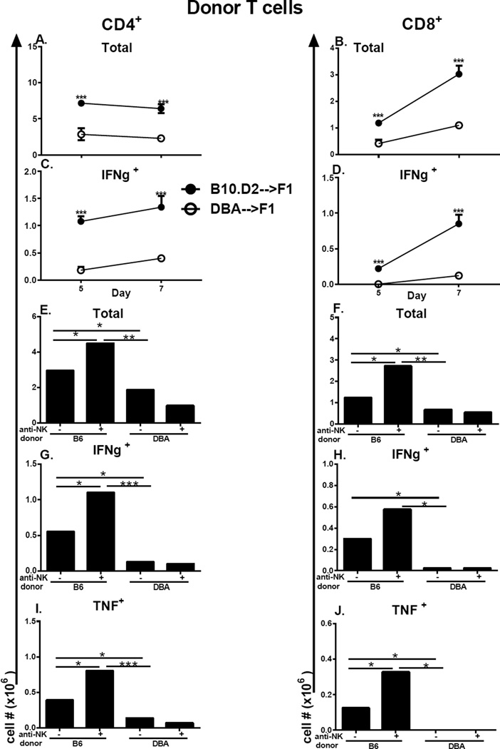
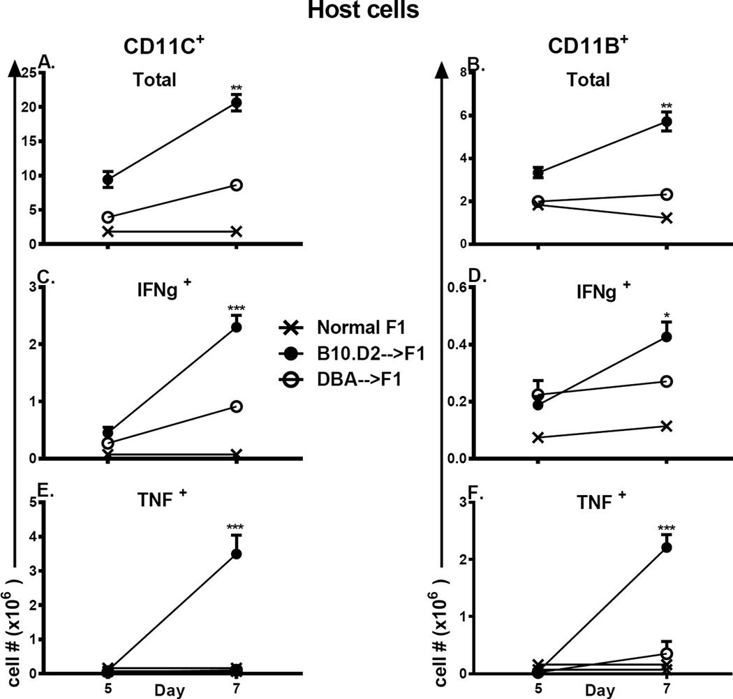
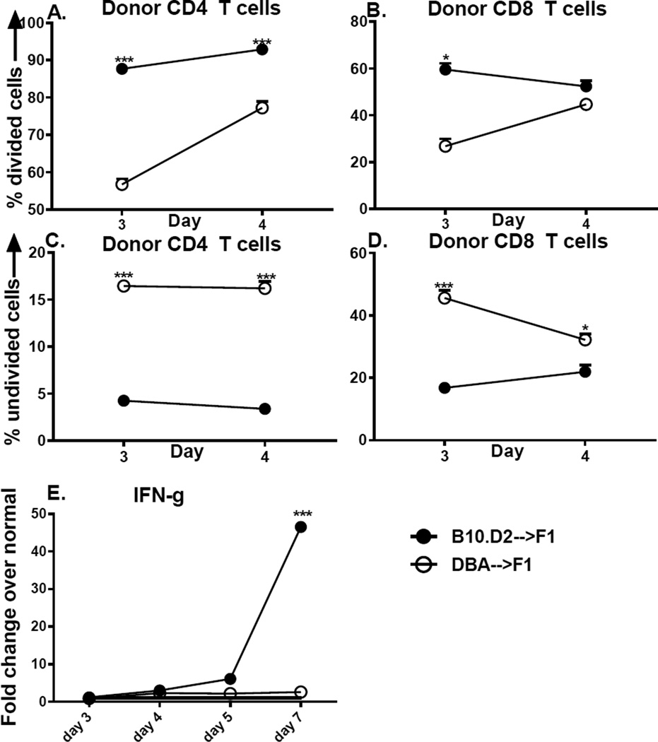
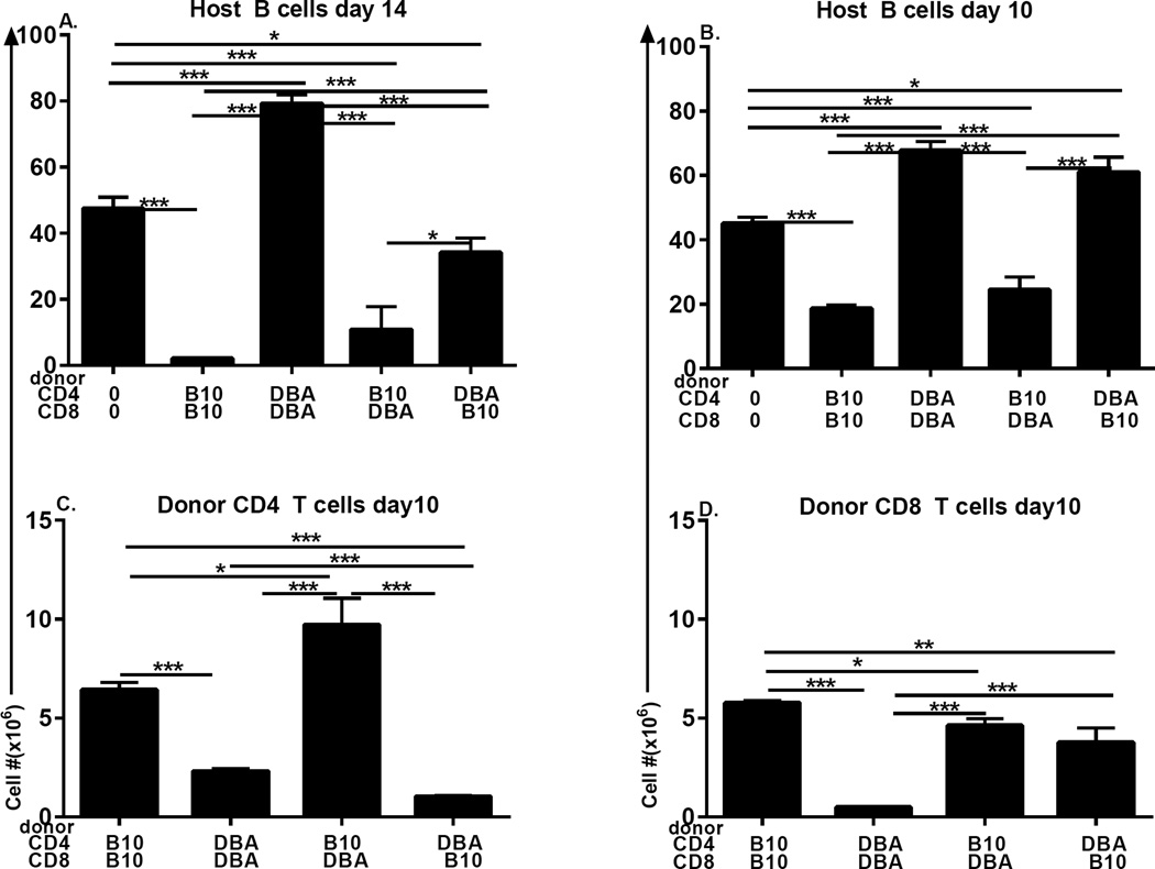
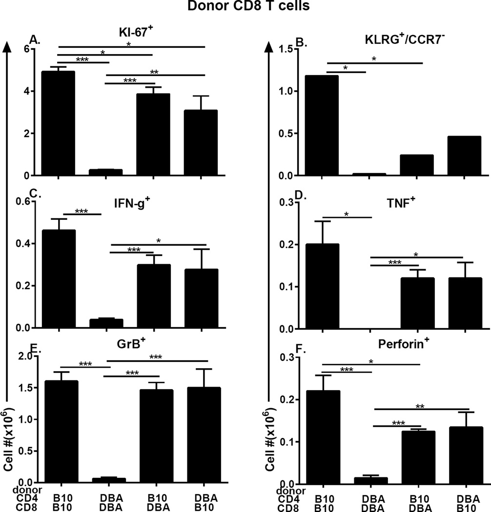
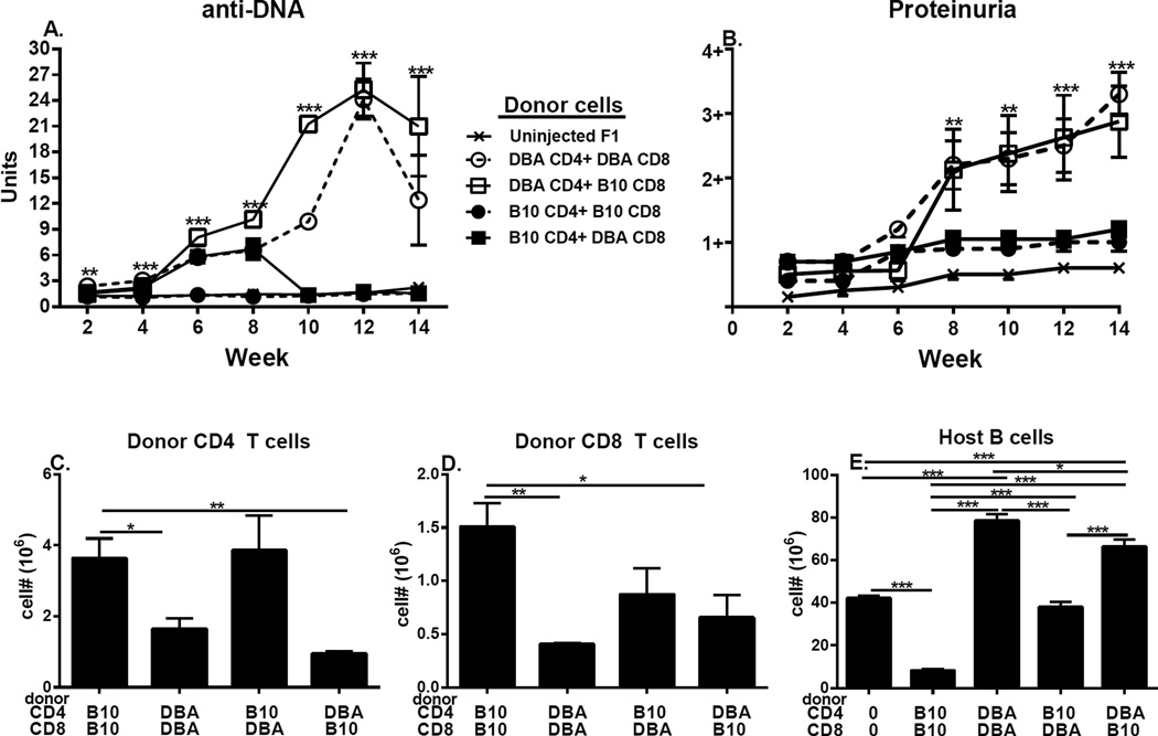
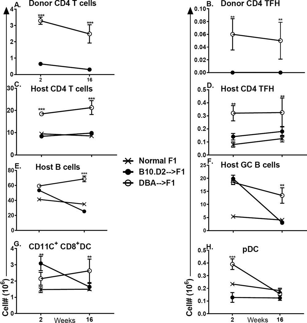
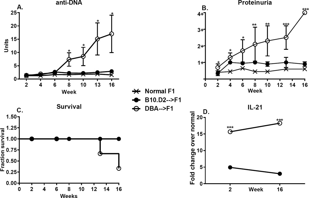
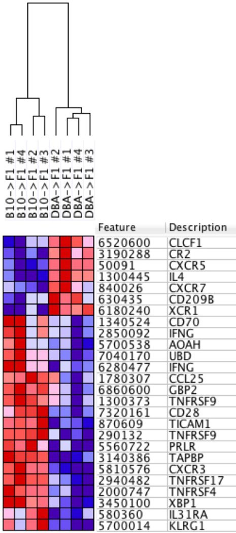
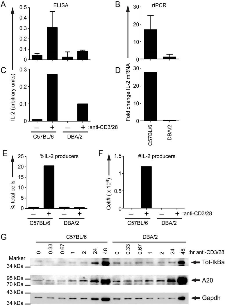
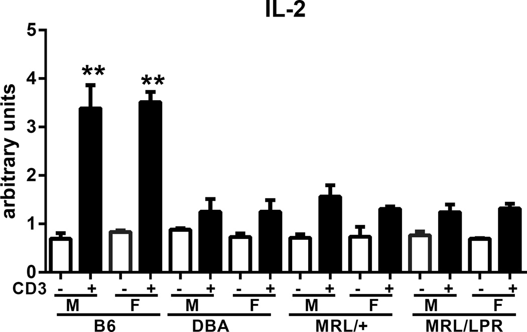
Using the parent-into-F1 model of induced lupus and (C57BL/6 × DBA2) F1 mice as hosts, we compared the inherent lupus-inducing properties of the two parental strain CD4 T cells. To control for donor CD4 recognition of alloantigen, we used H-2(d) identical DBA/2 and B10.D2 donor T cells. We demonstrate that these two normal, nonlupus-prone parental strains exhibit two different T cell activation pathways in vivo. B10.D2 CD4 T cells induce a strong Th1/CMI pathway that is characterized by IL-2/IFN-γ expression, help for CD8 CTLs, and skewing of dendritic cell (DC) subsets toward CD8a DCs, coupled with reduced CD4 T follicular helper cells and transient B cell help. In contrast, DBA/2 CD4 T cells exhibit a reciprocal, lupus-inducing pathway that is characterized by poor IL-2/IFN-γ expression, poor help for CD8 CTLs, and skewing of DC subsets toward plasmacytoid DCs, coupled with greater CD4 T follicular helper cells, prolonged B cell activation, autoantibody formation, and lupus-like renal disease. Additionally, two distinct in vivo splenic gene-expression signatures were induced. In vitro analysis of TCR signaling revealed defective DBA CD4 T cell induction of NF-κB, reduced degradation of IκBα, and increased expression of the NF-κB regulator A20. Thus, attenuated NF-κB signaling may lead to diminished IL-2 production by DBA CD4 T cells. These results indicate that intrinsic differences in donor CD4 IL-2 production and subsequent immune skewing could contribute to lupus susceptibility in humans. Therapeutic efforts to skew immune function away from excessive help for B cells and toward help for CTLs may be beneficial.


Conflict of interest statement

Disclosures

The authors have no financial conflicts of interest.

Figures

Figure 1
Figure 1. Defective in vivo Th1 cytokine expression by DBA T cells is not due to host NK function
BDF1 mice received a single injection of unfractionated splenocytes from either B10.D2 or DBA donors. On day 5 and 7 after donor cell transfer, host spleens were evaluated by flow cytometry for donor T cell numbers of CD4 T cells (A, C) and CD8 T cells (B, D) to include total numbers (A, B) and intracellular IFN-g expression (C, D) as described in Methods. In panels (E–J), host F1 mice were either untreated or were depleted of NK cells prior to receiving unfractionated B6 or DBA splenocytes as described in Methods. Mice were assessed at day 7 by flow cytometry for total numbers of donor CD4 and CD8 T cells (E, F) and for intracellular expression of IFN-g (E–F) or TNF (G–H) in donor CD4 (E, G) and CD8 (F, H) T cells as described in Methods. Values represent group mean ± SE (n= 4–5/grp). Donor cells were examined by flow cytometry prior to transfer and the inocula adjusted so that each contained 5 × 10⁶ CD8 T cells and 7.8–9.6 × 10⁶ CD4 T cells. For all figures, *p<0.05, **p<0.01, ***p<0.001.
Figure 2
Figure 2. Greater production IFN-g and TNF expression by host APC in B10.D2→F1 mice
The cohort described in Fig.1A–1D was also analyzed for host DC subsets by flow cytometry. Gating strategy is as described in Methods. Shown are: CD11c+ cells (A, C, E) or CD11b+ cells (B, D, F) to include: total numbers (A, B); IFN-g expressing (C, D); TNF expressing (E, F). Values represent group mean ±SE.
Fig. 3
Fig. 3. Initial DBA donor T cell proliferation is defective vs. B10.D2 T cells
BDF1 mice received CFSE labeled DBA or B10.D2 (unfractionated) donor splenocytes and were assessed at day 3 and day 4 in separate experiments by flow cytometry expression of KI-67 and CFSE (see Methods). Proliferating donor CD4 (A) and CD8 (B) T cells are shown as the percent of KI-67 positive, CFSE-dull cells. Non proliferating CD4 (C) and CD8 (D) cells are shown as per cent of KI-67 negative, CFSE-bright cells. Donor cells were examined by flow cytometry prior to transfer and the inocula adjusted so that each contained 5 × 10⁶ CD8 T cells and 7.8–9.6 × 10⁶ CD4 T cells. Spleens from the cohorts in Figs. 1A–D and Fig. 3 were also examined by RT-PCR for IFN-g expression as a function of time after donor cell injection (E). Values represent group mean ±SE. n= 5 mice/group.
Fig. 4
Fig. 4. Defective DBA CD8 CTL reflects both an intrinsic CD8 defect and an extrinsic CD4 helper defect
CD4 and CD8 T cells were each purified by negative isolation from DBA and B10.D2 spleens as described in Methods. BDF1 mice received either no donor cells or 7 × 10⁶ CD4 T cells and 4 × 10⁶ CD8 T cells from donor strains as designated on the x-axis. F1 mice were assessed by flow cytometry for (A) host B cells at day 14; (B) host B cells at day 10; and engraftment of donor CD4 (C) and CD8 (D) T cells at day 10. Values represent group mean ±SE. n= 3– 5 mice/group.
Fig. 5
Fig. 5. Intracellular markers of CD8 CTL effector maturation are defective in DBA CD8 T cells due to an intrinsic defect and extrinsic DBA CD4 helper defect
The cohort shown in Fig. 4B–4D was also examined by flow cytometry at day 10 for donor CD8 T cell: (A) KI-67+ proliferating cells; (B) SLEC (KLRG-1pos, CCR7neg) cells; and intracellular expression of (C) IFN-g, (D) TNF, (E) GrB, (F) perforin. Values represent group mean ±SE. n= 5 mice/group.
Fig. 6
Fig. 6. DBA CD4 T cells are lupus prone regardless of the strain of paired CD8 T cells
Experimental protocol is as described in Fig. 4. F1 mice were assessed serially for: (A) serum anti-DNA ab and (B) proteinuria. At 14 weeks, F1 spleens were assessed by flow cytometry for: (C) donor CD4 T cells, (D) donor CD8 T cells and (E) host B cells. Values represent group mean ±SE. n=3–5/group.
Fig. 7
Fig. 7. Purified DBA CD4 T cells induce greater lupus-like parameters than do purified B10.D2 CD4 T cells
F1 mice received 15 × 106 CD4 T cells negatively isolated from either DBA or B10.D2 donor spleens. In two separate experiments, mice were evaluated at either week 2 or week 16 for: (A) donor CD4 T cells (total); (B) donor CD4 Tfh cells (CXCR5+, PD-1+); (C) host CD4 T cells (total); (D) host CD4 Tfh cells; (E) host B cells (total); (F) host GC B cells (GL-7+); (G) host CD8a+, CD11c+ DC; (H) host pDC. Values represent group mean ±SE. n= 3– 5/group for week 2 and 5/group week 16.
Fig. 8
Fig. 8. Purified DBA CD4 T cells induce greater lupus-like disease than do purified B10.D2 CD4 T cells
The cohort described in Fig. 7 was serially evaluated for (A) serum anti-DNA ab, (B) proteinuria and (C) survival. Splenic IL-21 gene expression is shown in (D). Values represent group mean ±SE.
Fig. 9
Fig. 9. DBA CD4→F1 vs. B10.D2→F1 exhibit two different pathways of immune gene activation
Purified donor CD4 T cells were transferred into BDF1 mice as described for Figs. 7 & 8, and host spleens from B10.D2 CD4->F1 and DBA CD4->F1 mice (n=4/group) were profiled by whole-genome expression analysis at day 14 using Illumina BeadArray methodology (see Methods). Heat map of differentially expressed genes (red = higher, blue = lower expression) was generated by data output from GenomeStudio and analysis using GenePattern and Gene Set Enrichment Analysis.
Figure 10
Figure 10. Defective IL-2 production and intracellular IL-2 signaling in DBA CD4 T cells
B6 or DBA mice were tested for maximal IL-2 production using unfractionated splenocytes (A, B) or purified CD4 T cells (C–F) as described in Methods. At 48 hours supernatants were harvested and tested for IL-2 content by ELISA (A, C) and cells were harvested for IL-2 mRNA expression by PCR shown as fold increase over unstimulated (B, D) or intracellular IL-2 expression by flow cytometry as shown by per cent positive (E) or total number of cells (F). For all wells, the number of plated DBA CD4 T cells and B10.D2 CD4 T cells was equal. In preliminary studies, we were unable to detect IL-2 by ELISA before 48 hours. For (A, B), values represent group means ± SE, n=4 mice/group. For (C – E), spleens were pooled from 12 mice/group. (G) The same population of purified CD4 T cells used in (C–F) was stimulated for the indicated times with plate-bound anti-CD3 (145-2C11, 100 µg/mL) and anti-CD28 (37.51, 10 µg/mL). Whole cell lysates were separated by SDS-PAGE and probed with anti-IκBα (Cell Signaling Technology, 9246), anti-A20 (Santa Cruz Biotechnology, sc-166692), and anti-GAPDH (Santa Cruz Biotechnology, sc-32233).
Fig. 11
Fig. 11. Defective in vitro DBA IL-2 production is comparable to that of MRL spontaneous lupus mice
Splenocytes from 10-week-old male and female B6, DBA, MRL/+ and MRL/lpr were tested in vitro for anti-CD3 IL-2 production as described in Methods. Values represent the mean ± SE of 3 mice/group. Open bars are unstimulated and solid bars are stimulated.

Similar articles

Cited by

References

    1. Mohan C, Adams S, Stanik V, Datta SK. Nucleosome: a major immunogen for pathogenic autoantibody-inducing T cells of lupus. J Exp Med. 1993;177:1367–1381. - PMC - PubMed
    1. Linterman MA, Rigby RJ, Wong RK, Yu D, Brink R, Cannons JL, Schwartzberg PL, Cook MC, Walters GD, Vinuesa CG. Follicular helper T cells are required for systemic autoimmunity. J Exp Med. 2009;206:561–576. - PMC - PubMed
    1. Bubier JA, Sproule TJ, Foreman O, Spolski R, Shaffer DJ, Morse HC, 3rd, Leonard WJ, Roopenian DC. A critical role for IL-21 receptor signaling in the pathogenesis of systemic lupus erythematosus in BXSB-Yaa mice. Proc Natl Acad Sci U S A. 2009;106:1518–1523. - PMC - PubMed
    1. Shlomchik MJ, Aucoin AH, Pisetsky DS, Weigert MG. Structure and function of anti-DNA autoantibodies derived from a single autoimmune mouse. Proc Natl Acad Sci U S A. 1987;84:9150–9154. - PMC - PubMed
    1. Shlomchik MJ, Marshak-Rothstein A, Wolfowicz CB, Rothstein TL, Weigert MG. The role of clonal selection and somatic mutation in autoimmunity. Nature. 1987;328:805–811. - PubMed

Publication types

MeSH terms

Associated data