. 2015 Oct 6;23(10):1889-1899.
doi: 10.1016/j.str.2015.07.016. Epub 2015 Aug 27.

Crystal Structures of the Extracellular Domain from PepT1 and PepT2 Provide Novel Insights into Mammalian Peptide Transport

Affiliations

Crystal Structures of the Extracellular Domain from PepT1 and PepT2 Provide Novel Insights into Mammalian Peptide Transport

John H Beale et al. Structure. .

Abstract

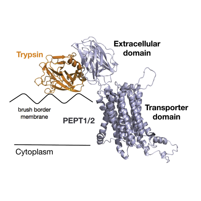
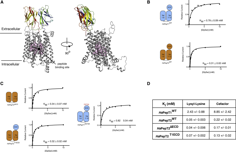
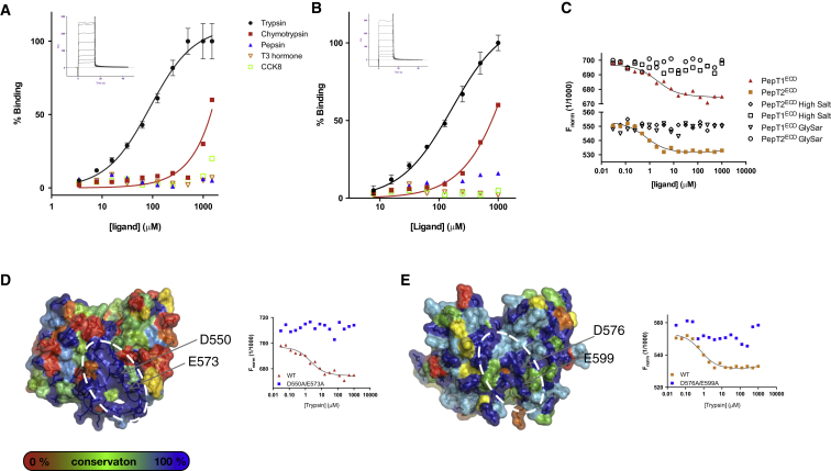
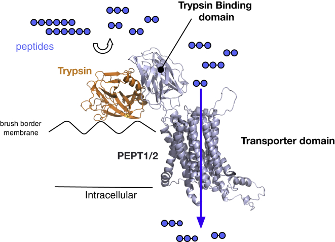
Mammals obtain nitrogen via the uptake of di- and tri-peptides in the gastrointestinal tract through the action of PepT1 and PepT2, which are members of the POT family of proton-coupled oligopeptide transporters. PepT1 and PepT2 also play an important role in drug transport in the human body. Recent crystal structures of bacterial homologs revealed a conserved peptide-binding site and mechanism of transport. However, a key structural difference exists between bacterial and mammalian homologs with only the latter containing a large extracellular domain, the function of which is currently unknown. Here, we present the crystal structure of the extracellular domain from both PepT1 and PepT2 that reveal two immunoglobulin-like folds connected in tandem, providing structural insight into mammalian peptide transport. Functional and biophysical studies demonstrate that these domains interact with the intestinal protease trypsin, suggesting a role in clustering proteolytic activity to the site of peptide transport in eukaryotic cells.


Figures

None
Graphical abstract
Figure 1
Figure 1
Topology of Mammalian Peptide Transporters Topology diagram of the human plasma membrane peptide transporter PepT1. Conserved PTR2/POT family signature motifs are indicated along with predicted N-linked glycosylation sites, three of which are in the extracellular domain. Inset: Crystal structure of the bacterial homolog PepTSt (PDB: 4D2C). The N- (light blue) and C-terminal (wheat) domains are shown as cylinders, with the bound peptide indicating the location of the central peptide-binding site conserved between mammalian and bacterial proteins.
Figure 2
Figure 2
Crystal Structure of the Extracellular Domain from PepT1 and PepT2 (A) The asymmetric unit of MmPepT1ECD containing two monomers related by a two-fold non-crystallographic symmetry axis (black oval). One monomer is rainbow colored from the N terminus to the C terminus; the second is shown in gray with the secondary structure labeled from β1 to β16. (B) Structure of the RnPepT2ECD colored from the N (blue) to the C terminus (red) and with the secondary structure components labeled as for (A). (C) The s020,W values of MmPepT1ECD and RnPepT2ECD from the AUC analysis are 2.16 and 2.22, respectively, consistent with both proteins migrating as a 20-kDa monomer in solution. Inset: the Lamm equation fit profiles for MmPepT1ECD and RnPepT2ECD.
Figure 3
Figure 3
Salt Bridges Stabilize the Interface between the Two Immunoglobulin-like Domains in PepT1ECD and PepT2ECD (A) Structure of PepT1ECD illustrating the two salt bridges, K398 and D574 and R490 and D476, that form an interaction between the lobes. (B) Comparative view in RnPepT2ECD, where a single salt bridge is observed between Asp505 and Arg518. (C) Size-exclusion chromatography traces from the MmPepT1ECD-3CX experiment. The cleaved MmPepT1ECD-3CX constructs elute at the same volume as wild-type, showing that the lobes still interact in solution even after the two lobes are separated. The cleaved MmPepT1ECD-3CX-D574A construct, however, elutes in a larger volume consistent with disruption of the interaction. (D) DAMMIN envelopes of MmPepT1ECD (dark purple) and RnPepT2ECD (light purple) calculated from the SAXS data, which show lengths of 48 and 61 Å, respectively, and illustrate the more dynamic behavior of PepT2ECD. For scale, a black and white outline of A is overlaid on the MmPepT1ECD envelope.
Figure 4
Figure 4
PepT1 and PepT2 are Modular Proteins with Functionally Distinct Domains (A) Homology model of the human PepT1 transporter generated using the crystal structure of MmPepT1ECD (colored blue to red as in Figure 1B) and the recently determined bacterial homolog PepTSo representing the transmembrane portion of the transporter (shown in gray). The peptide-binding site is highlighted (magenta). (B) Kinetic analysis of Gly-Sar uptake in human PepT1 and PepT2 using the TEVC method. (C) Kinetic analysis of Gly-Sar uptake in the PepT2ΔECD, PepT2T1ECD, and PepT1D573A constructs. (D) Ki values for the different constructs for lysyl-lysine and cefaclor are shown, indicating no effect of removing the ECD on peptide or drug uptake in PepT2.
Figure 5
Figure 5
Trypsin Interacts with a Di-acidic Motif on the Extracellular Domain of PepT1 and PepT2 (A) SPR analysis of the MmPepT1ECD interaction with trypsin. Inset: SPR sensorgram used to determine the binding constant. RU, response units. Error bars show the SEM (n = 3). (B) The binding experiment in (A) was repeated with the RnPepT2ECD protein. (C) MST binding analysis reveals no interaction with the Gly-Sar peptide and abolition of trypsin interaction in the presence of high salt. (D and E) Surface representation of (D) MmPepT1ECD and (E) RnPepT2ECD with the sequence conservation from cow, dog, chicken, human, mouse, and rat species mapped from blue to red. A highly conserved patch (indicated by the white dashed ellipse) was identified. Insets: MST binding analysis reveals an important role for D550 and E573 in MmPepT1ECD, and D576 and E599 in RnPepT2ECD, in mediating the electrostatic interaction with trypsin.
Figure 6
Figure 6
A Model for the Interaction between Trypsin and the Mammalian Peptide Transporters During protein digestion in the small intestine, trypsin transiently docks onto the conserved di-acidic motif on the trypsin-binding domain, localizing the protease to the main site of peptide import on the brush border membrane. Localization would create an increase in the local concentration of arginine- and lysine-containing peptides (shown here as blue circles), which would be expected to increase the efficiency of their uptake into the cell.

Comment in

Similar articles

Cited by

References

    1. Abrahams J.P., Leslie A.G.W. Methods used in the structure determination of bovine mitochondrial F1 ATPase. Acta Crystallogr. D Biol. Crystallogr. 1996;52:30–42. - PubMed
    1. Adams P.D., Afonine P.V., Bunkóczi G., Chen V.B., Davis I.W., Echols N., Headd J.J., Hung L.-W., Kapral G.J., Grosse-Kunstleve R.W. PHENIX: a comprehensive Python-based system for macromolecular structure solution. Acta Crystallogr. D Biol. Crystallogr. 2010;66:213–221. - PMC - PubMed
    1. Adibi S.A. The oligopeptide transporter (Pept-1) in human intestine: biology and function. Gastroenterology. 1997;113:332–340. - PubMed
    1. Adibi S.A. Renal assimilation of oligopeptides: physiological mechanisms and metabolic importance. Am. J. Physiol. 1997;272:E723–E736. - PubMed
    1. Ashida K., Katsura T., Motohashi H., Saito H., Inui K. Thyroid hormone regulates the activity and expression of the peptide transporter PEPT1 in Caco-2 cells. Am. J. Physiol. Gastrointest. Liver Physiol. 2002;282:G617–G623. - PubMed

Publication types

MeSH terms