. 2015 Sep 17;22(9):1174-84.
doi: 10.1016/j.chembiol.2015.07.017. Epub 2015 Aug 27.

Inflammatory Signaling by NOD-RIPK2 Is Inhibited by Clinically Relevant Type II Kinase Inhibitors

Affiliations

Inflammatory Signaling by NOD-RIPK2 Is Inhibited by Clinically Relevant Type II Kinase Inhibitors

Peter Canning et al. Chem Biol. .

Abstract

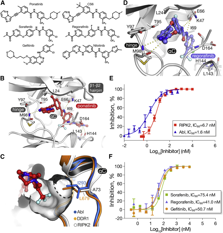
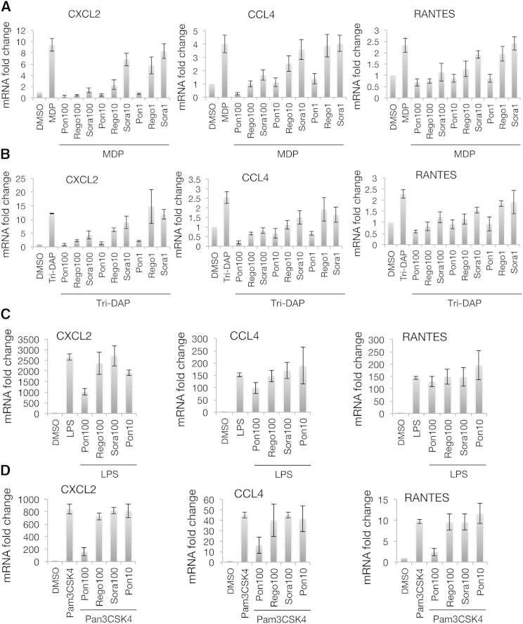
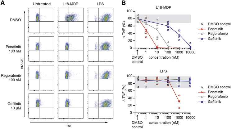
RIPK2 mediates pro-inflammatory signaling from the bacterial sensors NOD1 and NOD2, and is an emerging therapeutic target in autoimmune and inflammatory diseases. We observed that cellular RIPK2 can be potently inhibited by type II inhibitors that displace the kinase activation segment, whereas ATP-competitive type I inhibition was only poorly effective. The most potent RIPK2 inhibitors were the US Food and Drug Administration-approved drugs ponatinib and regorafenib. Their mechanism of action was independent of NOD2 interaction and involved loss of downstream kinase activation as evidenced by lack of RIPK2 autophosphorylation. Notably, these molecules also blocked RIPK2 ubiquitination and, consequently, inflammatory nuclear factor κB signaling. In monocytes, the inhibitors selectively blocked NOD-dependent tumor necrosis factor production without affecting lipopolysaccharide-dependent pathways. We also determined the first crystal structure of RIPK2 bound to ponatinib, and identified an allosteric site for inhibitor development. These results highlight the potential for type II inhibitors to treat indications of RIPK2 activation as well as inflammation-associated cancers.


Figures

None
Graphical abstract
Figure 1
Figure 1
Structural Features of the RIPK2 Kinase Domain (A) Crystal structure of the kinase domain of human RIPK2 showing the bound ponatinib molecule. See also Tables S1 and S2. (B) Superposition of the kinase domains of RIPK1 (pink, PDB: 4NEU), RIPK2 (white), and RIPK3 (blue, PDB: 4M69). The activation segment helix present in the structures of RIPK1 and RIPK3 is marked. (C) Sequence alignment of the kinase domains of human RIPK1–3. Residue numbers refer to the RIPK2 sequence, and secondary structure elements labeled in (A) are marked.
Figure 2
Figure 2
The RIPK2 Kinase Domain Is Dimeric The main panel shows the overall arrangement of the two monomers, and the inset panels show selected residues in the dimer interface. See also Figure S1.
Figure 3
Figure 3
Inhibition of Abl and RIPK2 Kinases In Vitro (A) Chemical structures of inhibitors used in this study. (B) Binding mode of ponatinib to RIPK2. (C) The DFG-out hydrophobic pocket in RIPK2 is almost uniquely large due to the small Ala73, which replaces the Ile/Leu residues of Abl/DDR2. A dashed line highlights the expanded pocket area in RIPK2, which may accommodate larger substitutions of the trifluoromethyl group for improved selectivity. (D) Predicted binding mode of regorafenib. Docking was performed with ICM-Pro (Molsoft). See also Figure S2. (E) Dose-response curves showing ponatinib inhibition of RIPK2 and Abl. (F) Dose-response curves for RIPK2 inhibition by sorefanib, regorafenib, and gefitinib. In (E) and (F), experiments were performed in duplicate; error bars indicate SD values. Kinase activity was measured using the ADPGlo assay. Non-linear curve fitting to calculate IC50 values was performed using Prism software. See also Figure S5.
Figure 4
Figure 4
Inhibition of RIPK2 Activation in HEKBlue and U2OS Cells (A) Phosphorylation changes in HEKBlue cells. Cells were treated with indicated concentrations of inhibitors, followed 30 min later by stimulation with 1 μg/ml L18-MDP. Cells were harvested after 30 min, and changes in protein phosphorylation were analyzed by western blotting. Levels of tubulin and total RIPK2 were used as loading controls. (B) Inhibition of NF-κB activation in HEKBlue cells. HEKBlue reporter cells, expressing NOD2 and NF-κB-SEAP reporter, were treated with 6–8 concentrations of each inhibitor in triplicate followed by stimulation with 1 μg/ml L18-MDP for 8 hr. SEAP activity was detected using HEKBlue media with detection of absorbance at 620 nM in a Wallac3V plate reader. Non-linear curve fitting to calculate EC50 values was performed using Prism software. Experiments were performed in triplicate, error bars indicate SD values. See also Figure S5. (C) Interaction of RIPK2 with inducibly overexpressed HA-NOD2 in U2OS cells in the presence of ponatinib. The immunoprecipitation was performed using anti-HA agarose after 24 hr of HA-NOD2 induction and ponatinib treatment. Dox, doxycycline. Representative result of the experiment performed three times. (D) NF-κB dual luciferase reporter assay in U2OS cells inducibly overexpressing HA-NOD2 and treated with ponatinib for 24 hr. Dox, doxycycline. Experiment performed three times in three technical replicates. Error bars represent ±SEM. ∗∗∗∗p < 0.0001.
Figure 5
Figure 5
Inhibition of NOD2-Dependent Ubiquitination and Signaling (A–C) THP-1 cells were pre-treated with kinase inhibitors or DMSO for 30 min and stimulated with 200 ng/ml L18-MDP (A, B) or TNF (C) as indicated. At the indicated time points, cells were lysed and ubiquitinated proteins were isolated using TUBE reagent. The isolated ubiquitinated proteins and input material were analyzed by immunoblotting. See also Figure S6.
Figure 6
Figure 6
Inhibition of NOD-Dependent Inflammatory Gene Expression in RAW264.7 Cells (A and B) Cells were pre-treated with 1, 10, or 100 nM inhibitors for 30 min and stimulated with 10 μg/ml MDP (A) or Tri-DAP (B) for 18–24 hr in triplicate. RNA samples were isolated and changes in gene expression were analyzed using gene-specific primers using SYBR qRT-PCR. All values were normalized to the levels of GAPDH. (C and D) Lack of inhibition of Toll-like receptor-dependent inflammatory gene expression in RAW264.7 cells. Experiments were performed as described above, except cells were stimulated with 10 ng/ml E. coli LPS (C) or 500 ng/ml Pam3CSK4 (D). All experiments were performed in triplicate; error bars indicate SD values.
Figure 7
Figure 7
Dose-Dependent Inhibitory Effect of Ponatinib, Regorafenib, and Gefitinib on MDP-Induced TNF in Primary Human Monocytes Intracellular TNF production was determined by flow cytometry in rested monocytes of healthy blood donors cultured in the presence or absence of L18-MDP (200 ng/ml) or LPS (200 ng/ml). Cells were pre-treated with the indicated concentrations of inhibitors for 60 min before receptor activation. (A) Representative fluorescence-activated cell sorting density blots of TNF-positive monocytes among all single, live, HLA-DR+, and CD14+ cells. (B) Induction of TNF in monocytes after L18-MDP or LPS stimulation is calculated as ΔTNF, subtracting the frequency of TNF-producing monocytes cultured in medium alone from the percentage of TNF-positive monocytes following activation. Experimental conditions are measured in 4–5 healthy donors. Individual replicates and the mean connected by a line are shown. Gray background indicates range without inhibitors. See also Figure S7.

References

    1. Ammann S., Elling R., Gyrd-Hansen M., Duckers G., Bredius R., Burns S.O., Edgar J.D., Worth A., Brandau H., Warnatz K. A new functional assay for the diagnosis of X-linked inhibitor of apoptosis (XIAP) deficiency. Clin. Exp. Immunol. 2014;176:394–400. - PMC - PubMed
    1. Andraos R., Qian Z., Bonenfant D., Rubert J., Vangrevelinghe E., Scheufler C., Marque F., Regnier C.H., De Pover A., Ryckelynck H. Modulation of activation-loop phosphorylation by JAK inhibitors is binding mode dependent. Cancer Discov. 2012;2:512–523. - PMC - PubMed
    1. Argast G.M., Fausto N., Campbell J.S. Inhibition of RIP2/RIck/CARDIAK activity by pyridinyl imidazole inhibitors of p38 MAPK. Mol. Cell. Biochem. 2005;268:129–140. - PubMed
    1. Bertrand M.J., Lippens S., Staes A., Gilbert B., Roelandt R., De Medts J., Gevaert K., Declercq W., Vandenabeele P. cIAP1/2 are direct E3 ligases conjugating diverse types of ubiquitin chains to receptor interacting proteins kinases 1 to 4 (RIP1-4) PLoS One. 2011;6:e22356. - PMC - PubMed
    1. Caruso R., Warner N., Inohara N., Nunez G. NOD1 and NOD2: signaling, host defense, and inflammatory disease. Immunity. 2014;41:898–908. - PMC - PubMed

Publication types

MeSH terms