. 2015 Sep 21;25(18):2411-6.
doi: 10.1016/j.cub.2015.07.056. Epub 2015 Aug 27.

CNS Myelin Sheath Lengths Are an Intrinsic Property of Oligodendrocytes

Affiliations

CNS Myelin Sheath Lengths Are an Intrinsic Property of Oligodendrocytes

Marie E Bechler et al. Curr Biol. .

Abstract

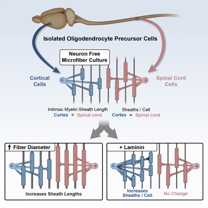
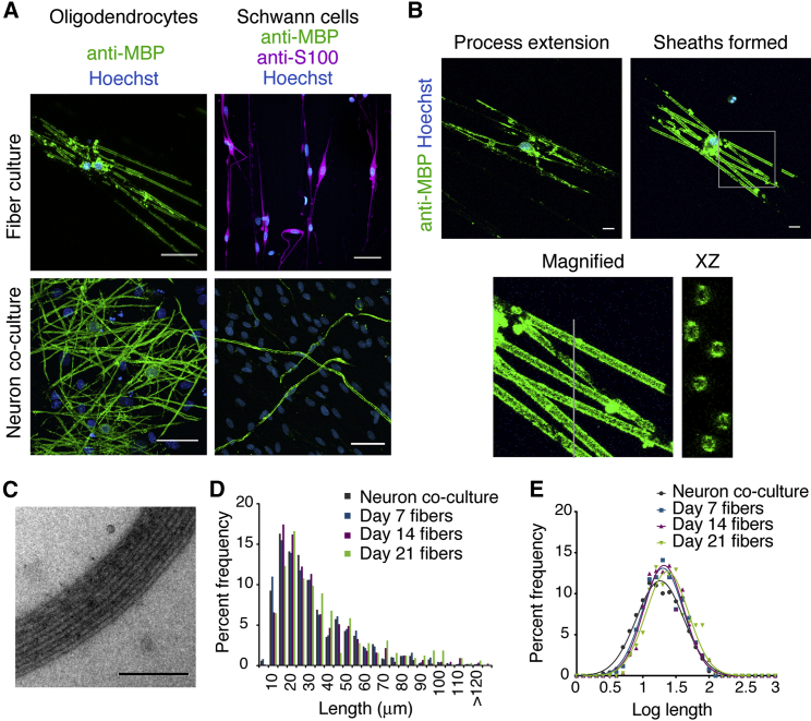
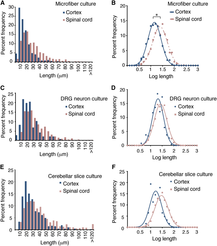
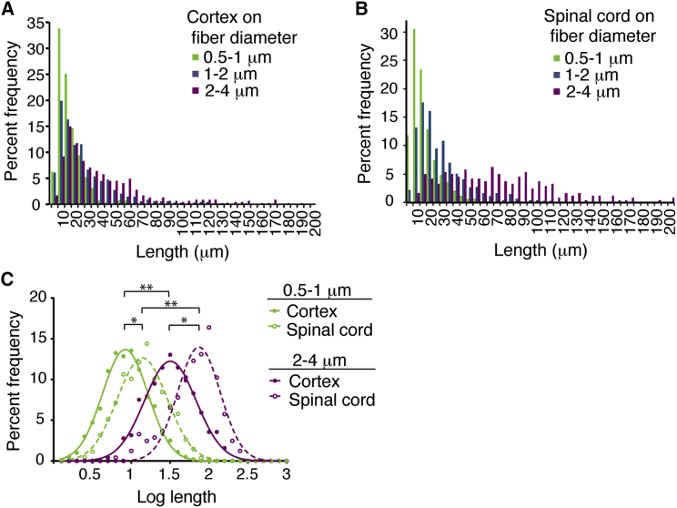
Since Río-Hortega's description of oligodendrocyte morphologies nearly a century ago, many studies have observed myelin sheath-length diversity between CNS regions. Myelin sheath length directly impacts axonal conduction velocity by influencing the spacing between nodes of Ranvier. Such differences likely affect neural signal coordination and synchronization. What accounts for regional differences in myelin sheath lengths is unknown; are myelin sheath lengths determined solely by axons or do intrinsic properties of different oligodendrocyte precursor cell populations affect length? The prevailing view is that axons provide molecular cues necessary for oligodendrocyte myelination and appropriate sheath lengths. This view is based upon the observation that axon diameters correlate with myelin sheath length, as well as reports that PNS axonal neuregulin-1 type III regulates the initiation and properties of Schwann cell myelin sheaths. However, in the CNS, no such instructive molecules have been shown to be required, and increasing in vitro evidence supports an oligodendrocyte-driven, neuron-independent ability to differentiate and form initial sheaths. We test this alternative signal-independent hypothesis--that variation in internode lengths reflects regional oligodendrocyte-intrinsic properties. Using microfibers, we find that oligodendrocytes have a remarkable ability to self-regulate the formation of compact, multilamellar myelin and generate sheaths of physiological length. Our results show that oligodendrocytes respond to fiber diameters and that spinal cord oligodendrocytes generate longer sheaths than cortical oligodendrocytes on fibers, co-cultures, and explants, revealing that oligodendrocytes have regional identity and generate different sheath lengths that mirror internodes in vivo.


Figures

None
Graphical abstract
Figure 1
Figure 1
Oligodendrocytes Have the Unique, Intrinsic Capability to Generate Compact Membrane Sheaths and Physiological Internode Lengths on Microfibers (A) Confocal stacks of rat primary cortical oligodendrocytes or Schwann cells cultured 14 or 21 days, respectively, on 1–2 μm microfibers or neurons. The scale bars represent 40 μm. (B) Representative confocal images showing the distinction between process extension and sheath formation. Magnified and cross-section (xz) images are shown on the bottom. The scale bars represent 10 μm. (C) Electron micrographs of multi-layered oligodendrocyte membranes around microfibers by 14 days. The scale bar represents 200 nm. (D) Sheath length histogram showing percent frequency in 5 μm bins. More than 700 sheaths were measured from three experiments, each with pooled cells from greater than six animals. (E) Log transformation of lengths shows Gaussian distributions with no significant difference in mean (one-way ANOVA). See also Figure S1 and Table S1.
Figure 2
Figure 2
Oligodendrocytes Have Regional Identity that Determines Relative Internode Lengths (A) Histogram of sheath lengths formed by spinal cord- or cortex-isolated oligodendrocytes cultured 14 days on 1–2 μm microfibers. (B) Log sheath lengths on 1–2 μm fibers. indicates p < 0.01; two-tailed t test of mean log lengths. More than 700 sheaths were measured from five experiments, each with pooled cells from greater than three animals. (C) Histogram of sheath lengths formed by either spinal cord- or cortex-isolated oligodendrocytes cultured 14 days on DRG neurons. For each region, more than 250 sheaths were measured from two DRG cultures with a pool of cells from greater than five animals. (D) Log sheath length plot for cultures on DRG neurons. (E) Histogram of sheath lengths formed by either EGFP-expressing spinal cord or cortical oligodendrocytes added to shiverer mouse cerebellar slice cultures for 14 days. For each region, more than 400 sheaths were measured from two slices with a pool of cells from greater than five animals. (F) Log sheath length plot for oligodendrocytes cultured on shiverer cerebellar slices. See also Figure S2 and Table S2.
Figure 3
Figure 3
Regional Identity and Physical Cues Explain Differences in Internode Lengths between Cortex and Spinal Cord (A) Sheath lengths of cortical oligodendrocytes increase with increased diameter microfibers. (B) Spinal cord oligodendrocyte sheath lengths also increase with increased diameter microfibers. (C) Log sheath lengths show significant differences between spinal cord and cortical oligodendrocytes on all diameter ranges of microfibers ( indicates p < 0.03; two-tailed t test for mean log lengths) and across microfiber diameters for cells from the same region (∗∗ indicates p < 0.01; one-way ANOVA for mean log lengths). More than 250 sheaths were measured from four experiments, each with pooled cells from greater than three animals. See also Figure S3 and Table S3.
Figure 4
Figure 4
Cortical, but Not Spinal Cord, Oligodendrocytes Respond to Laminin by Producing More Sheaths per Cell (A) Laminin, dependent on Fyn activity, increases the mean sheaths per single cortical oligodendrocyte 2-fold. Cortical oligodendrocytes were cultured 14 days on poly-D-lysine (PDL)-coated 1–2 μm microfibers with or without laminin or laminin + L1 coating and addition of the Fyn inhibitor PP2. Sheath number was analyzed for individual cells. Average with SD is indicated by bars. indicates p < 0.01, Kruskal-Wallis with Dunn’s post-test, with a minimum 42 single cells analyzed for each condition from three experiments, each with pooled cells from greater than three animals. (B) Log transformation of cortical oligodendrocyte sheath length shows no significant difference in the mean (one-way ANOVA) in the presence of laminin. (C) The number of sheaths per individual spinal cord oligodendrocyte does not increase in the presence of laminin. At least 42 individual cells were analyzed from three experiments, each with pooled cells from greater than three animals. See also Figure S4 and Table S4.

References

    1. Hildebrand C., Remahl S., Persson H., Bjartmar C. Myelinated nerve fibres in the CNS. Prog. Neurobiol. 1993;40:319–384. - PubMed
    1. Del Rio-Hortega P. Estudios sobre la neuroglia. La glia de escasas radiaciones oligodendroglia. Bol. Real Soc. Esp. Hist. Nat. 1921;21:63–92.
    1. Del Río-Hortega P. Tercera aportación al conocimiento morfológico e interpretación funcional de la oligodendroglía. Mem. Real Soc. Esp. Hist. Nat. 1928;14:40–122.
    1. Pajevic S., Basser P.J., Fields R.D. Role of myelin plasticity in oscillations and synchrony of neuronal activity. Neuroscience. 2014;276:135–147. - PMC - PubMed
    1. Murray J.A., Blakemore W.F. The relationship between internodal length and fibre diameter in the spinal cord of the cat. J. Neurol. Sci. 1980;45:29–41. - PubMed

Publication types