. 2015 Sep 3;17(3):287-99.
doi: 10.1016/j.stem.2015.08.007. Epub 2015 Aug 27.

The Isl1/Ldb1 Complex Orchestrates Genome-wide Chromatin Organization to Instruct Differentiation of Multipotent Cardiac Progenitors

Affiliations

The Isl1/Ldb1 Complex Orchestrates Genome-wide Chromatin Organization to Instruct Differentiation of Multipotent Cardiac Progenitors

Luca Caputo et al. Cell Stem Cell. .

Abstract

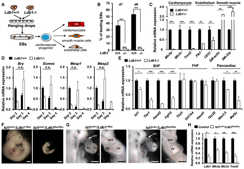
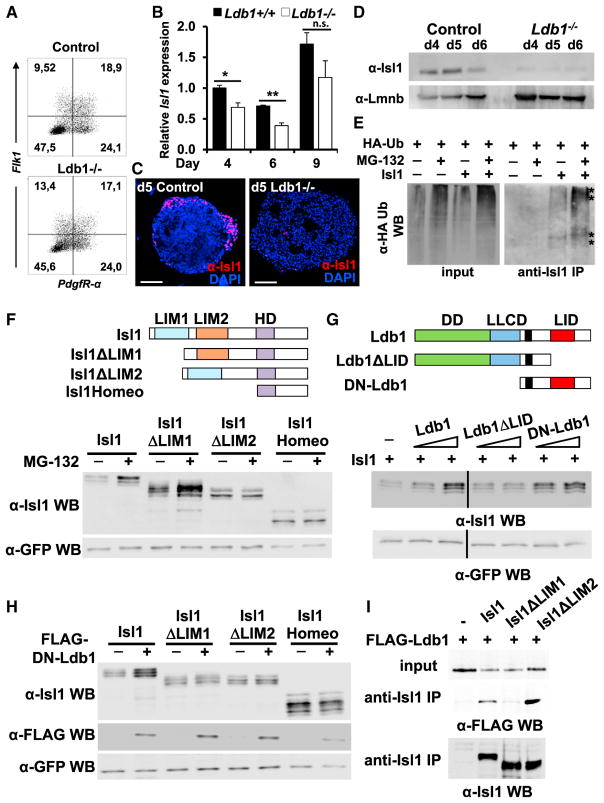
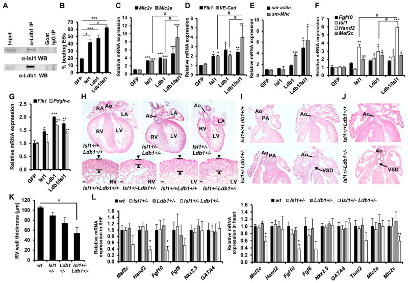
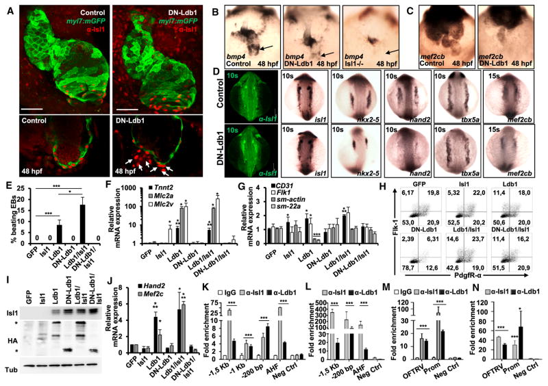
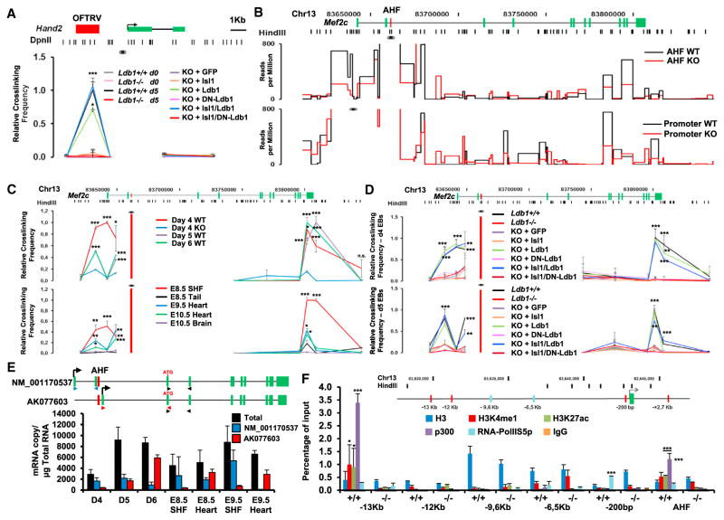
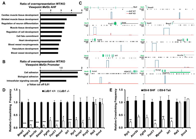
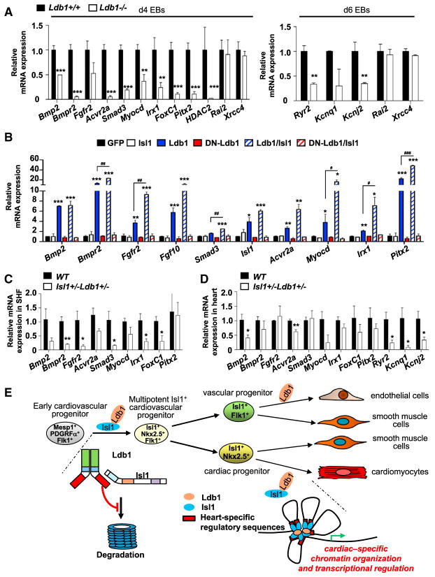
Cardiac stem/progenitor cells hold great potential for regenerative therapies; however, the mechanisms regulating their expansion and differentiation remain insufficiently defined. Here we show that Ldb1 is a central regulator of genome organization in cardiac progenitor cells, which is crucial for cardiac lineage differentiation and heart development. We demonstrate that Ldb1 binds to the key regulator of cardiac progenitors, Isl1, and protects it from degradation. Furthermore, the Isl1/Ldb1 complex promotes long-range enhancer-promoter interactions at the loci of the core cardiac transcription factors Mef2c and Hand2. Chromosome conformation capture followed by sequencing identified specific Ldb1-mediated interactions of the Isl1/Ldb1 responsive Mef2c anterior heart field enhancer with genes that play key roles in cardiac progenitor cell function and cardiovascular development. Importantly, the expression of these genes was downregulated upon Ldb1 depletion and Isl1/Ldb1 haplodeficiency. In conclusion, the Isl1/Ldb1 complex orchestrates a network for heart-specific transcriptional regulation and coordination in three-dimensional space during cardiogenesis.

Keywords: AHF enhancer; Hand2; Isl1; Ldb1; Mef2c; cardiac progenitors; cardiomyocyte differentiation; enhancer-promoter interactions; genome organization; second heart field.


Figures

Figure 1
Figure 1. Ablation of Ldb1 Results in Defects in Cardiac Progenitor Cell Differentiation and SHF Development
(A) Schematic diagram of the experimental setup. (B) Percentage of beating EBs in WT and Ldb1−/− ESCs. (C) Relative mRNA expression of cardiomyocyte (Mlc2a, Mlc2v, and Tnnt2), endothelial (Flk-1 and CD31), and smooth muscle (SM-actin, SM-22α) genes in d9 EBs differentiated from WT and Ldb1−/− ESCs. (D) Relative mRNA expression of mesodermal markers (Eomes, Bry, Mesp1, and Mesp2) in EBs differentiated from control and Ldb1−/− ESCs at different days. (E) Relative mRNA expression of cardiac progenitor marker genes in d4 EBs differentiated from Ldb1+/+ and Ldb1−/− ESCs. (F) Gross appearance of control (Isl1cre/+/Ldb1+/flox) and Isl1cre/+/Ldb1flox/flox embryos at E10.5. Scale bars, 500 μm. (G) Higher magnification of E9.5 embryos viewed from the right (left panels) and the front (right panels). Scale bars, 200 μm. OFT, outflow tract; RV, right ventricle; LV, left ventricle. (H) Relative mRNA expression analysis of Ldb1 and cardiomyocyte genes in dissected outflow tract and right ventricle of E9.25 WT and Isl1cre/+/Ldb1flox/flox embryos. Data are mean ± SEMs, n = 3 for each genotype. Data in (B)–(E) are mean ± SEMs, n = 3. *p < 0.05, **p < 0.01, ***p < 0.005. See also Figure S1.
Figure 2
Figure 2. Ldb1 Binds to Isl1 and Protects It from Proteasomal Degradation
(A) FACS analysis of Flk-1 and PdgfR-α expression in d3.75 EBs differentiated from control and Ldb1−/− ESCs. (B) Relative mRNA expression of Isl1 in EBs differentiated from control and Ldb1−/− ESCs at different days. Data are mean ± SEMs, n = 3. *p < 0.05, **p < 0.01. (C) Isl1 immunostaining on vibratome sections from d5 EBs differentiated from control and Ldb1−/− ESCs. Scale bars, 100 μm. (D) Western blot analysis of total protein extracts of day 4, 5, and 6 EBs differentiated from control and Ldb1−/− ESCs. Lamin B1 served as loading control. (E) HA-tagged ubiquitin and Isl1 were transiently expressed in HEK293T cells. Cells were either treated with DMSO or with MG-132 6 hr before being harvested. Equivalent amounts of total cellular protein were immunoprecipitated with an anti-Isl1 antibody and detected with an anti-HA antibody. (F) Schematic representation of Isl1 and Isl1 deletion constructs lacking either LIM1 or LIM2 or harboring only the homeodomain (top). Western blot analysis of whole-cell lysates of HEK293T cells transfected with Isl1, Isl1ΔLIM1, Isl1ΔLIM2, and Isl1Homeo expression plasmids, treated with DMSO or MG-132 is shown. (G) Schematic representation of Ldb1 and Ldb1 deletion constructs. Ldb1 harbors three domains important for its function: dimerization domain (DD), the Ldb1/Chip conserved domain (LLCD), and the LIM-interaction domain (LID) (top panel). HEK293T cells were transfected with constant amounts of Isl1 (10 μg) and GFP (1 μg, used as a control of transfection efficiency) expression plasmids and increasing amounts of Ldb1, Ldb1ΔLID, and DN-Ldb1 (5 and 9 μg). Immunoblot analysis of equal amounts of total protein extracts was performed using either anti-Isl1 or anti-GFP antibodies. (H) Western blot analysis of whole-cell lysates from HEK293T cells transfected with Isl1, Isl1ΔLIM1, Isl1ΔLIM2 and Isl1Homeo alone or together with DN-Ldb1 and GFP using anti-Isl1, anti-FLAG, and anti-GFP antibodies. (I) FLAG-HA-Ldb1 and Isl1 or Isl1 deletion constructs were transiently expressed in HEK293T cells and immunoprecipitation with an anti-Isl1 antibody was followed by immunoblot analysis with an anti-FLAG antibody.
Figure 3
Figure 3. Ldb1 and Isl1 Interact to Regulate Cardiogenesis
(A) Co-immunoprecipitation of nuclear extracts from d5 EBs using anti-Ldb1 antibody and detected with anti-Isl1 antibody. (B) Percentage of beating d7 EBs derived from ESCs overexpressing either GFP alone (control) or GFP together with Isl1, Ldb1, or both. (C–E) Relative mRNA expression of cardiomyocyte (C), endothelial (D), and smooth muscle marker genes (E) in d7 EBs differentiated from ESCs overexpressing either GFP alone or GFP together with Isl1, Ldb1, or Ldb1+Isl1. (F and G) Relative mRNA expression of cardiac progenitor marker genes in d4 EBs (F) and Flk-1 and PdgfR-α in d3.75 EBs (G) differentiated from ESCs over-expressing either GFP alone or GFP together with Isl1, Ldb1, or Ldb1+Isl1. For qPCR analysis of endogenous Isl1 levels, primers located in the 5′ UTR were utilized. (H–J) H&E staining of representative paraffin sections of E16.5 hearts of WT and Isl1+/−Ldb1+/− embryos (H, top panels), and higher magnification of right and left ventricles showing thinner compact myocardium of the right ventricle in Isl1+/−Ldb1+/− embryos (H, bottom panels), DORV (I), or OA in E18.5 hearts (J) with VSD (I and J). Ao, Aorta; PA, pulmonary artery; LA, left atrium; LV, left ventricle; RA, right atrium; RV, right ventricle; DORV, double outlet right ventricle; OA, overriding aorta; VSD, ventricular septal defect. (K) Morphometric analysis of right ventricle compact myocardial thickness. Data are mean ± SEMs, n = 4. (L) Relative mRNA expression analysis of cardiac progenitor and cardiomyocyte genes in dissected hearts and SHF of E9.25 WT, Isl1+/−, Ldb1+/− (controls), and Isl1+/−Ldb1+/− embryos. Data are mean ± SEMs, n = 4 for each genotype. *p < 0.05, **p < 0.01, ***p < 0.005. Data in (B)–(G) are mean ± SEMs, n = 3. *p < 0.05, **p < 0.01, ***p < 0.005. See also Figure S2.
Figure 4
Figure 4. The Dimerization Domain of Ldb1 Is Required for Cardiomyocyte Differentiation
(A) Confocal images of control and DN-Ldb1-overexpressing Tg(myl7:EGFP-HsHRAS)s883 zebrafish embryos at 48 hr post fertilization (hpf) stained with anti-GFP and anti-Isl1 (red) antibodies. Arrows indicate Isl1+ cells outside of the heart, which do not express the cardiomyocyte marker myl7. Scale bars, 50 μm. (B and C) In situ hybridization of control, DN-Ldb1-overexpressing, and isl1 mutant embryos at 48 hpf with bmp4 (B) and mef2cb (C) probes. The arrows indicate bmp4 expression at the sinus venosus. (D) Confocal images of control and DN-Ldb1-overexpressing embryos stained with anti-Isl1 antibody (leftmost panels) and in situ hybridization of control and DN-Ldb1-overexpressing embryos at 10–15 somite stages with isl1, nkx2.5, hand2, tbx5a, and mef2cb probes. (E) Percentage of beating d7 EBs derived from Ldb1−/− ESCs overexpressing either GFP alone (control) or GFP together with Isl1, Ldb1, DN-Ldb1, or these in different combinations. (F and G) Relative mRNA expression analysis of cardiomyocyte (F), smooth muscle, and endothelial marker (G) genes in d7 EBs. (H) FACS analysis of Flk-1 and PdgfR-α expression in d3.75 Ldb1−/− EBs overexpressing either GFP alone or GFP together with Isl1, Ldb1, DN-Ldb1, or these in different combinations. (I) Western blot analysis of total protein extracts of d5 EBs. (J) Relative mRNA expression of Mef2c and Hand2 in d4 EBs. (K and L) ChIP of nuclear extracts from d5 EBs (K) and pools of dissected SHF from E8–9 embryos (L) using anti-Isl1 and anti-Ldb1 antibodies or IgG as control. PCRs were performed using primers flanking conserved Isl1 binding sites in the Mef2c promoter and the AHF enhancer. Fold enrichment values for EBs were calculated relative to IgG control and for embryos relative to a genomic region that does not contain conserved Isl1 binding sites. Data are mean ± SEMs, n = 3. (M and N) ChIP of nuclear extracts from d5 EBs (M) and pools of dissected SHF from E8–9 embryos (N) using anti-Isl1 and anti-Ldb1 antibodies or IgG as a control. Data are mean ± SEMs, n = 3. Data in (E)–(G) and (J) are mean ± SEMs, n = 3. *p < 0.05, **p < 0.01, ***p < 0.005. See also Figures S3–S7.
Figure 5
Figure 5. Ldb1 Facilitates Enhancer-Promoter Interactions within the Hand2 and Mef2c Loci
(A) Schematic representation of the Hand2 genomic locus and the position of the DpnII restriction sites used in the 3C assay (top). 3C-qPCR relative crosslinking frequency in WT, Ldb1−/− ESCs, WT and Ldb1−/− ESCs, or d5 EBs derived from Ldb1−/− ESCs overexpressing either GFP alone or GFP together with Isl1, Ldb1, DN-Ldb1, or these in different combinations is shown. The Hand2 promoter was used as the viewpoint. Values were normalized to the β-actin locus and the value for the OFTRV enhancer in d5 WT EBs was set as one. Data are mean ± SEMs, n = 3. (B) Schematic representation of the Mef2c genomic locus and the position of the restriction sites of HindIII, used in the 3C assay (top). 3C-seq analysis of Mef2c AHF- (middle panel) and Mef2c promoter-associated regions (bottom panel) in d5 WT and Ldb1−/− EBs is shown. The viewpoint is indicated with an eye symbol. Only statistically significant interactions identified by the r3Cseq package (Thongjuea et al., 2013) are presented (p < 0.05). (C) 3C-qPCR relative crosslinking frequency in EBs at different days (top panel) and in microdissected SHF region, heart, brain, or tail from embryos at different developmental stages (bottom panel). Data are mean ± SEMs, n = 3. (D) 3C-qPCR relative crosslinking frequency observed in d4 (top) and d5 (bottom) EBs derived from WT and Ldb1−/− ESCs or Ldb1−/− ESCs overexpressing either GFP alone or GFP together with Isl1, Ldb1, and DN-Ldb1 in different combinations. Data are mean ± SEMs, n = 3. For the 3C-qPCR in (C) and (D) the HindIII fragment containing the Mef2c-AHF was used as the viewpoint (red bar, eye symbol). (E) Schematic representation of alternative Mef2c transcripts (top) and absolute quantification of these transcripts using primers indicated in the scheme (bottom). (F) ChIP of d4 EBs derived from WT and Ldb1−/− ESCs using antibodies against H3, H3K4me1, H3K27Ac, p300, RNA-PolIIS5p, and IgG as a control. Data are mean ± SEMs, n = 3. *p < 0.05, **p < 0.01, ***p < 0.005. See also Figure S7.
Figure 6
Figure 6. Ldb1 Promotes Chromatin Looping between the AHF Enhancer and Genes That Play Key Roles in Cardiovascular Development
(A and B) Gene ontology analysis of genes interacting with the Mef2c AHF (A) or the Mef2c promoter (B), overrepresented in WT versus Ldb1−/− cells. (C) Schematic representation of 3C-seq results showing specific interactions of the Mef2c AHF with genes that play key roles in cardiovascular development. The HindIII restriction sites are shown as black bars. y axes: reads per million. (D and E) 3C-qPCR validation of the interactions observed using the 3C-seq approach in WT or Ldb1−/−d5 EBs (D) or in microdissected SHF regions and tails of E8–9 embryos (E) using the Mef2c-AHF as viewpoint. Data are mean ± SEMs, n = 3. *p < 0.05, **p < 0.01, ***p < 0.005.
Figure 7
Figure 7. The Isl1/Ldb1 Complex Orchestrates a Network for Transcriptional Regulation and Coordination in Three-Dimensional Space, Driving Cardiogenesis
(A) Relative mRNA expression analysis in d4 and d6 WT and Ldb1−/− EBs of selected genes identified in the 3C-seq analysis to specifically interact with the Mef2c-AHF in WT, but not in Ldb1−/− EBs. (B) Relative mRNA expression analysis of selected genes in d5 Ldb1−/−EBs overexpressing either GFP alone or GFP together with Isl1, Ldb1, DN-Ldb1, or these in different combinations. (C and D) Relative mRNA expression analysis of selected genes in SHF (C) or heart (D) of WT or Isl1+/− Ldb1+/− E9.25 embryos. Data are shown as mean ± SEMs, n = 4. *p < 0.05, **p < 0.01, ***p < 0.005. (E) Model of the role of Ldb1 in cardiac lineage differentiation. Ldb1 binds to Isl1 and protects it from proteasomal degradation. The stabilized Isl1/Ldb1 complex orchestrates a network for transcriptional regulation and coordination in three-dimensional space, driving cardiac progenitor cell differentiation and heart development.

References

    1. Aguirre A, Sancho-Martinez I, Izpisua Belmonte JC. Reprogramming toward heart regeneration: stem cells and beyond. Cell Stem Cell. 2013;12:275–284. - PubMed
    1. Bach I, Carrière C, Ostendorff HP, Andersen B, Rosenfeld MG. A family of LIM domain-associated cofactors confer transcriptional synergism between LIM and Otx homeodomain proteins. Genes Dev. 1997;11:1370–1380. - PubMed
    1. Bach I, Rodriguez-Esteban C, Carrière C, Bhushan A, Krones A, Rose DW, Glass CK, Andersen B, Izpisúa Belmonte JC, Rosenfeld MG. RLIM inhibits functional activity of LIM homeodomain transcription factors via recruitment of the histone deacetylase complex. Nat Genet. 1999;22:394–399. - PubMed
    1. Becker T, Ostendorff HP, Bossenz M, Schlüter A, Becker CG, Peirano RI, Bach I. Multiple functions of LIM domain-binding CLIM/NLI/ Ldb cofactors during zebrafish development. Mech Dev. 2002;117:75–85. - PubMed
    1. Cai CL, Liang X, Shi Y, Chu PH, Pfaff SL, Chen J, Evans S. Isl1 identifies a cardiac progenitor population that proliferates prior to differentiation and contributes a majority of cells to the heart. Dev Cell. 2003;5:877–889. - PMC - PubMed

Publication types

MeSH terms

Associated data

LinkOut - more resources