. 2015 Sep 8;12(10):1606-17.
doi: 10.1016/j.celrep.2015.07.058. Epub 2015 Aug 28.

TMC1 and TMC2 Localize at the Site of Mechanotransduction in Mammalian Inner Ear Hair Cell Stereocilia

Affiliations

TMC1 and TMC2 Localize at the Site of Mechanotransduction in Mammalian Inner Ear Hair Cell Stereocilia

Kiyoto Kurima et al. Cell Rep. .

Abstract

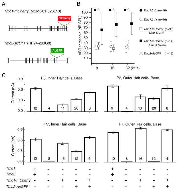
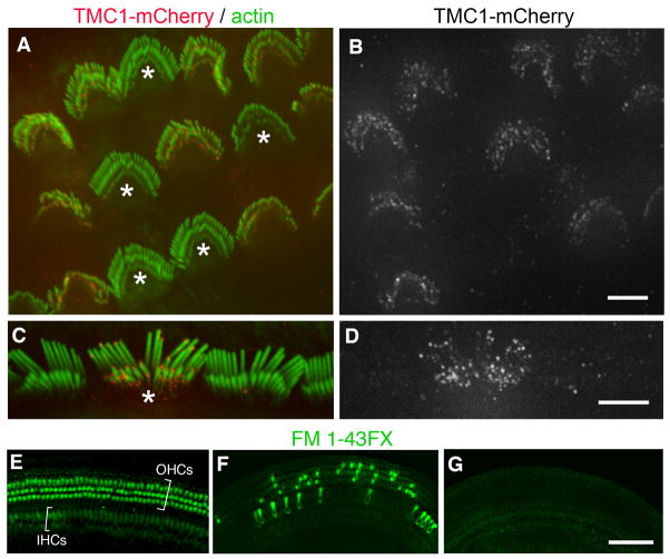
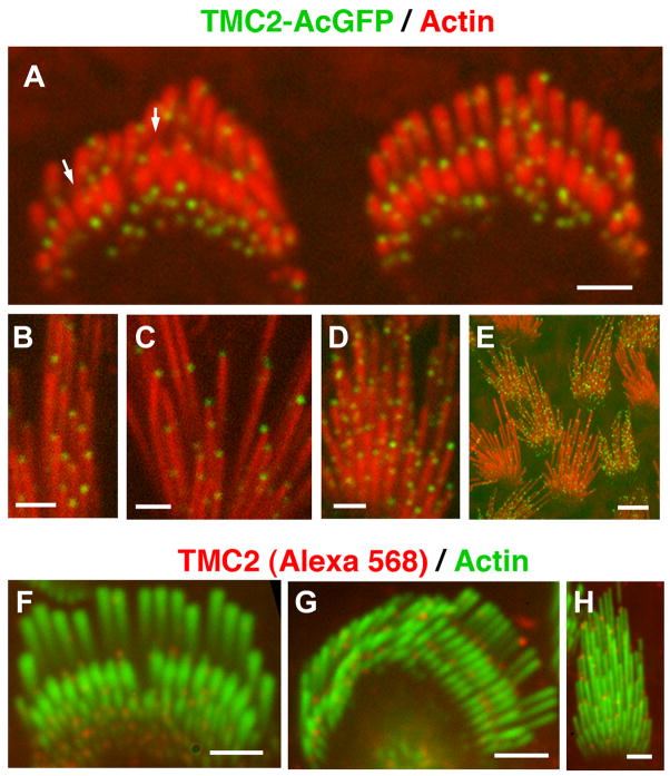
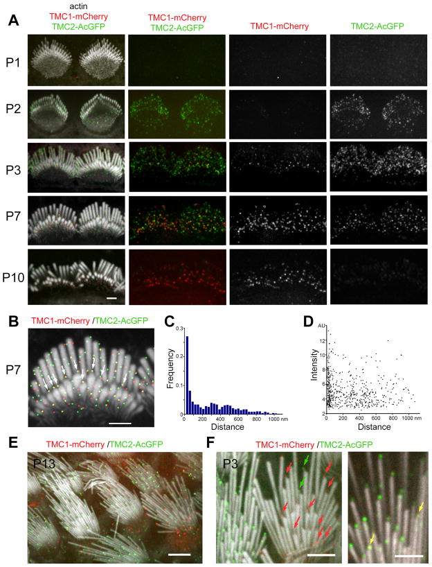
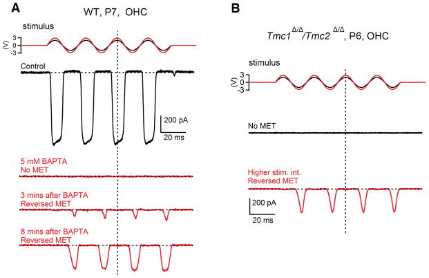
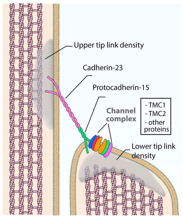
Mechanosensitive ion channels at stereocilia tips mediate mechanoelectrical transduction (MET) in inner ear sensory hair cells. Transmembrane channel-like 1 and 2 (TMC1 and TMC2) are essential for MET and are hypothesized to be components of the MET complex, but evidence for their predicted spatiotemporal localization in stereocilia is lacking. Here, we determine the stereocilia localization of the TMC proteins in mice expressing TMC1-mCherry and TMC2-AcGFP. Functionality of the tagged proteins was verified by transgenic rescue of MET currents and hearing in Tmc1(Δ/Δ);Tmc2(Δ/Δ) mice. TMC1-mCherry and TMC2-AcGFP localize along the length of immature stereocilia. However, as hair cells develop, the two proteins localize predominantly to stereocilia tips. Both TMCs are absent from the tips of the tallest stereocilia, where MET activity is not detectable. This distribution was confirmed for the endogenous proteins by immunofluorescence. These data are consistent with TMC1 and TMC2 being components of the stereocilia MET channel complex.


Figures

Figure 1
Figure 1. Rescue of hair cell function inTmc1Δ/Δ;Tmc2Δ/Δ mice expressing TMC1-mCherry and TMC2-AcGFP
(A) Bacterial artificial chromosome (BAC) transgenes. The stop codons of Tmc1 or Tmc2 were replaced with cDNA encoding mCherry or AcGFP, respectively. (B) Mean (±SD) ABR thresholds of 6 to 8-week-old mice for tone-burst stimuli of 8, 16 or 32 kHz. The hearing in mice lacking functional TMC1 was rescued by Tmc1-mCherry fully (line 1, 2 and 4) or partially (line 3 females), but not by Tmc2-AcGFP. (C) Mean (±SEM) maximal mechanotransduction currents recorded from hair cells excised from mice of the indicated genotypes. Mice with transgenes are on Tmc1Δ/Δ;Tmc2Δ/Δ background. Transduction currents were evoked using saturating hair bundle deflections and were recorded at −84 mV using the whole-cell, tight-seal technique. Number of hair cells is indicated for each genotype excised from 3 (wild-type), 1 (Tmc1Δ/Δ;Tmc2Δ/Δ), 3 (Tmc1-mCherry), 5 (Tmc2-AcGFP) and 2 (Tmc1-mCherry:Tmc2-AcGFP) P3 mice and 3 (wild type), 2 (Tmc1Δ/Δ;Tmc2Δ/Δ), 4 (Tmc1-mCherry), 5 (Tmc2-AcGFP) and 1 (Tmc1-mCherry:Tmc2-AcGFP) P7 mice.
Figure 2
Figure 2. Localization of TMC1-mCherry in hair cell stereocilia
(A) Confocal fluorescence image of IHCs from Tmc1-mCherry transgenic mice at P8 (Line 4). TMC1-mCherry (red) localizes predominantly to the tips of the shorter rows of stereocilia, counterstained in green with Alexa Fluor488-phalloidin. (B) TMC1-mCherry (red) distribution in mouse OHC stereocilia (green) at P8 (Line 2). (C and D) TMC1-mCherry (red) localizes to the tips of mouse vestibular stereocilia (green) at P7 (Line 2). (E) Immunofluorescence confocal image of anti-TMC1 antibody (PB277), in P9 mouse OHCs. (F) Close-up view of OHC stereocilia labeled with PB277 shows that TMC1 labeling (red) is present at the tips of second and third rows of stereocilia. Lining up of TMC1 immunofluorescence puncta at the tips of stereocilia is clearly visualized in the boxed region in F, and magnified below. (G and H) Immunofluorescence confocal images of anti-TMC1 antibody (PB277), in P15 rat IHC (G) and OHC (H) stereocilia also shows TMC1 labeling (red) at the tips of second and third rows. (I) TMC1 (red) is consistently detected at the tips of P15 rat vestibular hair cell stereocilia (green) using antibody PB612. [Scale bars: 2 μm]. See also Figure S1, S2 and S3.
Figure 3
Figure 3. Mosaic expression of TMC1-mCherry in mouse line 3
(A) Confocal image of OHC stereocilia (green) from P10 mice, showing mosaic expression of TMC1-mCherry fluorescent puncta (red). Cells that are not expressing TMC1-mCherry are highlighted with asterisks. (B) Corresponding red channel (TMC1-mCherry) shown in gray. (C) IHC stereocilia (green), showing only one cell (asterisk) expressing TMC1-mCherry. (D) Corresponding red channel (TMC1-mCherry) shown in gray. (E–G) Confocal images of P10 mouse cochlear hair cells exposed to 5 μM FM1-43 FX. From left to right, IHCs and OHCs from maleTmc1-mCherry line 3 mouse, from female Tmc1-mCherry line 3 mouse, and from Tmc1Δ/Δ;Tmc2Δ/Δ mouse. The Tmc1-mCherry line 3 mice were on a Tmc1Δ/Δ;Tmc2Δ/Δ background. [Scale bars: AD = 5 μm; EG= 100 μm]. See also Figure S1.
Figure 4
Figure 4. Localization of TMC2-AcGFP in hair cell stereocilia
(A) Confocal images of IHC stereocilia (labeled in red with AlexaFluor568 phalloidin) from Tmc2-AcGFP transgenic mice at P4 (Line 4), showing localization of TMC2-AcGFP (green). (B, C) TMC2-AcGFP (green) localizes at the tips of vestibular hair cell stereocilia (red) at P7 (Line 2). (D) In addition to stereocilia tip labeling, some hair bundles at P7 also show abundant TMC2-AcGFP puncta along the length of stereocilia. (E) The expression of TMC2-AcGFP (green) within stereocilia (red) was seen to vary from cell to cell in vestibular organs. (Line 4, P8). (F and G) Immunofluorescence confocal images of rat IHC and OHC stereocilia (green) at P7, showing immunofluorescence of TMC2 (red) using anti-TMC2 antibody PB361. (H) Immunofluorescence staining of TMC2 (red) in stereocilia (green) of rat vestibular hair cells at P3. [Scale bars: AD, FH = 2μm, E = 5 μm]. See also Figure S3.
Figure 5
Figure 5. Simultaneous localization of TMC1-mCherry and TMC2-AcGFP in mouse cochlear and vestibular stereocilia
(A) Confocal images of IHC stereocilia from transgenic mice expressing TMC1-mCherry and TMC2-AcGFP at P1, P2, P3, P7 and P10 (Tmc1-mCherry Line 2; Tmc2-AcGFP Line 3). All cochlear stereocilia bundles are from the mid-apical region of the cochlea (located between 30–50% from the apex of the cochlea). Stereocilia, (labeled with AlexaFluor405 phalloidin) are shown in white (left). Right panels show, respectively, merge of red and green channels, only red channel and only green channel. (B) IHC stereocilia at P7, with calculated centroids for TMC1-mCherry and TMC2-AcGFP puncta represented as red and green dots, respectively, to show relative localization of both signals. (C) Histogram showing the frequency distribution of TMC1-mCherry puncta centroids according to their distance to the centroid of the corresponding closest neighboring TMC2-AcGFP punctum (histogram bin size = 30 nm; number of puncta = 709; number of hair cells =12). (D) Scatter plot of fluorescence intensity (in arbitrary units, AU) of each TMC1-mCherry puncta as a function of the distance to their corresponding closest neighboring TMC2-AcGFP punctum. (E) Confocal images of vestibular hair cells from P13 mice expressing TMC1-mCherry and TMC2-AcGFP. Stereocilia, labeled with AlexaFluor405 conjugated phalloidin, are shown in white. (Tmc1-mCherry Line 2; Tmc2-AcGFP Line 4). (F) Close up views of vestibular stereocilia bundles from P3 mouse expressing both TMC1-mCherry and TMC2-AcGFP showing presence of only TMC1-mCherry (red arrows), only TMC2-AcGFP (green arrows), or both (yellow arrows) at stereocilia tips. (Tmc1-mCherry Line 2; Tmc2-AcGFP Line 4). [Scale bars: A–D, F = 2μm; E = 5μm]. See also Figure S4–6.
Figure 6
Figure 6. Reverse-polarity currents in WT andTmc1Δ/Δ;Tmc2Δ/Δ hair cells
(A) Example of MET current (black trace) recorded from a WT outer hair cell (from mid-apical turn of the cochlea, P7 mouse) in response to a sine wave stimulus (black) with a fluid jet device. Upon application of 5 mM BAPTA the MET current of the same cell is abolished (red trace) and followed with the progressive appearance of the reverse polarity currents. (B) Representative MET current recordings from a Tmc1Δ/Δ;Tmc2Δ/Δouter hair cell. No conventional MET currents were detected during regular stimulation (black trace). A higher stimulus (red sine wave) evoked reverse-polarity currents (red trace). All recordings in panels A and B are averages of 3–8 individual traces (n=5 cells).
Figure 7
Figure 7. Schematic diagram of stereocilia MET channel complex proteins
Diagram of the stereocilia MET channel complex illustrating the localization of known associated proteins. Localization of TMC1 and TMC2 is consistent with these proteins being components of the complex.

Comment in

References

    1. Assad JA, Shepherd GM, Corey DP. Tip-link integrity and mechanical transduction in vertebrate hair cells. Neuron. 1991;7:985–994. - PubMed
    1. Barr-Gillespie PG, Nicolson T. Who needs tip links? Backwards transduction by hair cells. J Gen Physiol. 2013;142:481–486. - PMC - PubMed
    1. Beurg M, Fettiplace R, Nam JH, Ricci AJ. Localization of inner hair cell mechanotransducer channels using high-speed calcium imaging. Nat Neurosci. 2009;12:553–558. - PMC - PubMed
    1. Beurg M, Kim KX, Fettiplace R. Conductance and block of hair-cell mechanotransducer channels in transmembrane channel-like protein mutants. J Gen Physiol. 2014;144:55–69. - PMC - PubMed
    1. Beurg M, Xiong W, Zhao B, Muller U, Fettiplace R. Subunit determination of the conductance of hair-cell mechanotransducer channels. Proceedings of the National Academy of Sciences of the United States of America. 2015;112:1589–1594. - PMC - PubMed

Publication types