

ABCB5 Identifies Immunoregulatory Dermal Cells
- PMID: 26321644
- PMCID: PMC4565759
- DOI: 10.1016/j.celrep.2015.08.010
ABCB5 Identifies Immunoregulatory Dermal Cells
Abstract
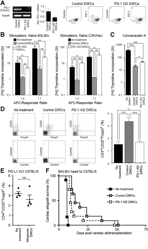
Cell-based strategies represent a new frontier in the treatment of immune-mediated disorders. However, the paucity of markers for isolation of molecularly defined immunomodulatory cell populations poses a barrier to this field. Here, we show that ATP-binding cassette member B5 (ABCB5) identifies dermal immunoregulatory cells (DIRCs) capable of exerting therapeutic immunoregulatory functions through engagement of programmed cell death 1 (PD-1). Purified Abcb5(+) DIRCs suppressed T cell proliferation, evaded immune rejection, homed to recipient immune tissues, and induced Tregs in vivo. In fully major-histocompatibility-complex-mismatched cardiac allotransplantation models, allogeneic DIRCs significantly prolonged allograft survival. Blockade of DIRC-expressed PD-1 reversed the inhibitory effects of DIRCs on T cell activation, inhibited DIRC-dependent Treg induction, and attenuated DIRC-induced prolongation of cardiac allograft survival, indicating that DIRC immunoregulatory function is mediated, at least in part, through PD-1. Our results identify ABCB5(+) DIRCs as a distinct immunoregulatory cell population and suggest promising roles of this expandable cell subset in cellular immunotherapy.
Copyright © 2015 The Authors. Published by Elsevier Inc. All rights reserved.
Figures


Similar articles
-
A novel in vivo regulatory role of P-glycoprotein in alloimmunity.Biochem Biophys Res Commun. 2010 Apr 9;394(3):646-52. doi: 10.1016/j.bbrc.2010.03.040. Epub 2010 Mar 15. Biochem Biophys Res Commun. 2010. PMID: 20230790 Free PMC article.
-
ABCB5 is a limbal stem cell gene required for corneal development and repair.Nature. 2014 Jul 17;511(7509):353-7. doi: 10.1038/nature13426. Epub 2014 Jul 2. Nature. 2014. PMID: 25030174 Free PMC article.
-
Differential effects of B and T lymphocyte attenuator and programmed death-1 on acceptance of partially versus fully MHC-mismatched cardiac allografts.J Immunol. 2005 Nov 1;175(9):5774-82. doi: 10.4049/jimmunol.175.9.5774. J Immunol. 2005. PMID: 16237069
-
ATP-binding cassette member B5 (ABCB5) promotes tumor cell invasiveness in human colorectal cancer.J Biol Chem. 2018 Jul 13;293(28):11166-11178. doi: 10.1074/jbc.RA118.003187. Epub 2018 May 22. J Biol Chem. 2018. PMID: 29789423 Free PMC article.
-
P-glycoprotein--a novel therapeutic target for immunomodulation in clinical transplantation and autoimmunity?Curr Drug Targets. 2003 Aug;4(6):469-76. doi: 10.2174/1389450033490894. Curr Drug Targets. 2003. PMID: 12866661 Review.
Cited by
-
Kinetics of Wound Development and Healing Suggests a Skin-Stabilizing Effect of Allogeneic ABCB5+ Mesenchymal Stromal Cell Treatment in Recessive Dystrophic Epidermolysis Bullosa.Cells. 2023 May 24;12(11):1468. doi: 10.3390/cells12111468. Cells. 2023. PMID: 37296590 Free PMC article.
-
Translational perspectives to treat Epidermolysis bullosa-Where do we stand?Exp Dermatol. 2020 Nov;29(11):1112-1122. doi: 10.1111/exd.14194. Exp Dermatol. 2020. PMID: 33043517 Free PMC article.
-
Clinical trial of ABCB5+ mesenchymal stem cells for recessive dystrophic epidermolysis bullosa.JCI Insight. 2021 Nov 22;6(22):e151922. doi: 10.1172/jci.insight.151922. JCI Insight. 2021. PMID: 34665781 Free PMC article.
-
The Pathobiology of Skin Aging: New Insights into an Old Dilemma.Am J Pathol. 2020 Jul;190(7):1356-1369. doi: 10.1016/j.ajpath.2020.03.007. Epub 2020 Apr 1. Am J Pathol. 2020. PMID: 32246919 Free PMC article. Review.
-
Mesenchymal stromal cells in wound healing applications: role of the secretome, targeted delivery and impact on recessive dystrophic epidermolysis bullosa treatment.Cytotherapy. 2021 Nov;23(11):961-973. doi: 10.1016/j.jcyt.2021.06.004. Epub 2021 Aug 8. Cytotherapy. 2021. PMID: 34376336 Free PMC article. Review.
References
-
- Asahara T, Murohara T, Sullivan A, Silver M, van der Zee R, Li T, Witzenbichler B, Schatteman G, Isner JM. Isolation of putative progenitor endothelial cells for angiogenesis. Science. 1997;275:964–967. - PubMed
-
- Bartholomew A, Sturgeon C, Siatskas M, Ferrer K, McIntosh K, Patil S, Hardy W, Devine S, Ucker D, Deans R, et al. Mesenchymal stem cells suppress lymphocyte proliferation in vitro and prolong skin graft survival in vivo. Exp Hematol. 2002;30:42–48. - PubMed
-
- Casiraghi F, Azzollini N, Cassis P, Imberti B, Morigi M, Cugini D, Cavinato RA, Todeschini M, Solini S, Sonzogni A, et al. Pretransplant infusion of mesenchymal stem cells prolongs the survival of a semiallogeneic heart transplant through the generation of regulatory T cells. J Immunol. 2008;181:3933–3946. - PubMed
-
- Clark RA, Chong B, Mirchandani N, Brinster NK, Yamanaka K, Dowgiert RK, Kupper TS. The vast majority of CLA+ T cells are resident in normal skin. J Immunol. 2006;176:4431–4439. - PubMed
-
- Devine SM, Cobbs C, Jennings M, Bartholomew A, Hoffman R. Mesenchymal stem cells distribute to a wide range of tissues following systemic infusion into nonhuman primates. Blood. 2003;101:2999–3001. - PubMed
