

Salivary Gland Dysplasia in Fgf10 Heterozygous Mice: A New Mouse Model of Xerostomia
- PMID: 26321752
- PMCID: PMC5405808
- DOI: 10.2174/1566524015666150831141307
Salivary Gland Dysplasia in Fgf10 Heterozygous Mice: A New Mouse Model of Xerostomia
Abstract
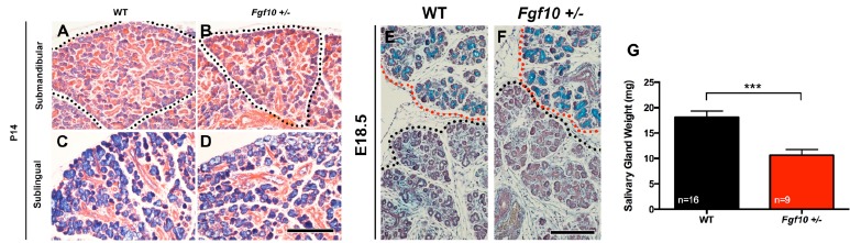
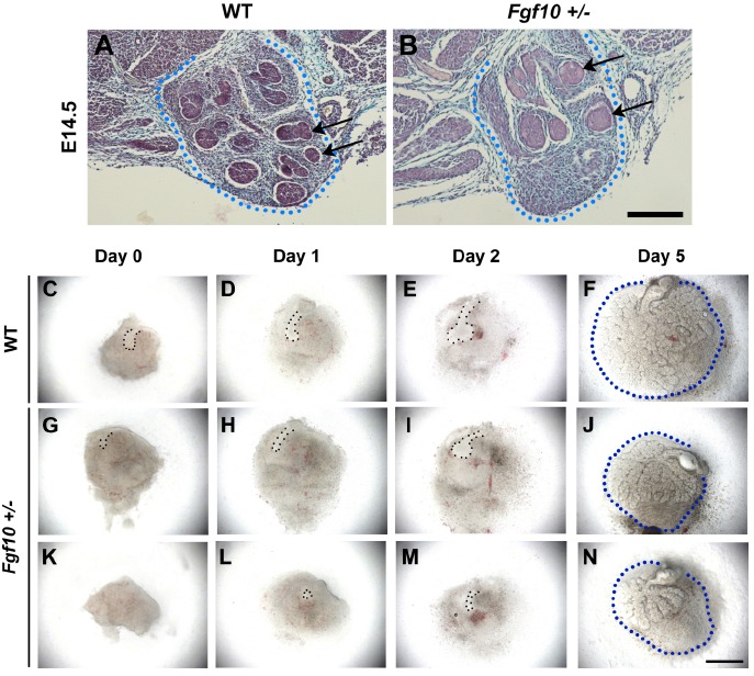
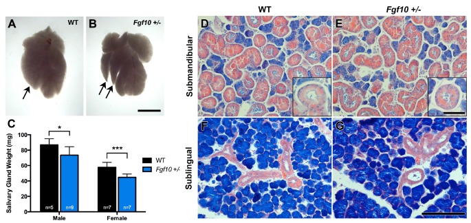
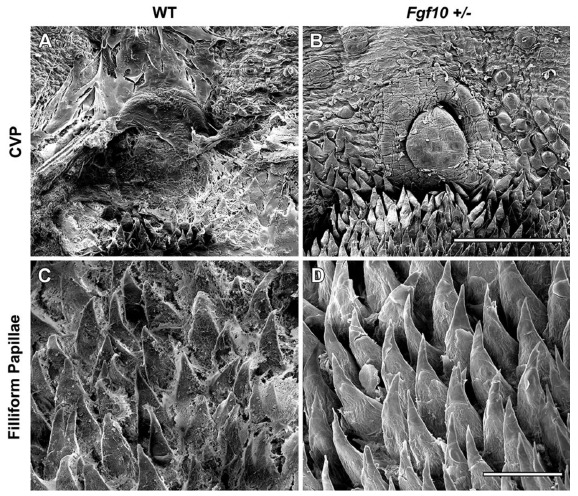
Xerostomia, or chronic dry mouth, is a common syndrome caused by a lack of saliva that can lead to severe eating difficulties, dental caries and oral candida infections. The prevalence of xerostomia increases with age and affects approximately 30% of people aged 65 or older. Given the large numbers of sufferers, and the potential increase in incidence given our aging population, it is important to understand the complex mechanisms that drive hyposalivation and the consequences for the dentition and oral mucosa. From this study we propose the Fgf10 +/- mouse as a model to investigate xerostomia. By following embryonic salivary gland development, in vivo and in vitro, we show that a reduction in Fgf10 causes a delay in branching of salivary glands. This leads to hypoplasia of the glands, a phenotype that is not rescued postnatally or by adulthood in both male and female Fgf10 +/- mice. Histological analysis of the glands showed no obvious defect in cellular differentiation or acini/ductal arrangements, however there was a significant reduction in their size and weight. Analysis of saliva secretion showed that hypoplasia of the glands led to a significant reduction in saliva production in Fgf10 +/- adults, giving rise to a reduced saliva pellicle in the oral cavity of these mice. Mature mice were shown to drink more and in many cases had severe tooth wear. The Fgf10 +/- mouse is therefore a useful model to explore the causes and effects of xerostomia.
Conflict of interest statement
The authors confirm that this article content has no conflict of interest.
Figures

Similar articles
-
New pathogenic variant in the FGF10 gene in the agenesis of lacrimal and salivary gland syndrome: Ophthalmological and genetic study.Ophthalmic Genet. 2018 Jan-Feb;39(1):125-128. doi: 10.1080/13816810.2017.1381976. Epub 2017 Oct 20. Ophthalmic Genet. 2018. PMID: 29053399
-
An intronic alteration of the fibroblast growth factor 10 gene causing ALSG-(aplasia of lacrimal and salivary glands) syndrome.BMC Med Genet. 2008 Dec 22;9:114. doi: 10.1186/1471-2350-9-114. BMC Med Genet. 2008. PMID: 19102732 Free PMC article.
-
Associations between xerostomia, histopathological alterations, and autonomic innervation of labial salivary glands in men in late midlife.Exp Gerontol. 2014 Sep;57:211-7. doi: 10.1016/j.exger.2014.06.004. Epub 2014 Jun 4. Exp Gerontol. 2014. PMID: 24905142
-
Established and novel approaches for the management of hyposalivation and xerostomia.Curr Pharm Des. 2012;18(34):5515-21. doi: 10.2174/138161212803307509. Curr Pharm Des. 2012. PMID: 22632391 Review.
-
Xerostomia: diagnosis and management.Oncology (Williston Park). 1996 Mar;10(3 Suppl):7-11. Oncology (Williston Park). 1996. PMID: 8723427 Review.
Cited by
-
Loss of Hs3st3a1 or Hs3st3b1 enzymes alters heparan sulfate to reduce epithelial morphogenesis and adult salivary gland function.Matrix Biol. 2021 Sep;103-104:37-57. doi: 10.1016/j.matbio.2021.10.002. Epub 2021 Oct 12. Matrix Biol. 2021. PMID: 34653670 Free PMC article.
-
Cell signaling regulation in salivary gland development.Cell Mol Life Sci. 2021 Apr;78(7):3299-3315. doi: 10.1007/s00018-020-03741-2. Epub 2021 Jan 15. Cell Mol Life Sci. 2021. PMID: 33449148 Free PMC article. Review.
-
Magnetic bioassembly platforms towards the generation of extracellular vesicles from human salivary gland functional organoids for epithelial repair.Bioact Mater. 2022 Feb 16;18:151-163. doi: 10.1016/j.bioactmat.2022.02.007. eCollection 2022 Dec. Bioact Mater. 2022. PMID: 35387159 Free PMC article.
-
Autologous mesenchymal stem cells offer a new paradigm for salivary gland regeneration.Int J Oral Sci. 2023 May 10;15(1):18. doi: 10.1038/s41368-023-00224-5. Int J Oral Sci. 2023. PMID: 37165024 Free PMC article. Review.
-
FGFR2b is essential for salivary gland duct homeostasis and MAPK-dependent seromucous acinar cell differentiation.Res Sq [Preprint]. 2023 Feb 16:rs.3.rs-2557484. doi: 10.21203/rs.3.rs-2557484/v1. Res Sq. 2023. Update in: Nat Commun. 2023 Oct 14;14(1):6485. doi: 10.1038/s41467-023-42243-0. PMID: 36824936 Free PMC article. Updated. Preprint.
References
-
- Hand A.R., Pathmanathan D., Field R.B. Morphological features of the minor salivary glands. Arch. Oral Biol. 1999;44(Suppl. 1):S3–S10. - PubMed
-
- Rothova M., Thompson H., Lickert H., Tucker A.S. Lineage tracing of the endoderm during oral development. Dev. Dyn. 2012;241:1183–1191. - PubMed
-
- Jaskoll T., Zhou Y.M., Trump G., Melnick M. Ectodysplasin receptor-mediated signaling is essential for embryonic submandibular salivary gland development. Anat. Rec. A Discov. Mol. Cell. Evol. Biol. 2003;271:322–331. - PubMed
-
- Hall H. Protective and maintenance functions of human saliva. Quintessence Int. 1993;24:813–816. - PubMed
-
- Ship J.A., Pillemer S.R., Baum B.J. Xerostomia and the geriatric patient. J. Am. Geriatr. Soc. 2002;50:535–543. - PubMed
Publication types
MeSH terms
Substances
Grants and funding
LinkOut - more resources
Full Text Sources
Medical
Molecular Biology Databases