

β-ESTRADIOL INDUCES CYTOTOXIC EFFECTS TO HUMAN T-LYMPHOMA (JURKAT) CELLS THROUGH OXIDATIVE STRESS
- PMID: 26321773
- PMCID: PMC4550313
β-ESTRADIOL INDUCES CYTOTOXIC EFFECTS TO HUMAN T-LYMPHOMA (JURKAT) CELLS THROUGH OXIDATIVE STRESS
Abstract
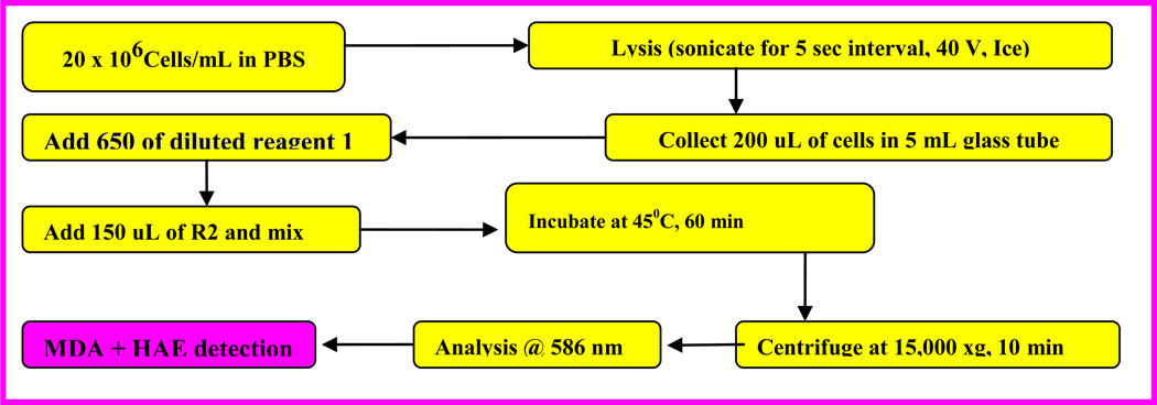
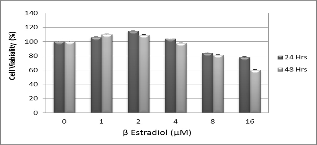
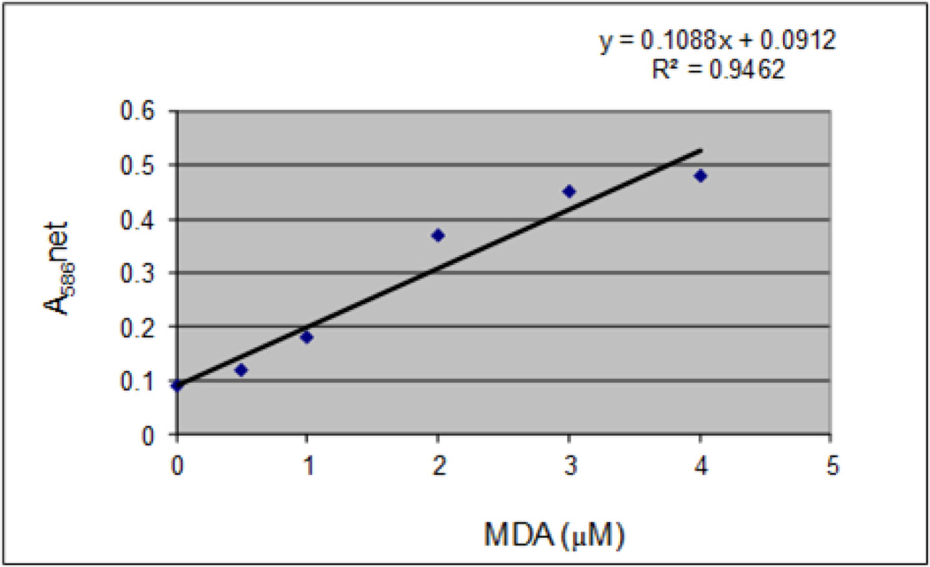
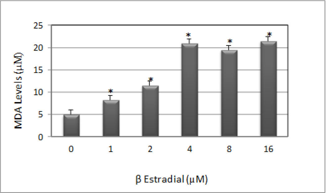
β-estradiol is the most potent estrogen of a group of endogenous estrogen steroids which includes estrone and estriol. This steroid hormone is the most potent natural estrogen, produced mainly by the ovary, placenta, and in smaller amounts by the adrenal cortex, and the male testes. Although β-estradiol protects the renal and cardiovascular systems, the mechanisms involved remain unclear. In this research, we performed the MTT [3-(4, 5-dimethylthiazol-2-yl)-2,5-diphenyl tetrazolium bromide] assay to evaluate the effect of β-estradiol on human T-lymphoma (Jurkat) cells upon 24 and 48 hours, respectively. Lipid peroxidation assay was also performed to estimate the levels of malondialdehyde (MDA) production in β-estradiol-treated cells. The results of MTT assay demonstrated that low, physiological levels of β-estradiol induce cellular proliferation in Jurkat T-cells. At higher dose of exposure, β-estradiol decreases the viability of Jurkat T-cells compared to the control cells. Data generated from lipid peroxidation assay resulted in a significant increase (p < 0.05) in MDA production in β-estradiol treated sample. Upon 48 h of exposure, MDA concentrations in the sample [µM] (mean ±SE, n = 3) compared to untreated control were 4.9 ± 1.7, 8.1 ± 1,6 11.5 ± 2.2, 21.1 ± 2.3, 19.5 ± 1.4, and 21.5 ± 2.6 in 0, 1, 2, 4, 8, and 16 µM β-estradiol, respectively. In summary, findings from this study demonstrated that high dose of β-estradiol is cytotoxic to Jurkat T-cells. This cytotoxicity is found to be associated with oxidative stress.
Keywords: Jurkat T-cells; Lipid peroxidation assay; MTT assay; β-estradiol.
Figures
Similar articles
-
Conversion of estrone to 17 beta-estradiol in Jurkat acute T cell leukemia Hut-78 T- and Raji B lymphoma cell lines in vitro.Biomed Pharmacother. 2013 May;67(4):299-303. doi: 10.1016/j.biopha.2012.11.003. Epub 2012 Nov 20. Biomed Pharmacother. 2013. PMID: 23540283
-
OXIDATIVE STRESS IN HUMAN LEUKEMIA (HL-60), HUMAN LIVER CARCINOMA (HepG2), AND HUMAN (JURKAT-T) CELLS EXPOSED TO ARSENIC TRIOXIDE.Met Ions Biol Med. 2006;9:298-303. Met Ions Biol Med. 2006. PMID: 26435679 Free PMC article.
-
Effects of pharmacological concentrations of estrogens on proliferation and cell cycle kinetics of human breast cancer cell lines in vitro.Cancer Res. 1987 Oct 15;47(20):5323-9. Cancer Res. 1987. PMID: 3652038
-
Enzymatic control of estrogen production in human breast cancer: relative significance of aromatase versus sulfatase pathways.Ann N Y Acad Sci. 1986;464:126-37. doi: 10.1111/j.1749-6632.1986.tb16000.x. Ann N Y Acad Sci. 1986. PMID: 3524346 Review.
-
Importance of estrogen sulfates in breast cancer.J Steroid Biochem. 1989;34(1-6):155-63. doi: 10.1016/0022-4731(89)90077-0. J Steroid Biochem. 1989. PMID: 2560511 Review.
Cited by
-
Oxidative Stress Is Increased in Combined Oral Contraceptives Users and Is Positively Associated with High-Sensitivity C-Reactive Protein.Molecules. 2021 Feb 18;26(4):1070. doi: 10.3390/molecules26041070. Molecules. 2021. PMID: 33670593 Free PMC article.
-
The G-Protein-Coupled Estrogen Receptor Agonist G-1 Inhibits Proliferation and Causes Apoptosis in Leukemia Cell Lines of T Lineage.Front Cell Dev Biol. 2022 Feb 14;10:811479. doi: 10.3389/fcell.2022.811479. eCollection 2022. Front Cell Dev Biol. 2022. PMID: 35237599 Free PMC article.
-
Functional characterization of Clonorchis sinensis sodium-bile acid co-transporter (CsSBAT) as a steroid sulfate transporter.Parasitol Res. 2022 Jan;121(1):217-224. doi: 10.1007/s00436-021-07393-4. Epub 2021 Nov 26. Parasitol Res. 2022. PMID: 34825261
-
Oxidative Stress in Female Athletes Using Combined Oral Contraceptives.Sports Med Open. 2016 Dec;2(1):40. doi: 10.1186/s40798-016-0064-x. Epub 2016 Sep 21. Sports Med Open. 2016. PMID: 27747795 Free PMC article.
-
A meta-analysis of the relationship between glycaemic variability and the mortality of patients with heart failure.ESC Heart Fail. 2024 Jun;11(3):1305-1316. doi: 10.1002/ehf2.14627. Epub 2024 Jan 19. ESC Heart Fail. 2024. PMID: 38243645 Free PMC article. Review.
References
-
- Acke E, Money CT, Jones BR. Estrogen toxicity in a dog. Ir Vet J. 2003;56:465–468.
-
- Athreya BH, Pletcher J, Zulian F, Weiner DB, Williams WV. Subset-specific effects of sex hormones and pituitary gonadotropins on human lymphocyte proliferation in vitro. Clinical Immunology and Immunopathology. 1993;66:201–211. - PubMed
-
- Hall EJ. Use of lithium for treatment of estrogen-induced bone marrow hypoplasia in a dog. J Am Vet Med Assoc. 1992;200:814–816. - PubMed
Grants and funding
LinkOut - more resources
Full Text Sources