. 2015 Aug 12:9:303.
doi: 10.3389/fncel.2015.00303. eCollection 2015.

Spiral ganglion cells and macrophages initiate neuro-inflammation and scarring following cochlear implantation

Affiliations

Spiral ganglion cells and macrophages initiate neuro-inflammation and scarring following cochlear implantation

Esperanza Bas et al. Front Cell Neurosci. .

Abstract

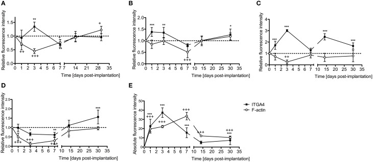
Conservation of a patient's residual hearing and prevention of fibrous tissue/new bone formation around an electrode array are some of the major challenges in cochlear implant (CI) surgery. Although it is well-known that fibrotic tissue formation around the electrode array can interfere with hearing performance in implanted patients, and that associated intracochlear inflammation can initiate loss of residual hearing, little is known about the molecular and cellular mechanisms that promote this response in the cochlea. In vitro studies in neonatal rats and in vivo studies in adult mice were performed to gain insight into the pro-inflammatory, proliferative, and remodeling phases of pathological wound healing that occur in the cochlea following an electrode analog insertion. Resident Schwann cells (SC), macrophages, and fibroblasts had a prominent role in the inflammatory process in the cochlea. Leukocytes were recruited to the cochlea following insertion of a nylon filament in adult mice, where contributed to the inflammatory response. The reparative stages in wound healing are characterized by persistent neuro-inflammation of spiral ganglion neurons (SGN) and expression of regenerative monocytes/macrophages in the cochlea. Accordingly, genes involved in extracellular matrix (ECM) deposition and remodeling were up-regulated in implanted cochleae. Maturation of scar tissue occurs in the remodeling phase of wound healing in the cochlea. Similar to other damaged peripheral nerves, M2 macrophages and de-differentiated SC were observed in damaged cochleae and may play a role in cell survival and axonal regeneration. In conclusion, the insertion of an electrode analog into the cochlea is associated with robust early and chronic inflammatory responses characterized by recruitment of leukocytes and expression of pro-inflammatory cytokines that promote intracochlear fibrosis and loss of the auditory hair cells (HC) and SGN important for hearing after CI surgery.

Keywords: Schwann cells; cochlea; cochlear implant; fibrosis; macrophages; neuro-inflammation; pathology; spiral ganglion neurons.


Figures

Figure 1
Figure 1
Analysis of tracked cells for direct leukocyte and cochlear tissue co-cultures. (A) Leukocyte trajectory and (B) leukocyte distance covered. Comparison are between EIT-cochlear tissues and undamaged controls and with EIT pre-treated with Dexamethasone (DXM, 20 μg/ml). N = 239−250 cells/group, One-Way ANOVA and Tukey's Multiple Comparison Test was used for statistical analysis ***p < 0.001.
Figure 2
Figure 2
Cell-cell interactions analysis for direct leukocyte and cochlear tissue co-cultures. (A) Number of leukocyte-leukocyte interactions in leukocytes and cochlear tissues co-cultures; (B) number of leukocyte-cochlear cells interactions in co-cultures and (C) time average that leukocytes interact. Comparisons are between EIT-cochlear tissues and undamaged controls and with EIT pre-treated with dexamethasone (DXM, 20 μg/ml). In upper and middle graphs, the number of cells interacting was averaged from 9 to 11 quadrants or regions of interest. For the bottom panel, N = 278 cells/group. One-Way ANOVA and Tukey's Multiple Comparison Test was used for statistical analysis ***p < 0.001, ns non-significant p-value.
Figure 3
Figure 3
Gene expression studies in vitro. The experiments were performed in indirect leukocyte and cochlear tissue co-cultures. The top panel shows the gene expression levels for leukocytes (white), where (A) Ccl2, (B) Sell, (C) Icam, and (D) Tgfb1. The bottom (gray) panel shows the gene expression levels in cochlear tissues, i.e., organ of Corti and lateral wall tissues, where (E) Ccl2, (F) Sele, (G) Vcam, and (H) Tgfb1. Comparisons are between EIT-cochlear tissues and undamaged controls and with EIT pre-treated with Dexamethasone (DXM, 20 μg/ml). Six animals per group were included in each set of experiments and a total of 4 independent experiments were carried out. N = 4, One-Way ANOVA and Tukey's Multiple Comparison Test was used for statistical analysis ***p < 0.001.
Figure 4
Figure 4
Time-course immunofluorescence analysis for integrin 4α and f-actin levels in different areas of adult mice implanted cochleae. (A) Lateral wall tissues (see Figure 6, spiral ligament, SL), (B) organ of Corti, (C) spiral ganglion, (D) cochlear nerve, and (E) wound site. Three to four specimens for each time point and were analyzed. ImageJ was used to analyze the images, histograms of red and green channels for each region of interest i.e., lateral wall, organ of Corti, spiral ganglion, cochlear nerve, and wound site images were recorded. N = 8 each time point, Two-Way ANOVA followed by Bonferroni posttests was used to analyze the relative and absolute fluorescence intensities. ***p < 0.001, **p < 0.01, *p < 0.05 and +++p < 0.001, ++p < 0.01, +p < 0.05 represent the p-values for ITGA4 and F-actin, respectively.
Figure 5
Figure 5
Immunofluorescence images for integrin α4 (ITGA4) in red and cell nuclei in blue. Cross sections corresponding to a contralateral cochlea (bottom), and cochleae at 1, 3, 7, 14, and 30 days post-implantation. The sections were observed under a confocal microscope. Mosaic images of different areas of the cochlea were acquired and later constructed. Three to four specimens were analyzed for each time point. Scale bars = 100 μm.
Figure 6
Figure 6
Immunofluorescence images for f-actin in green and cell nuclei in blue. Cross sections corresponding to a contralateral cochlea (bottom), and cochleae at 1, 3, 7, 14, and 30 days post-implantation. The sections were observed under a confocal microscope. Mosaic images of different areas of the cochlea were acquired and later constructed. Annotations for the different structures of the cochlea are in the contralateral photograph. Spiral ligament, SL; organ of Corti, OC; Scala tympanica, ST; Spiral ganglion, SG. In the implanted cochleae: electrode analog channel, ECh; an asterisk marks hyperproliferative tissue in the Scala tympanica. Three to four specimens were analyzed for each time point. Scale bars = 100 μm.
Figure 7
Figure 7
Time-course immunofluorescence analysis for IL-1β, Arg1, and presence of macrophages/microglia (F4/80 positive cells) levels in different areas of the implanted cochleae. (A) Lateral wall tissues (marked as SL, spiral ligament in the immunofluorescence images), (B) organ of Corti (OC, see Figure 8), (C) spiral ganglion (SG), (D) cochlear nerve, and (E) wound site (asterisk in Figure 8). ImageJ was used to analyze the images, histograms of red and green channels for each region of interest i.e., lateral wall, organ of Corti, spiral ganglion, cochlear nerve, and wound site images were recorded. N = 8 each time point, Two-Way ANOVA followed by Bonferroni posttests was used to analyze the relative and absolute fluorescence intensities. ***p < 0.001, **p < 0.01, *p < 0.05, +++p < 0.001, and ###p < 0.001, ##p < 0.01 represent the p values for F4/80, Arg1 and IL-1β, respectively.
Figure 8
Figure 8
Immunofluorescence images for F4/80 (monocytes/macrophages/microglia, green) and nuclei (blue). Representative micrographs of cross sections correspond to a contralateral cochlea (bottom), and cochleae at 1, 3, 7, 14, and 30 days post-implantation. A heavy influx of monocytes/macrophages/microglia can be observed at 3 days in the newly formed tissue en sheathing electrode analog as well as in the lateral wall, organ of Corti, and to a lessen extend to the spiral ganglion area. The sections were observed under a confocal microscope. Images of different areas were acquired and stitched together afterwards. Annotations for the different structures of the cochlea are in the contralateral photograph. Spiral ligament, SL; organ of Corti, OC; Scala tympanica, ST; Spiral ganglion, SG. In the implanted cochleae: electrode analog channel, ECh; an asterisk marks hyperproliferative tissue in the Scala tympanica. Four to five specimens were analyzed for each time point. Scale bars = 100 μm.
Figure 9
Figure 9
Immunofluorescence images for interleukin-1β (IL-1β, red), and nuclei (blue). Representative micrographs of cross sections correspond to a contralateral cochlea (bottom), and cochleae at 1, 3, 7, 14, and 30 days post-implantation. A strong red signal at 7 days post-implantation, especially in the spiral ganglion area and wound site, reveals a severe neuro-inflammatory response. The sections were observed under a confocal microscope. Images of different areas were acquired and stitched together afterwards. Four to five specimens were analyzed for each time point. Scale bars = 100 μm.
Figure 10
Figure 10
Immunofluorescence images for Arginase I (Arg1, red) and nuclei (blue). Representative micrographs of cross sections are from a contralateral cochlea (bottom), and cochleae at 1, 3, 7, 14, and 30 days post-implantation. Note that red blood cells are accumulated over the organ of Corti area, these cells are auto-fluorescent due to hemoglobin fluorescence. An increase in Arg1 levels at 7 days post- implantation, especially in the spiral ganglion area and wound site, indicates healing process. The sections were observed under a confocal microscope. Images of different areas were acquired and stitched together afterwards. Four to five specimens were analyzed for each time point. Scale bars = 100 μm.
Figure 11
Figure 11
Immunofluorescence images for the myofibroblast marker α-smooth muscle actin (α-SMA, red), Collagen type 1A (COL1A1, green) and nuclei (blue). Representative micrographs of cross sections correspond to a contralateral cochlea and a cochlea implanted for 1 month. Cells from the fibrotic tissue enclosing the electrode analog stain positive for α-SMA and COL1A1, asterisks indicate the presence of fibrotic tissue. The sections were observed under a confocal microscope. Images of different areas were acquired and stitched together afterwards. Scale bars = 100 μm.
Figure 12
Figure 12
Masson's trichrome staining for the presence of scar tissue. Representative micrographs of section of a contralateral non-implanted cochleae on the left, section of implanted cochlea on the right. Collagen fibers stained blue, nuclei stained black and cytoplasm and erythrocytes stained red. Images of different areas were acquired and stitched together afterwards. Four contralateral and four implanted cochleae were used. Scale bar = 100 μm.

References

    1. Bas E., Dinh C. T., Garnham C., Polak M., Van de Water T. R. (2012a). Conservation of hearing and protection of hair cells in cochlear implant patients' with residual hearing. Anat. Rec. (Hoboken) 295, 1909–1927. 10.1002/ar.22574 - DOI - PubMed
    1. Bas E., Gupta C., Van De Water T. R. (2012b). A novel organ of Corti explant model for the study of cochlear implantation trauma. Anat. Rec. (Hoboken) 295, 1944–1956. 10.1002/ar.22585 - DOI - PubMed
    1. Bas E., Van De Water T. R., Lumbreras V., Rajguru S., Goss G., Hare J. M., et al. . (2014). Adult human nasal mesenchymal-like stem cells restore cochlear spiral ganglion neurons after experimental lesion. Stem Cells Dev. 23, 502–514. 10.1089/scd.2013.0274 - DOI - PMC - PubMed
    1. Chen Y., Yu H., Zhang Y., Li W., Lu N., Ni W., et al. . (2013). Cotransfection of Pax2 and Math1 promote in situ cochlear hair cell regeneration after neomycin insult. Sci. Rep. 3:2996. 10.1038/srep02996 - DOI - PMC - PubMed
    1. Choi C. H., Oghalai J. S. (2005). Predicting the effect of post-implant cochlear fibrosis on residual hearing. Hear. Res. 205, 193–200. 10.1016/j.heares.2005.03.018 - DOI - PMC - PubMed

LinkOut - more resources