. 2015 Aug 11:9:315.
doi: 10.3389/fncel.2015.00315. eCollection 2015.

Inhibitory and excitatory axon terminals share a common nano-architecture of their Cav2.1 (P/Q-type) Ca(2+) channels

Affiliations

Inhibitory and excitatory axon terminals share a common nano-architecture of their Cav2.1 (P/Q-type) Ca(2+) channels

Daniel Althof et al. Front Cell Neurosci. .

Abstract

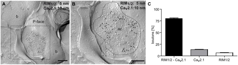
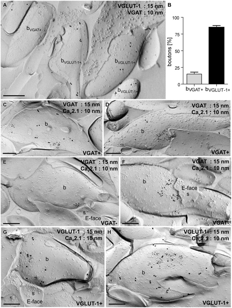
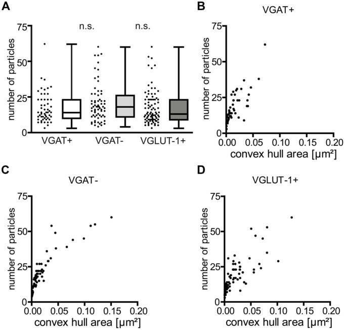
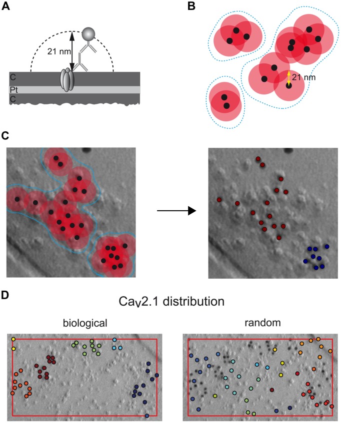
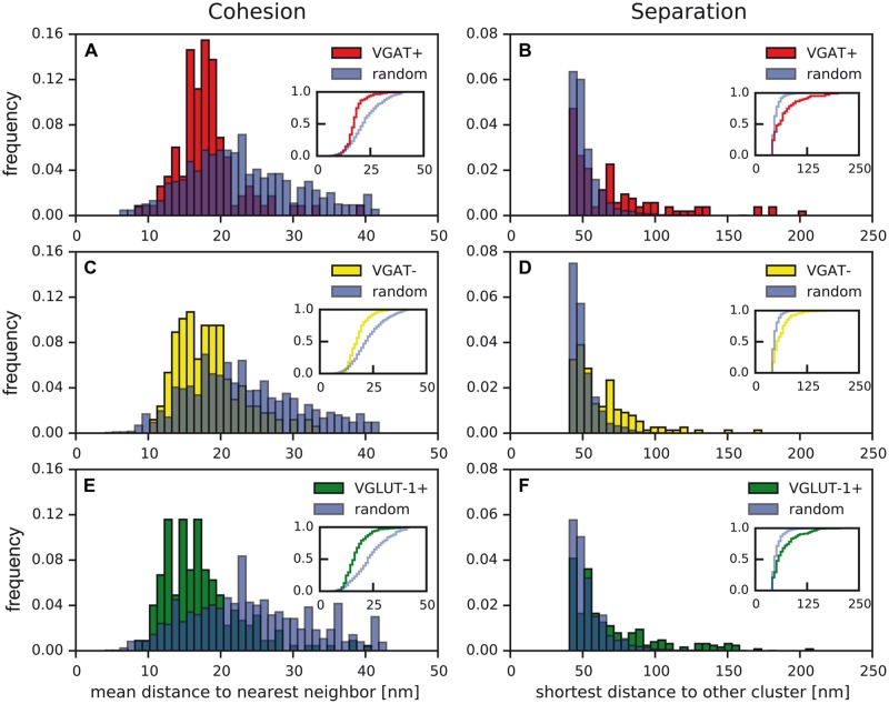
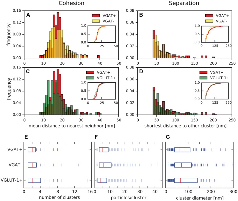
Tuning of the time course and strength of inhibitory and excitatory neurotransmitter release is fundamental for the precise operation of cortical network activity and is controlled by Ca(2+) influx into presynaptic terminals through the high voltage-activated P/Q-type Ca(2+) (Cav2.1) channels. Proper channel-mediated Ca(2+)-signaling critically depends on the topographical arrangement of the channels in the presynaptic membrane. Here, we used high-resolution SDS-digested freeze-fracture replica immunoelectron microscopy together with automatized computational analysis of Cav2.1 immunogold labeling to determine the precise subcellular organization of Cav2.1 channels in both inhibitory and excitatory terminals. Immunoparticles labeling the pore-forming α1 subunit of Cav2.1 channels were enriched over the active zone of the boutons with the number of channels (3-62) correlated with the area of the synaptic membrane. Detailed analysis showed that Cav2.1 channels are non-uniformly distributed over the presynaptic membrane specialization where they are arranged in clusters of an average five channels per cluster covering a mean area with a diameter of about 70 nm. Importantly, clustered arrangement and cluster properties did not show any significant difference between GABAergic and glutamatergic terminals. Our data demonstrate a common nano-architecture of Cav2.1 channels in inhibitory and excitatory boutons in stratum radiatum of the hippocampal CA1 area suggesting that the cluster arrangement is crucial for the precise release of transmitters from the axonal boutons.

Keywords: Ca2+ channels; cluster analysis; hippocampus; quantitative immunoelectron microscopy; rat.


Figures

FIGURE 1
FIGURE 1
Cav2.1 localized to the active zone of axon terminals of hippocampal cells. (A,B) Colocalization of Cav2.1 (10 nm gold particles) and the presynaptic marker proteins RIM1/2 (5 nm gold particles) in the active zone (az) of a bouton (b; inset, B) as assessed by the SDS-FRL method. The presynaptic active zone is indicated by the high density of intramembrane particles (IMPs) on the concave shape of the protoplasmic face (P-face) of the membrane (delineated by broken line). (C) Bar graph summarizing labeling and co-labeling of Cav2.1 and RIM1/2 in 303 axon terminals. Note co-labeling in the majority of terminals (81%). Scale bars, 0.2 μm.
FIGURE 2
FIGURE 2
Cav2.1 channels are organized in discrete groups in the presynaptic active zone of boutons in inhibitory GABAergic and excitatory glutamatergic cells in the stratum radiatum of the hippocampal CA1 area. (A) Electron micrograph of a replica double-labeled for vesicular GABA transporter (VGAT; 10 nm gold particles) and VGLUT-1 (15 nm) showing no overlap between the two subpopulations. (B) Quantification of gold particles further demonstrated that 15% of the examined axon terminals (n = 328) were VGAT+ (bVGAT+) and 85% were immunoreactive for VGLUT-1 (bVGLUT-1+). (C–H) Replica images showing aggregation of immunogold particles labeling Cav2.1 (10 nm) in small (C,E,G) and large (D,F,H) active zones of VGAT+ (15 nm; C,D), VGAT- (E,F) and VGLUT-1+ (15 nm; G,H) boutons (b). Note that VGAT- and VGLUT-1+ terminals make asmmetrical synapses with dendritic spines (s in E,F,G) that can be recognized by the high density of IMPs on the E-face of the plasma membrane. Scale bars, 0.2 μm.
FIGURE 3
FIGURE 3
The number of immunoparticles for Cav2.1 channels is highly variable and proportional to the active zone area of the boutons. (A) Summary plot (scatter plot: single values, box-, and whisker plots: median, interquartile range (iqr) as well as minimum and maximum) of Cav2.1 particles in the indicated terminals. Note the lack of differences between the distinct types of boutons (p = 0.22 and p = 0.11), Mann–Whitney test between VGAT+ and VGAT- and between VGAT- and VGLUT-1+ terminals. (B–D) The number of immunogold particles labeling Cav2.1 strongly correlated with the convex hull area of both GABAergic and glutamatergic boutons [Spearman correlation coefficient (rs) = 0.83 for VGAT+; rs = 0.92 for VGAT-; rs = 0.86 for VGLUT-1+].
FIGURE 4
FIGURE 4
Rational and operation of the automatized computational procedure used for quantitative assessment of immunoparticle distribution. (A) Spatial constraints arising from the Cav2.1 (embedded into the carbon (C) and platinum (Pt) layers of the replica) labeling by primary and secondary antibodies (8 nm each) and the gold particle (10 nm). (B) Agglomerative clustering of immunoparticles (black dots) using a maximal inter-particle distance of 42 nm (overlapping circles in red); blue broken lines frame individual clusters of immunoparticles derived by this distance constraint (overlapping vs non-overlapping circles). (C,D) Operation of the computational procedure: all immunoparticles (black dots) detected in an electron micrograph are evaluated for inter-particle distances based on their 2D-coordinates and grouped into clusters as shown in (B). (C) Application to a set of Cav2.1 particles (left image) resulting in the assignment of two distinct clusters (right image). (D) Comparison of a clustered distribution (‘biological’) determined by the algorithm for a set of Cav2.1 particles in an axon terminal (area given by box framed in red) and a random sample (‘random’) generated by randomly distributing the same number of particles on an area identical to that determined in the terminal.
FIGURE 5
FIGURE 5
Distinct distribution of Cav2.1 channels in active zones of presynaptic boutons and in random controls. (A–F) Distances between nearest Cav2.1 particles (‘cohesion,’ A,C,E) and shortest distances between distinct clusters (‘separation,’ B,D,F) determined in active zones of VGAT+ (A,B; red), VGAT- (C,D; yellow), VGLUT-1+ terminals (E,F; green) and in random control samples (blue). Insets: cumulative frequency distributions indicating significant differences between biological data (VGAT+, red; VGAT-, yellow; VGLUT-1+, green) and random controls (blue; two-sample Kolmogorov–Smirnov test; p < 0.05, exact p-values given in Table 1).
FIGURE 6
FIGURE 6
Properties of Cav2.1 clusters are similar in inhibitory and excitatory axon terminals. (A–D) Cohesion and separation of Cav2.1 clusters as determined in VGAT+ (red), VGAT- (yellow), and VGLUT-1+ (green) terminals. No significant differences were detected in (A,B,D), (p < 0.05, two-sample Kolmogorov–Smirnov test), while cohesion in (C) was significantly different. (E–G) Number of clusters per terminal (E), number of Cav2.1 particles per cluster (F), and cluster diameter (G) are determined in the boutons. Boxes indicate median (red) and iqr of the data (blue bars). No significant differences were detected between the data sets from distinct types of boutons (two-sample Kolmogorov–Smirnov test, exact p-values given in Table 1).

References

    1. Amiry-Moghaddam M., Ottersen O. P. (2013). Immunogold cytochemistry in neuroscience. Nat. Neurosci. 16 798–804. 10.1038/nn.3418 - DOI - PubMed
    1. Ariel P., Hoppa M. B., Ryan T. A. (2013). Intrinsic variability in Pv, RRP size, Ca2+ channel repertoire, and presynaptic potentiation in individual synaptic boutons. Front. Syn. Neurosci. 4:9 10.3389/fnsyn.2012.00009 - DOI - PMC - PubMed
    1. Arikkath J., Campbell K. P. (2003). Auxiliary subunits: essential components of the voltage-gated calcium channel complex. Curr. Opin. Neurobiol. 13 298–307. 10.1016/S0959-4388(03)00066-7 - DOI - PubMed
    1. Barber C. B., Dobkin D. P., Huhdanpaa H. (1996). The quickhull algorithm for convex hulls. ACM Trans. Math. Softw. 22 469–483. 10.1145/235815.235821 - DOI
    1. Baur D., Bornschein G., Althof D., Watanabe M., Kulik A., Eilers J., et al. (2015). Developmental tightening of cerebellar cortical synaptic influx-release coupling. J. Neurosci. 35 1858–1871. 10.1523/JNEUROSCI.2900-14.2015 - DOI - PMC - PubMed

LinkOut - more resources